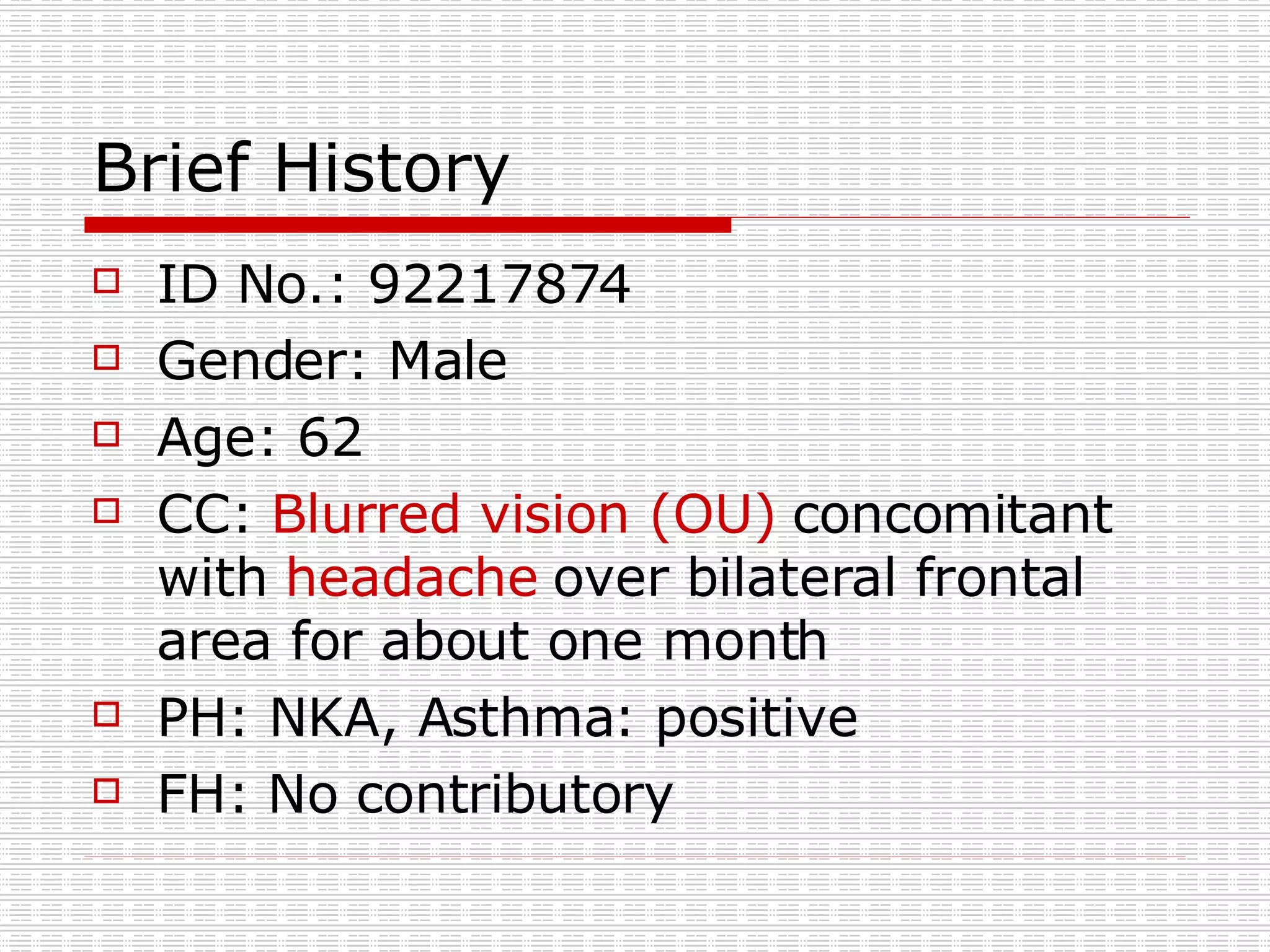
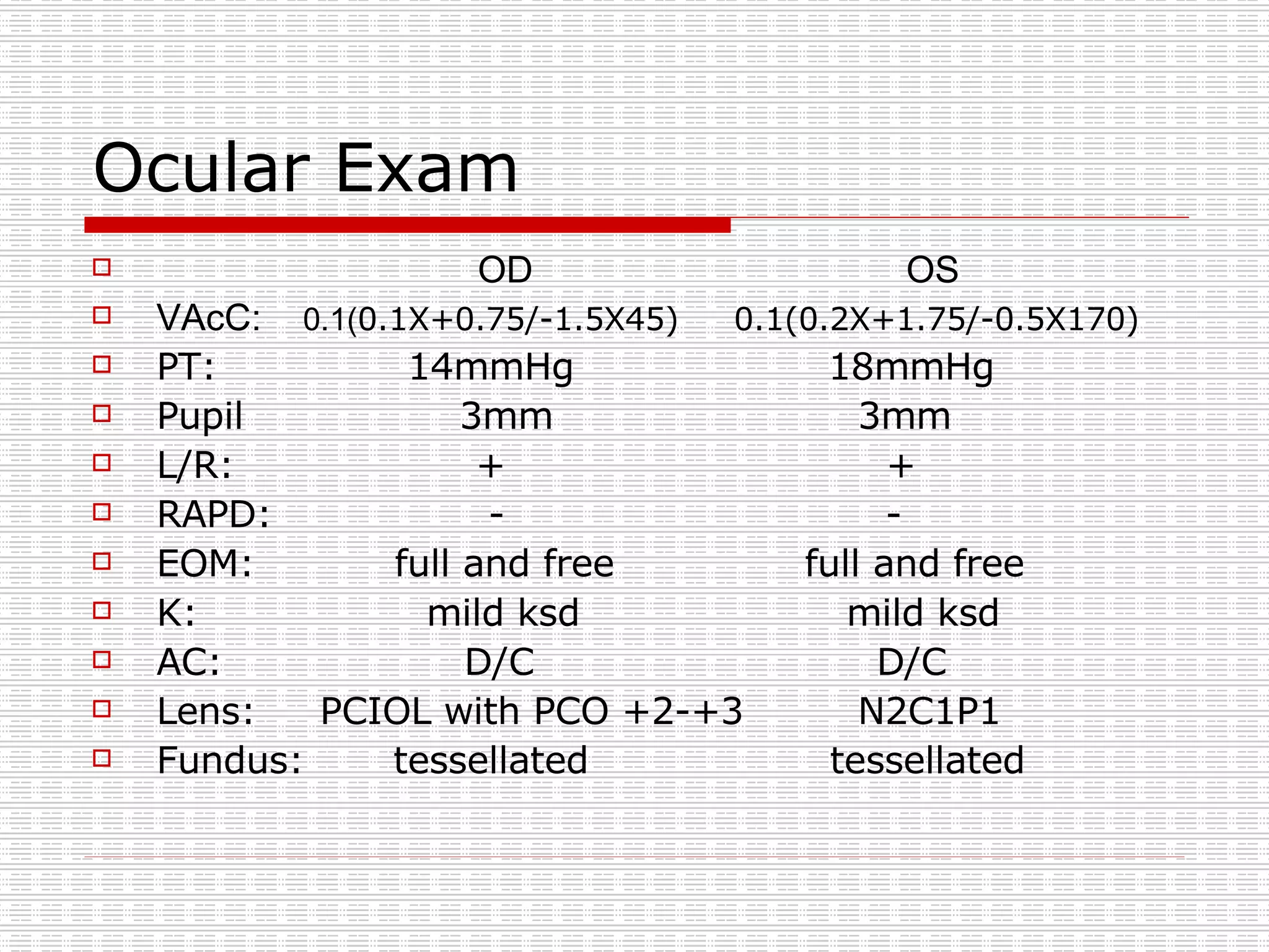
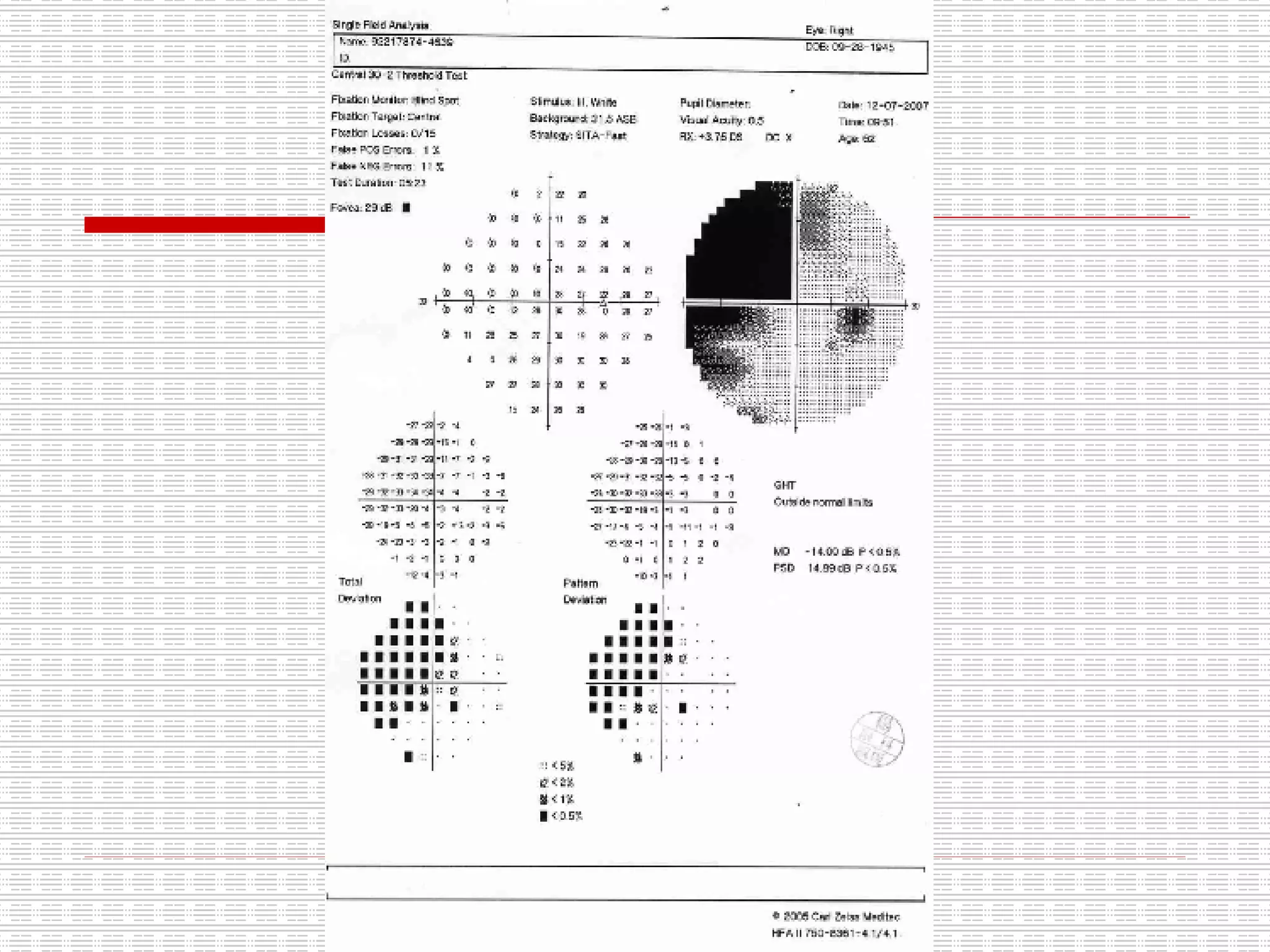
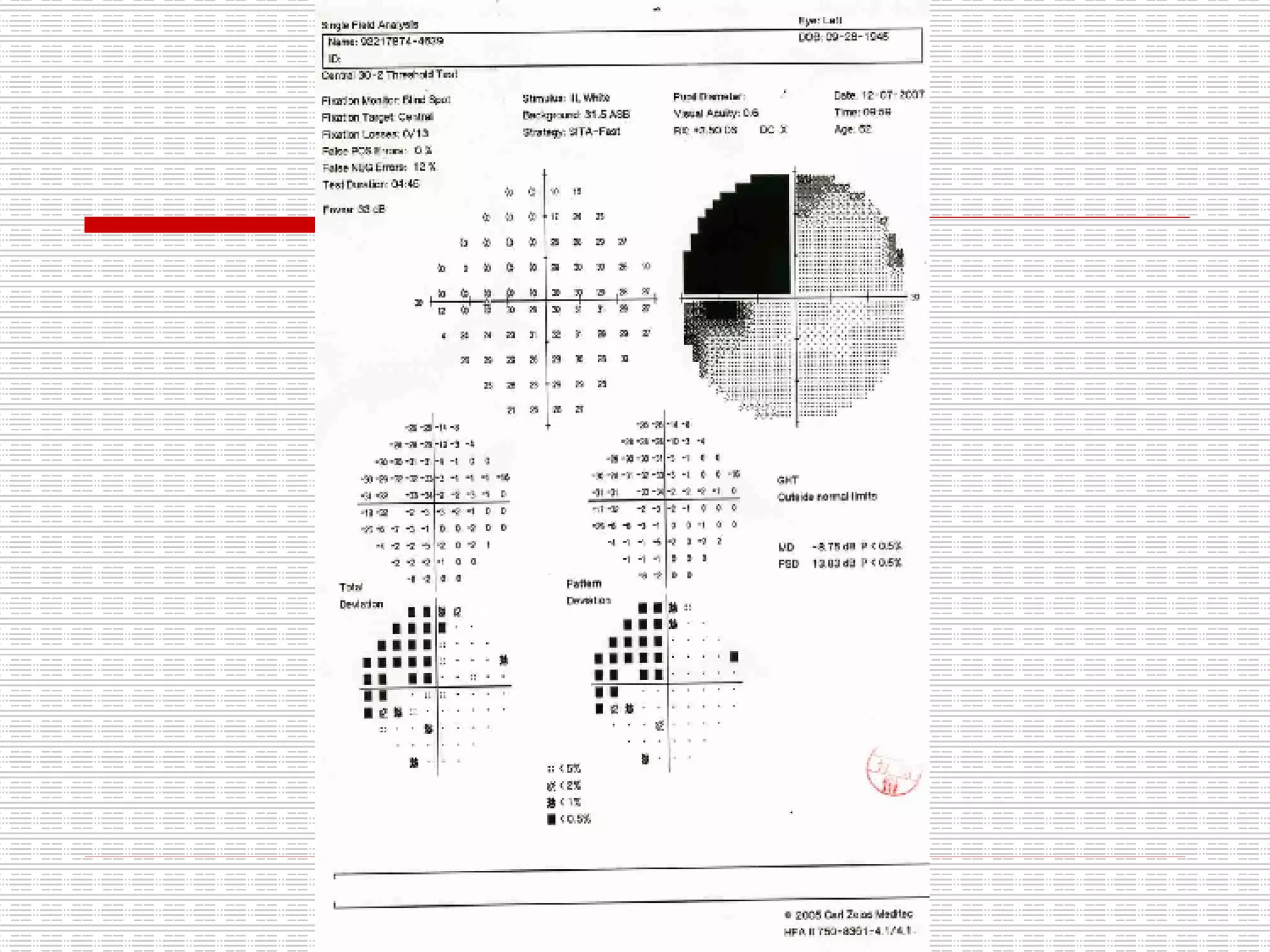
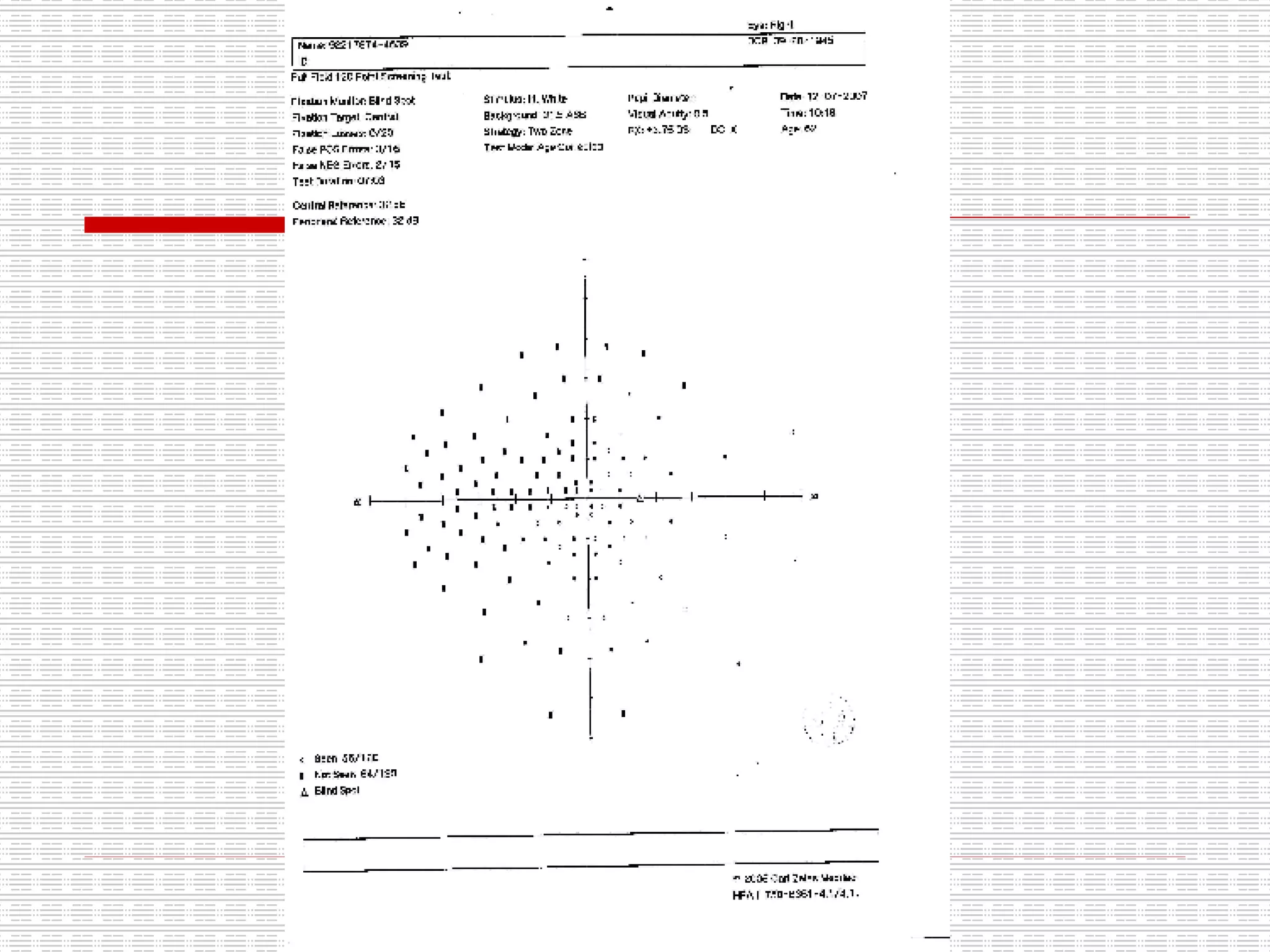
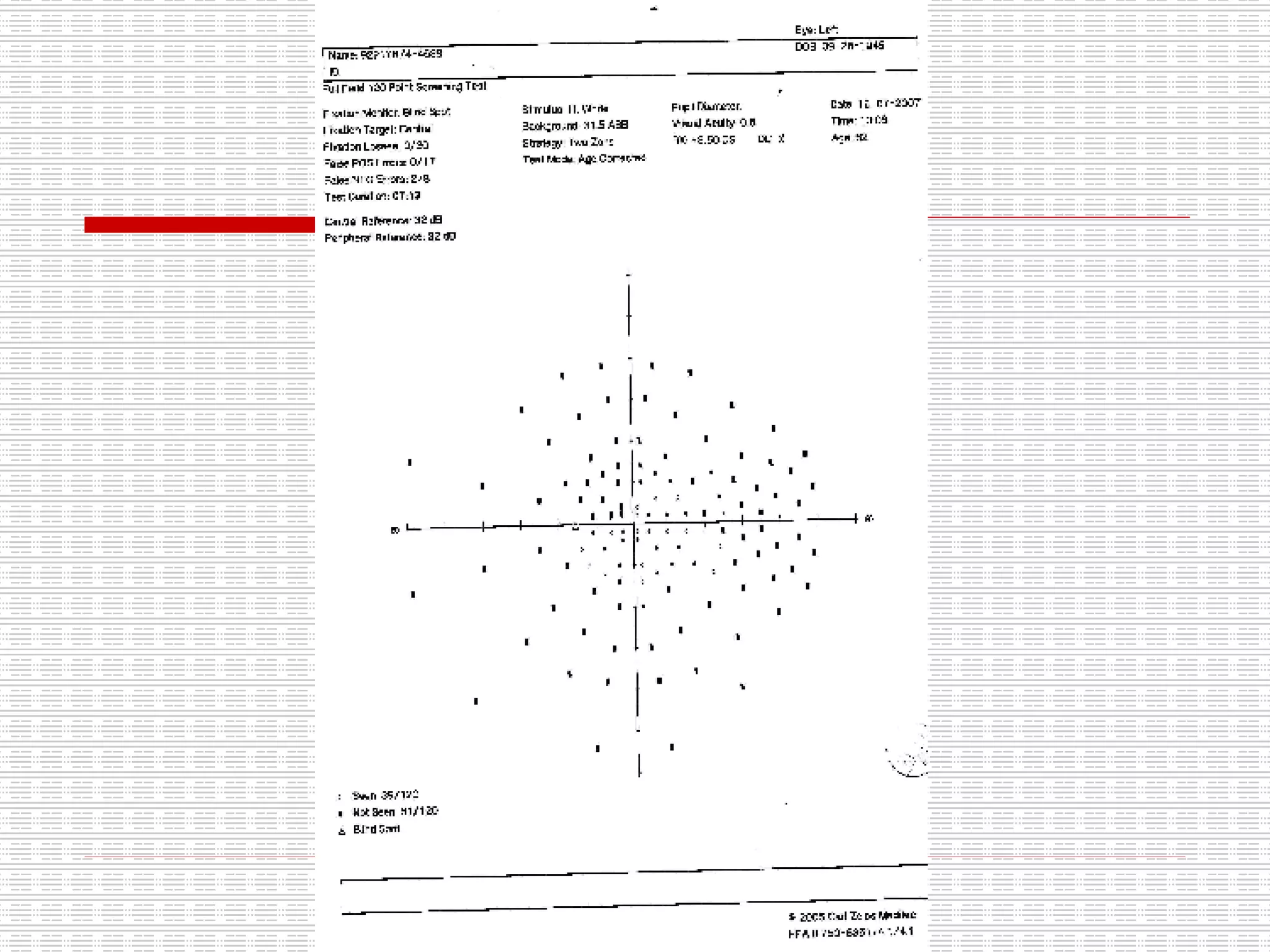
1. The patient is a 62-year-old male who presented with blurred vision and headaches for one month. Examination found mild cataracts and tessellated fundi in both eyes.
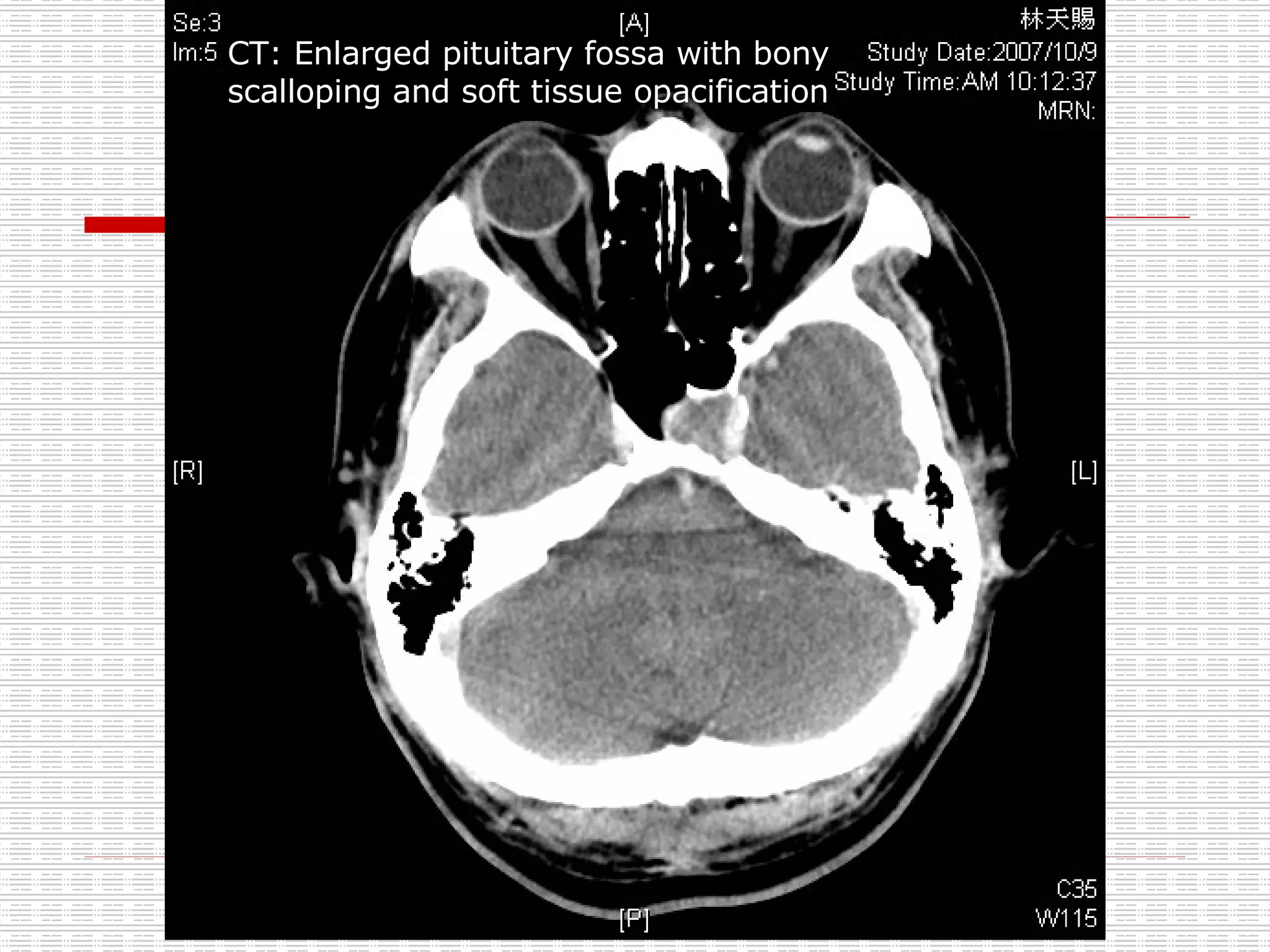
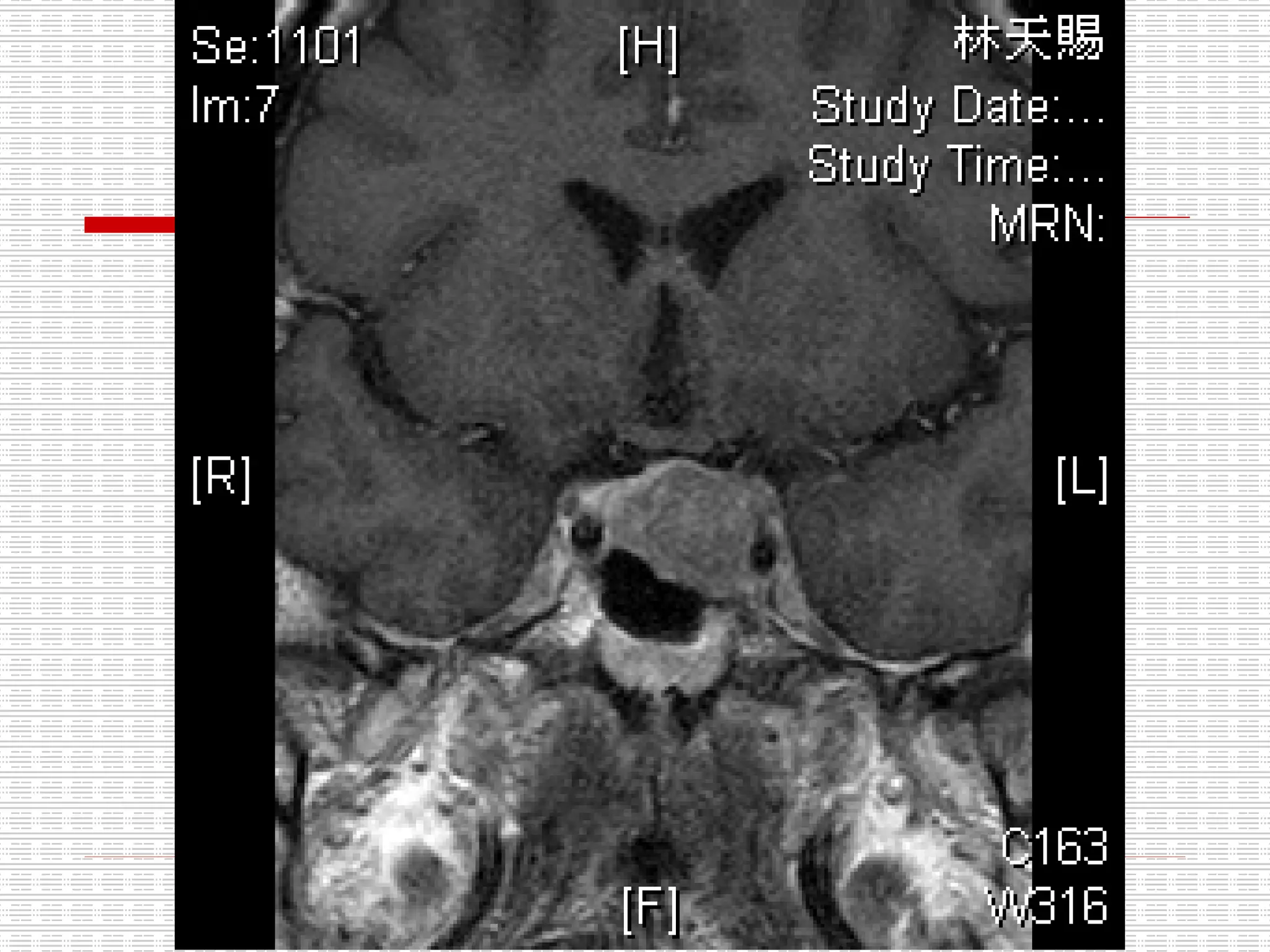
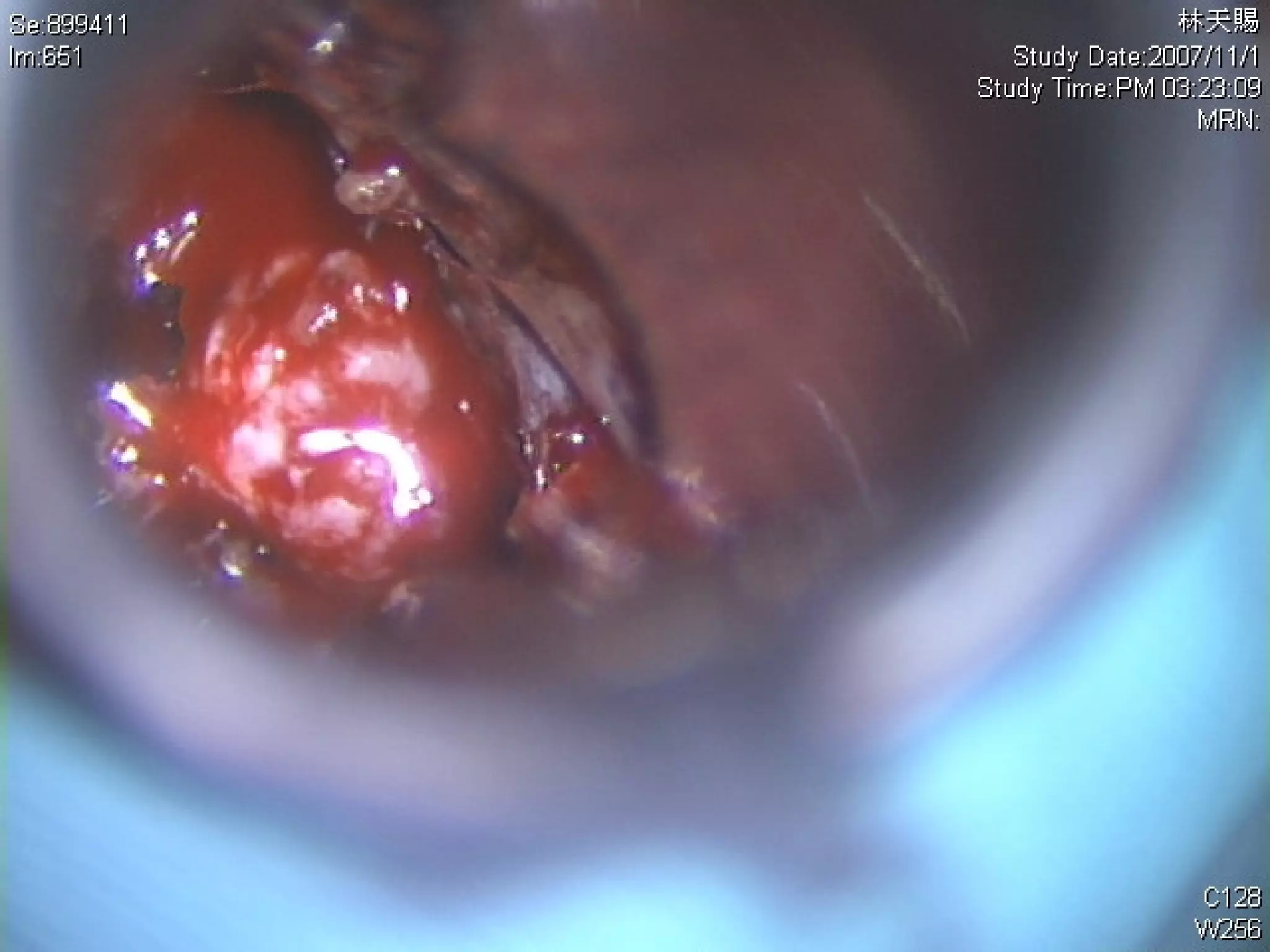
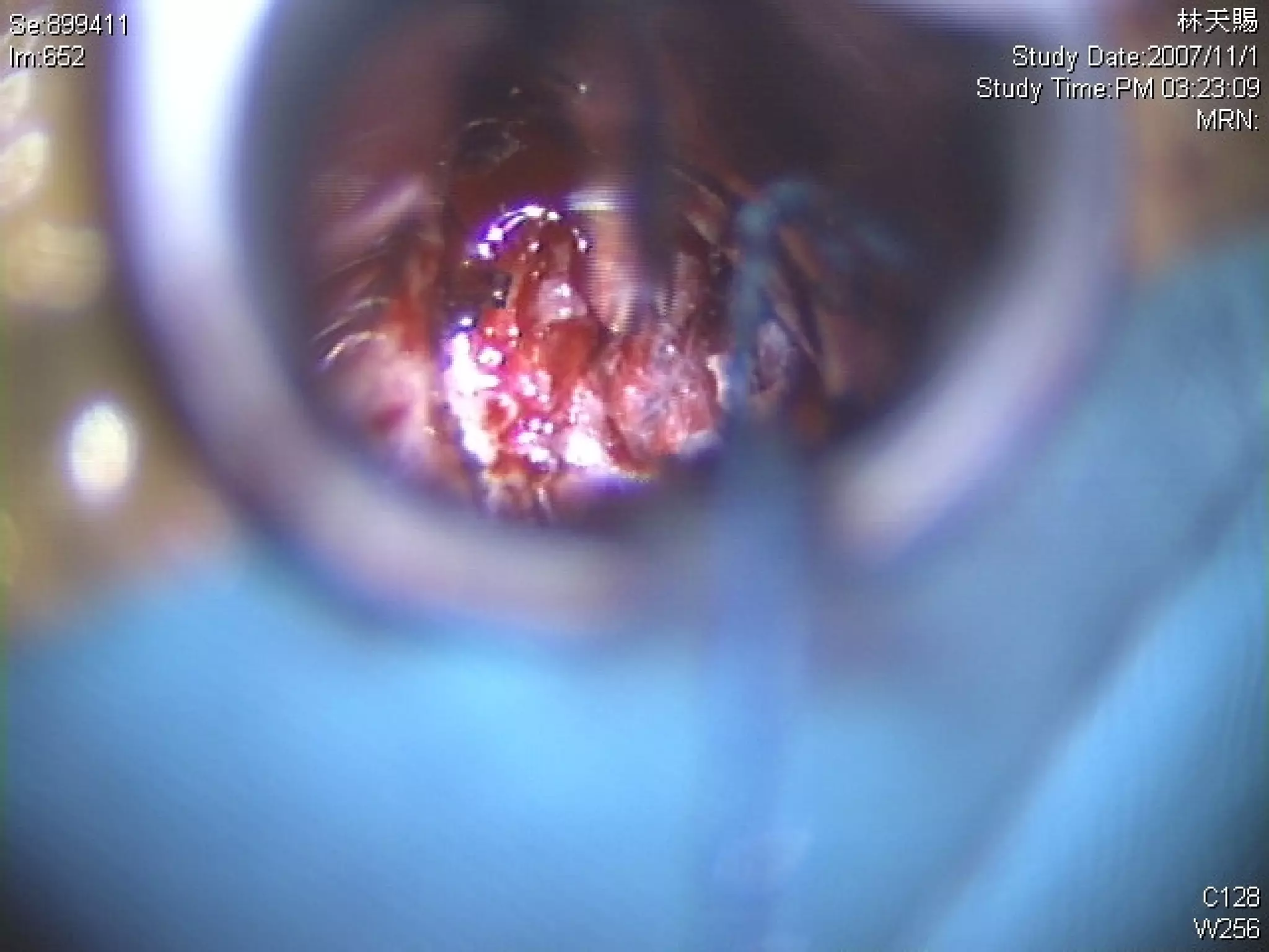
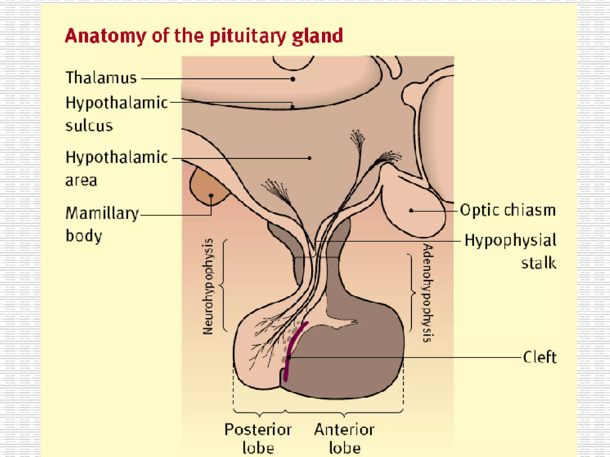
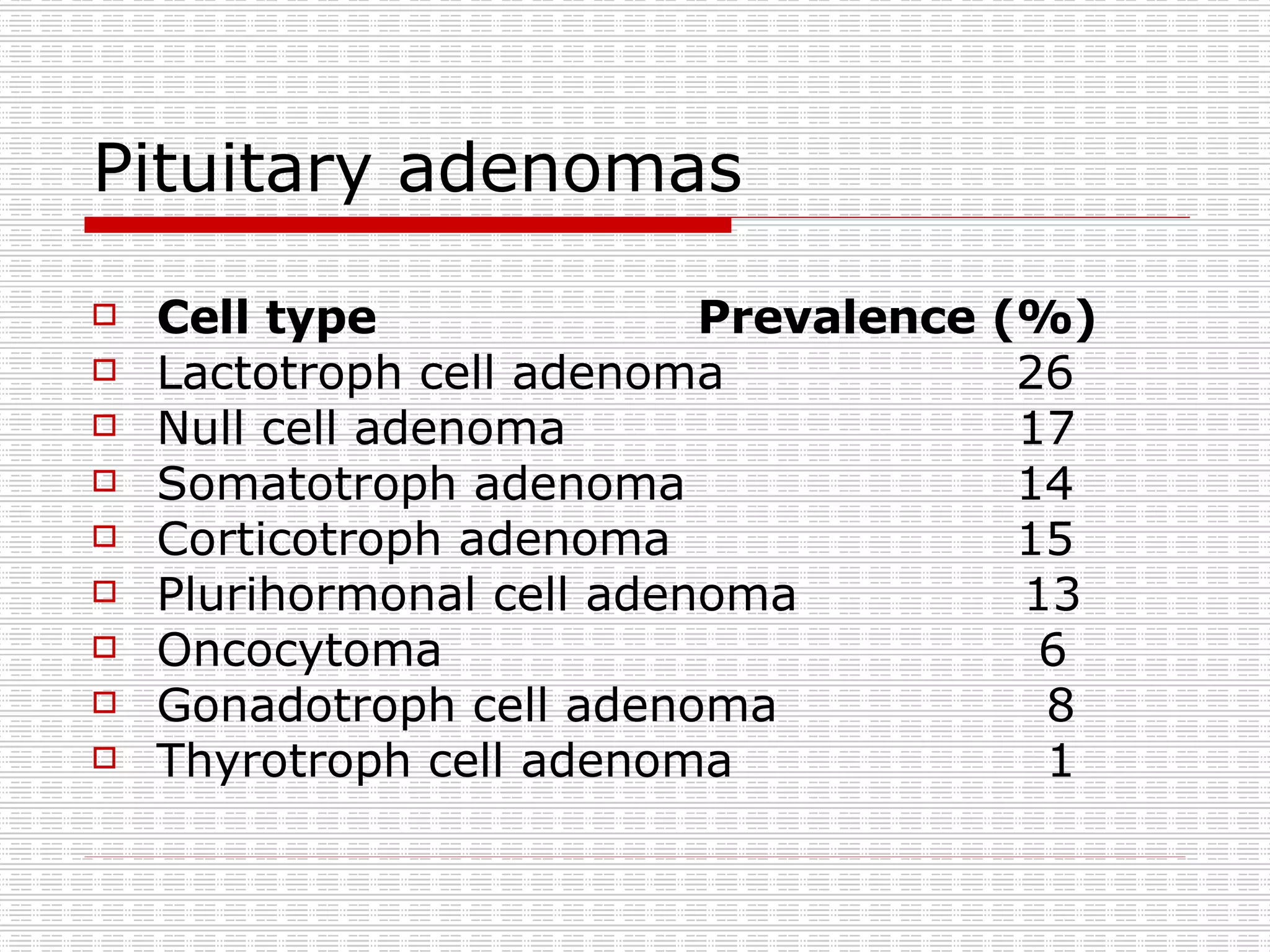
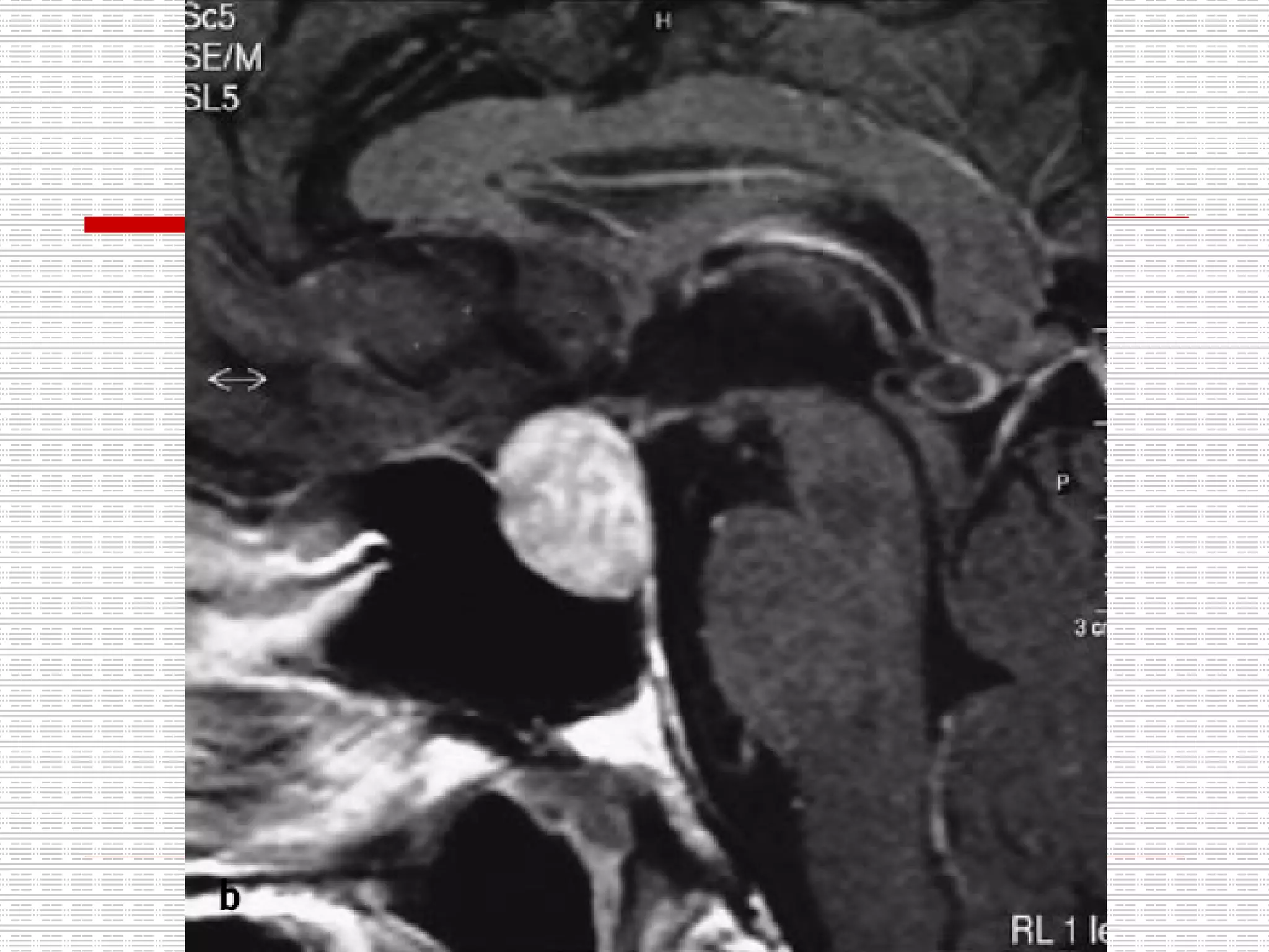
2. Imaging found an enlarged pituitary fossa with bony changes and soft tissue, suggestive of a pituitary tumor.
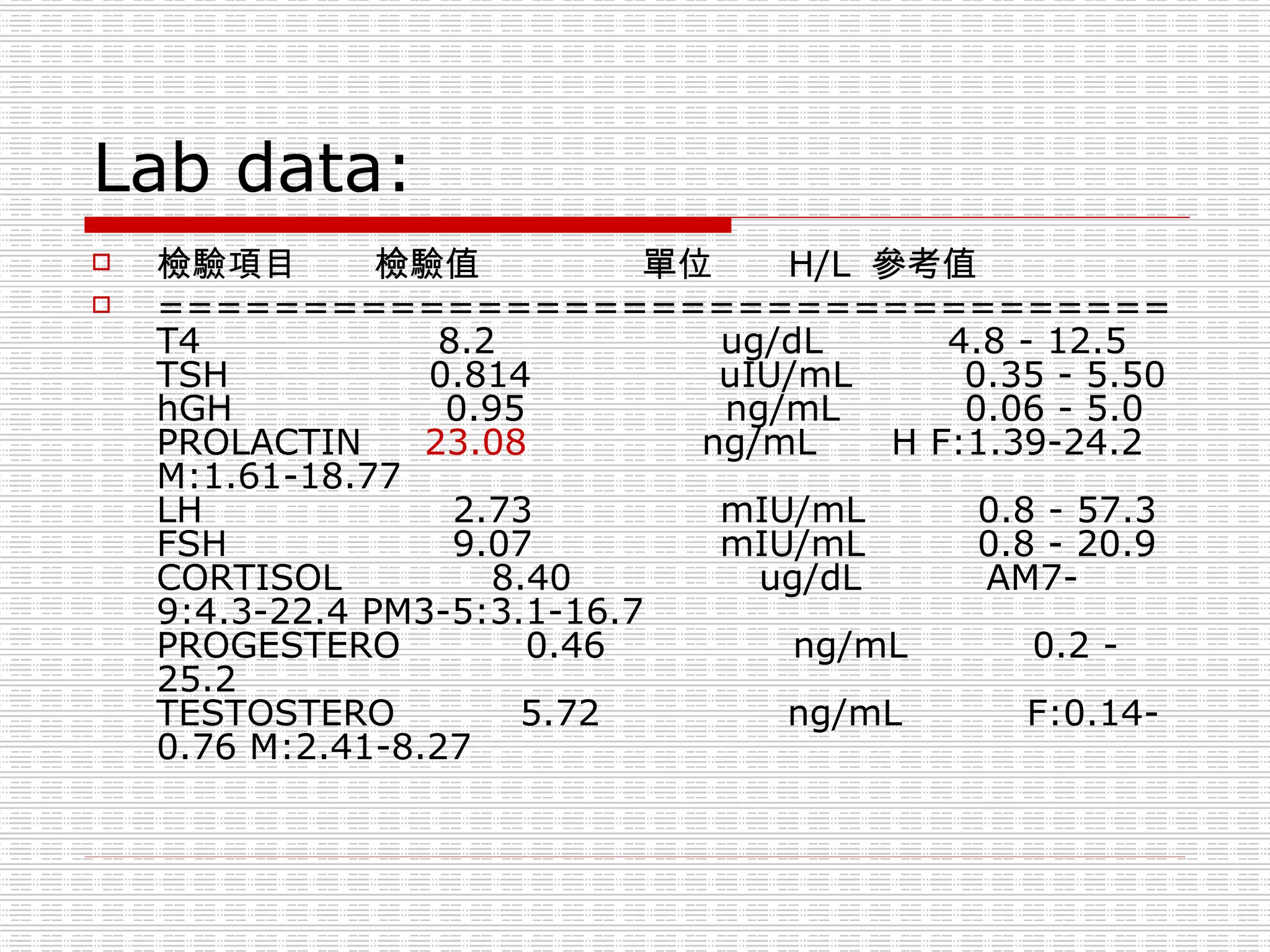
3. Laboratory tests found slightly elevated prolactin and slightly decreased TSH, consistent with a possible prolactinoma or non-functioning pituitary adenoma.
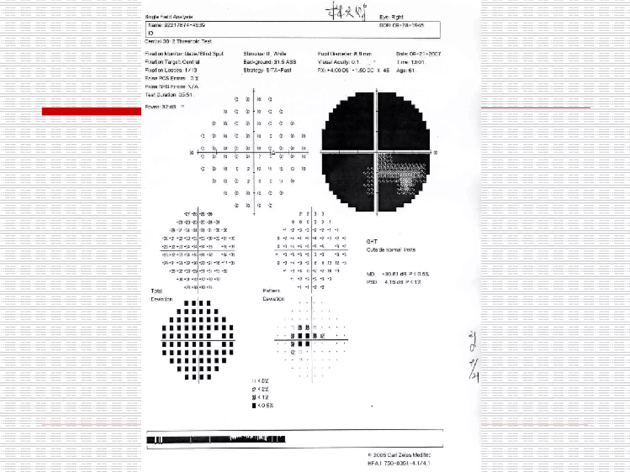
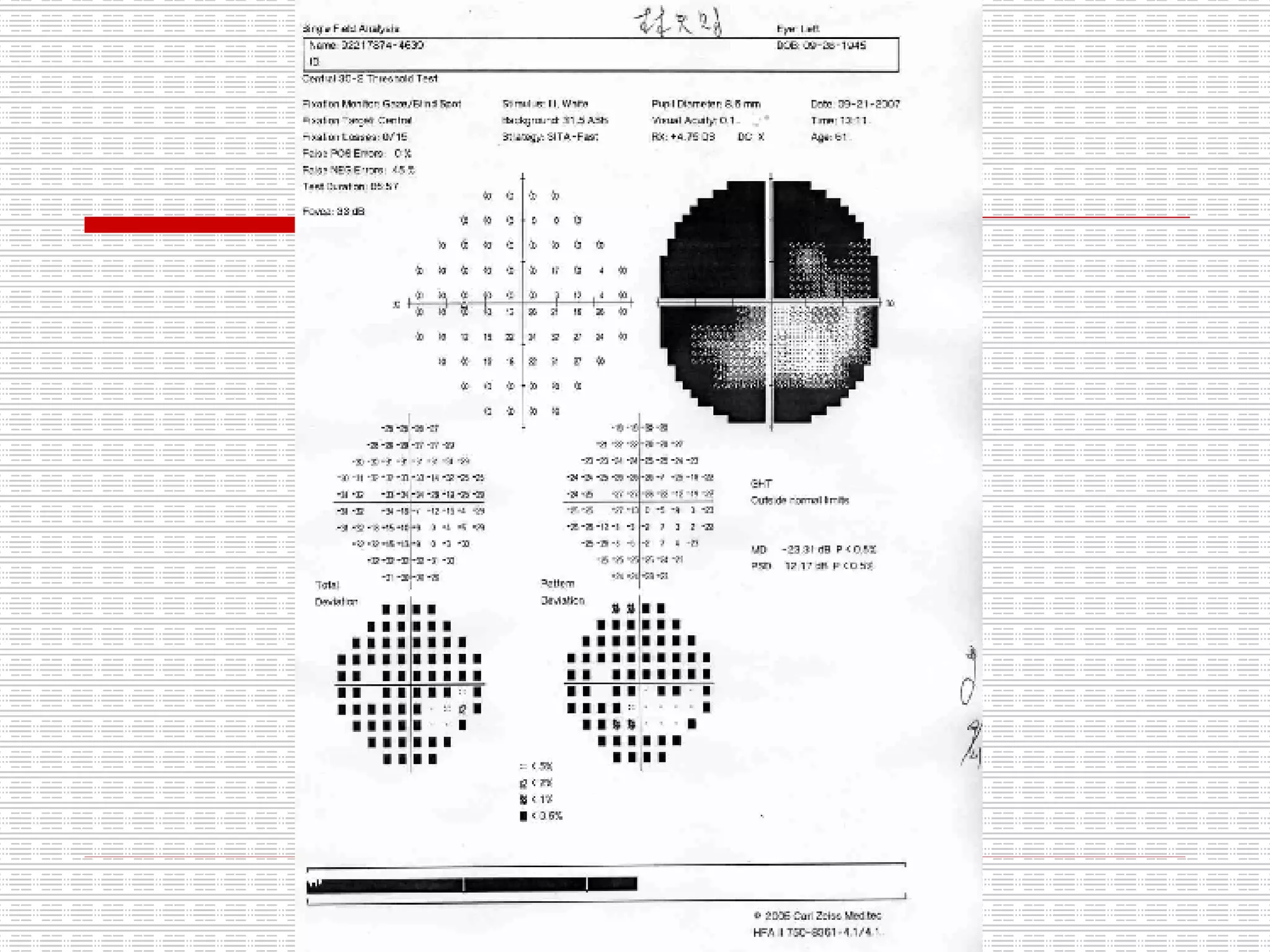
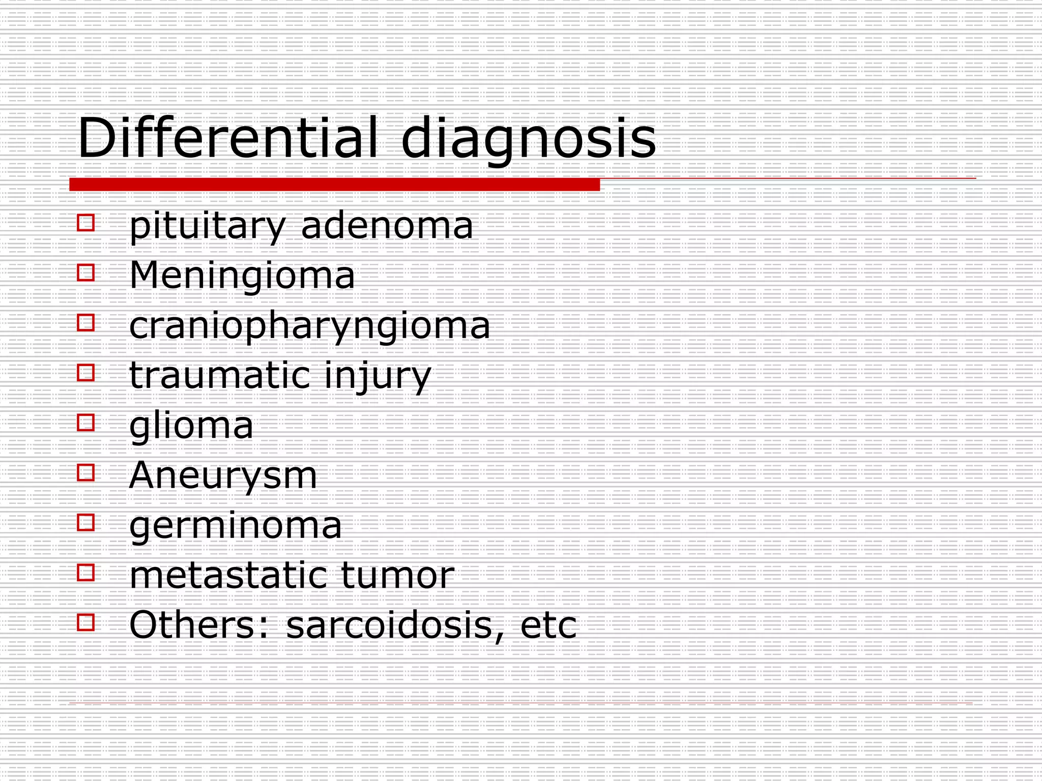
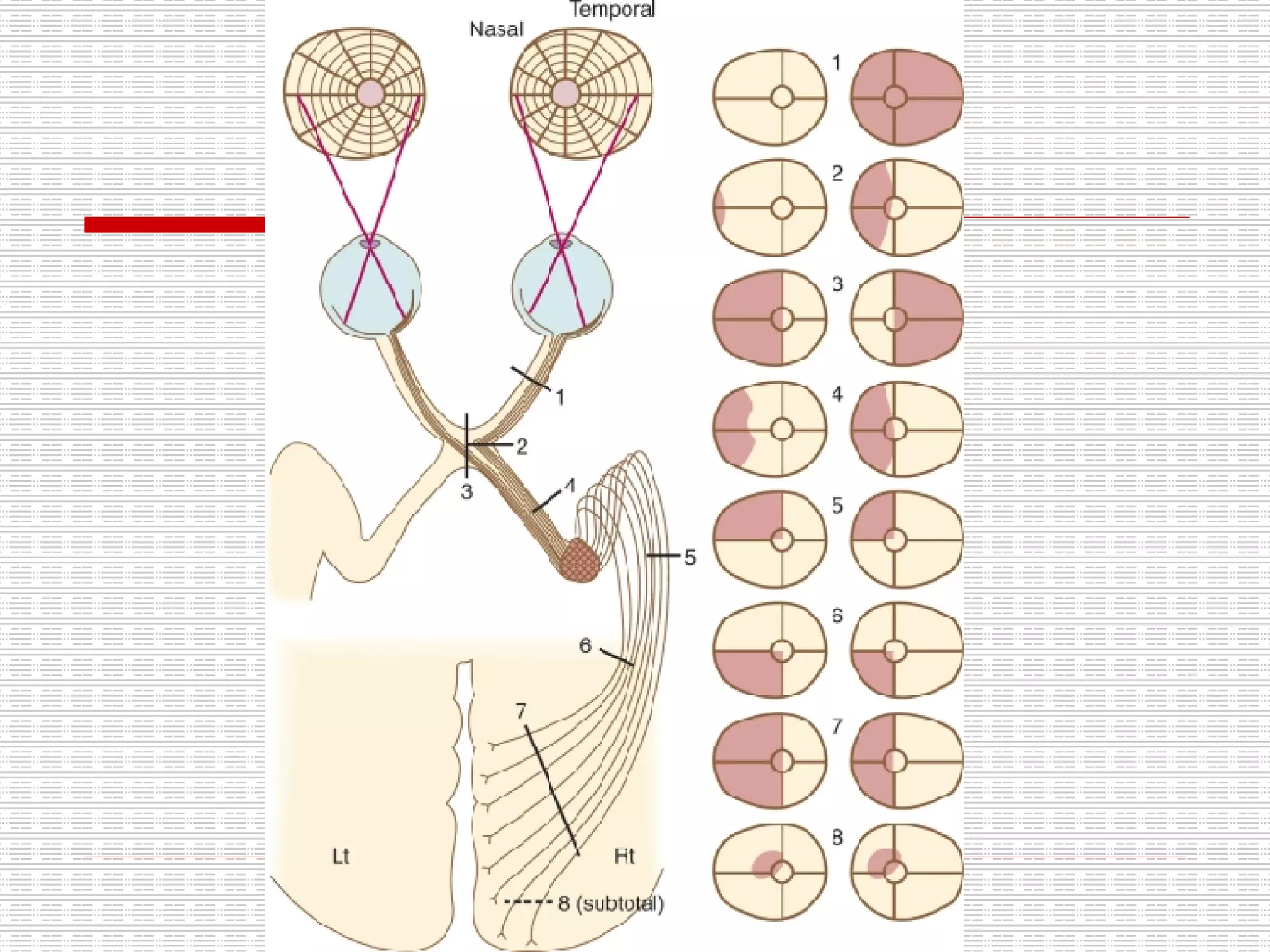
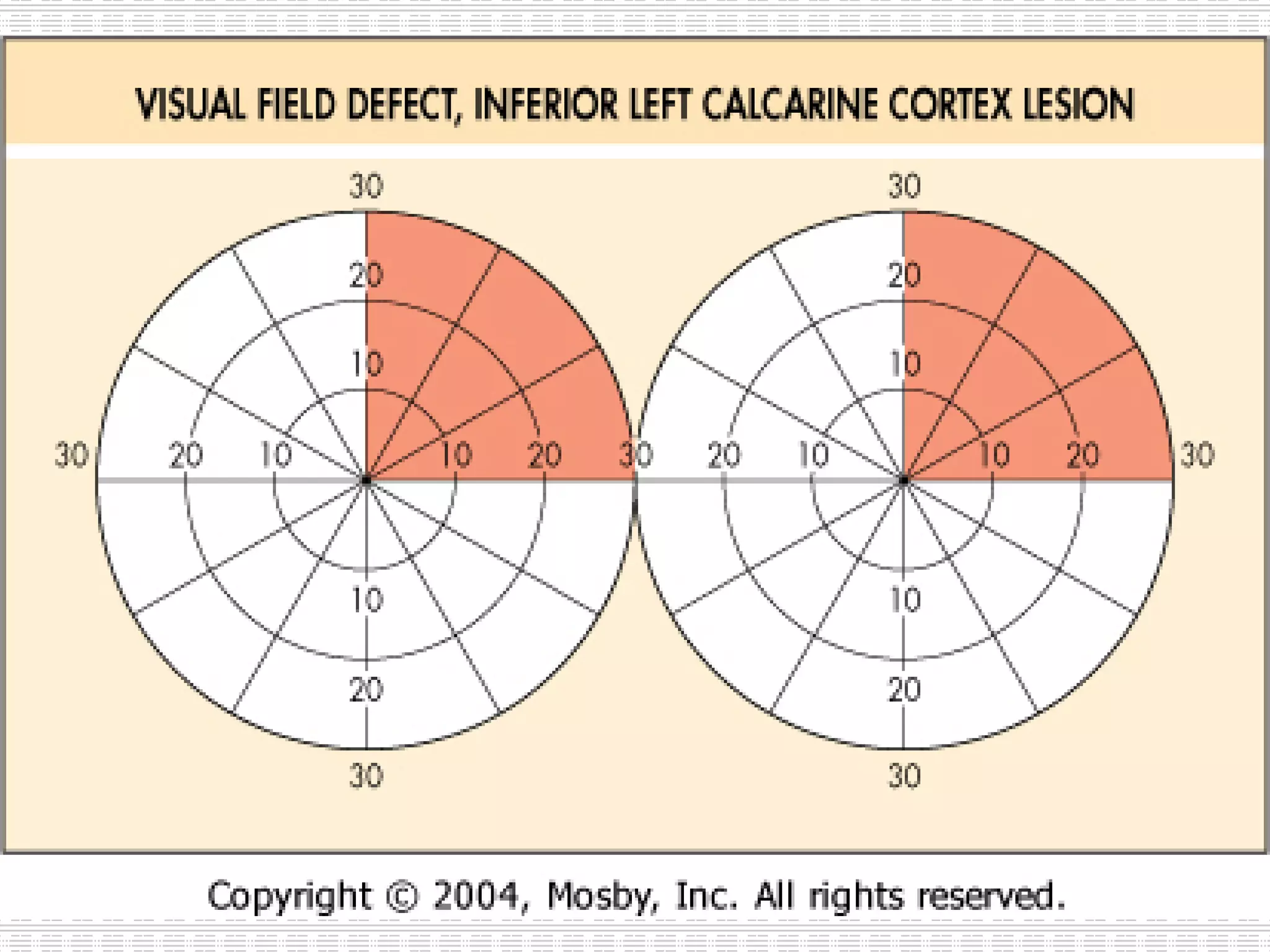
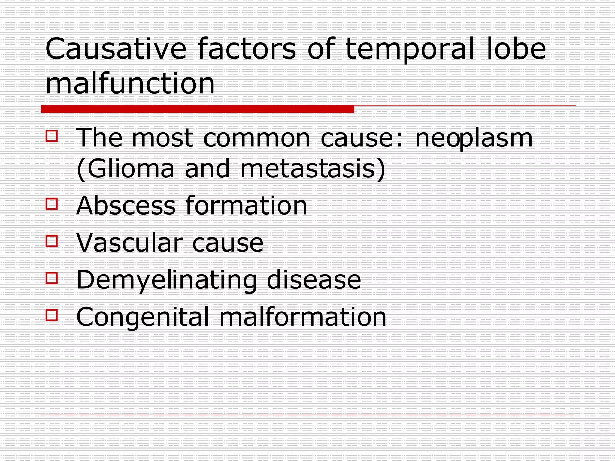
4. The differential diagnosis includes pituitary adenoma, meningioma, craniopharyngioma, or other etiologies. Follow-up with visual field testing and MRI is planned.


![Coded Agents – with UiPath SDK + LangGraph [Virtual Hands-on Workshop]](https://cdn.slidesharecdn.com/ss_thumbnails/codedagentsdeck-251215155422-5497c599-thumbnail.jpg?width=640&height=640&fit=bounds)


