Download to read offline


![*NUCLEAR IMAGING
Most commonly used methods is the cardiac
blood pool scan.
A small amount of patients blood is removed,
mixed with a radioactive isotope [cardiolite]
and reinjected. Using the ECG and other imaging
technology studies are done.](https://image.slidesharecdn.com/diagnosticscontinued2-241112045004-bc2441aa/85/CARDIOVASCULAR-SYSTEM-DIAGNOSTICS-CONTINUED-2-pptx-7-320.jpg)
![*Types
*Single photon emission computed tomography
[SPECT]
*Positron emission tomography [ PET]
*Stress perfusion imaging](https://image.slidesharecdn.com/diagnosticscontinued2-241112045004-bc2441aa/85/CARDIOVASCULAR-SYSTEM-DIAGNOSTICS-CONTINUED-2-pptx-9-320.jpg)

![COMPUTED TOMOGRAPHY
In CT scan the patient is placed in a
computerized field and electron beams [EBCT]
are used to take images to identify calcification
in coronary arteries and heart valves.](https://image.slidesharecdn.com/diagnosticscontinued2-241112045004-bc2441aa/85/CARDIOVASCULAR-SYSTEM-DIAGNOSTICS-CONTINUED-2-pptx-11-320.jpg)
![CARDIAC CATHETERIZATION
It is performed by insertion of a Radio opaque
catheter into the right or left side of the heart.
Types:
*Right sided cardiac catheterization [ARTERY ]
*Left sided cardiac catheterization [VEIN]](https://image.slidesharecdn.com/diagnosticscontinued2-241112045004-bc2441aa/85/CARDIOVASCULAR-SYSTEM-DIAGNOSTICS-CONTINUED-2-pptx-12-320.jpg)

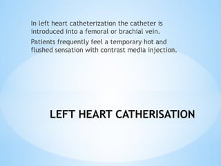
![RIGHT HEART CATHERISATION
The catheter is then inserted into the aorta then
to the pulmonary artery and pressures are
recorded. The catheter is then wedged or lodged
in position. [pulmonary artery wedge position]
This obstructs the flow and pressure from the
right side of the heart and looks forward through
the capillary bed to the pressure of the left side
of the heart.](https://image.slidesharecdn.com/diagnosticscontinued2-241112045004-bc2441aa/85/CARDIOVASCULAR-SYSTEM-DIAGNOSTICS-CONTINUED-2-pptx-14-320.jpg)


![Arterial Puncture
*Access is easiest from right side of patient due to
aortic bend
*Puncture is generally done via the femoral artery
*Alternative sites include the radial and brachial
arteries of the arm
*Catheter introduction After puncture of femoral,
radial or brachial artery (primarily on right side of
patient), a catheter is advanced into the aorta and
then the coronary arteries
*Contrast medium [Iodine] is introduced and Xray
images are taken
*If any blockage identified, it is removed and stents
are placed.[coronary angioplasty]](https://image.slidesharecdn.com/diagnosticscontinued2-241112045004-bc2441aa/85/CARDIOVASCULAR-SYSTEM-DIAGNOSTICS-CONTINUED-2-pptx-21-320.jpg)
![TMT- TREAD MILL TEST
Cardiac stress test is used to measure the hearts
ability to respond to external stress in a
clinically controlled status.
It compares the hearts activity during rest and
stress induced [exercise or drug induced]
Exercise is done using a tread mill [slope and
speed is adjusted]or stair climbing and in bed
ridden patients, drugs like Dobutamine,
Adenosine, Dipyridamole is used to increase the
heart rate.
[target heart rate= 220-person’s age]](https://image.slidesharecdn.com/diagnosticscontinued2-241112045004-bc2441aa/85/CARDIOVASCULAR-SYSTEM-DIAGNOSTICS-CONTINUED-2-pptx-23-320.jpg)

![*BRUCE PROTOCOL
*During a Bruce protocol Heart rate and rating of perceived
exertion [slope and speed] are taken every minute,
*BP is taken at the end of each stage. [every 3 minutes]
*In every 3 minutes the speed and angle of treadmill/ slope
gradient si adjusted.](https://image.slidesharecdn.com/diagnosticscontinued2-241112045004-bc2441aa/85/CARDIOVASCULAR-SYSTEM-DIAGNOSTICS-CONTINUED-2-pptx-26-320.jpg)
![*The test is continued
until:
• Reach the target heart rate [220-patients age ]
• Develop complications such as chest pain or a sudden rise or
fall in BP
• ECG evidence of myocardial ischemia
*Patient should be kept under observation for 10-15
minutes after exercising or until your heart rate returns to
baseline.](https://image.slidesharecdn.com/diagnosticscontinued2-241112045004-bc2441aa/85/CARDIOVASCULAR-SYSTEM-DIAGNOSTICS-CONTINUED-2-pptx-27-320.jpg)

The document provides an overview of various cardiac diagnostic procedures including echocardiography, cardiac catheterization, and coronary angiography. It details the types of tests, their purposes, contraindications, and potential complications. Additionally, it discusses preparations for these tests and the methodologies used, such as stress tests and imaging techniques.


![CARDIOVASCULAR SYSTEM new [Autosaved].pptx](https://cdn.slidesharecdn.com/ss_thumbnails/cardiovascularsystemnewautosaved-240711150130-2b5a8369-thumbnail.jpg?width=640&height=640&fit=bounds)


