Download to read offline

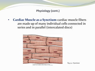
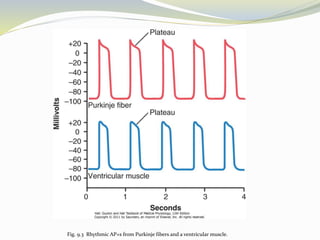
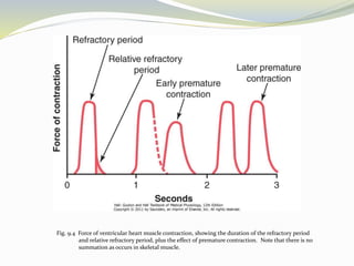
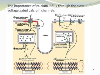
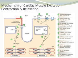
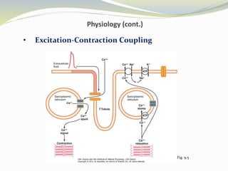

The document summarizes key aspects of cardiac muscle physiology and the function of the heart as a pump. It describes the structure of cardiac muscle fibers arranged in a syncytium connected by intercalated discs. It explains that the cardiac muscle action potential is caused by the opening of fast sodium and slow calcium channels, resulting in a long plateau phase. The refractory period prevents premature stimulation from causing contraction. Calcium influx through slow calcium channels triggers further calcium release from the sarcoplasmic reticulum, initiating muscle contraction.


