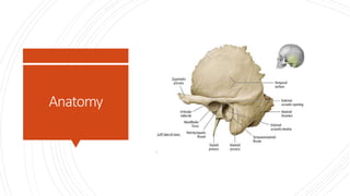
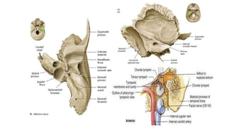
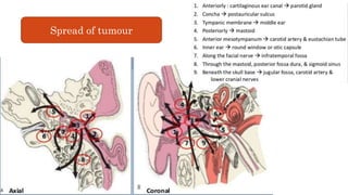
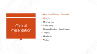
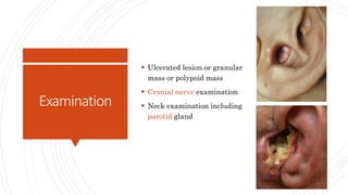
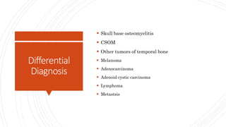
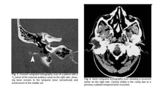
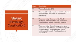
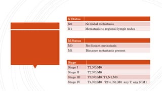
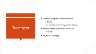
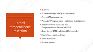
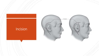
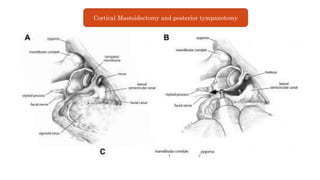
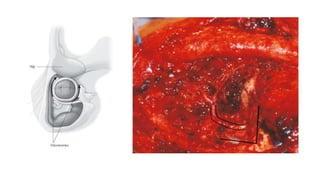
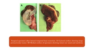
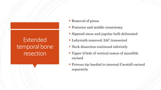
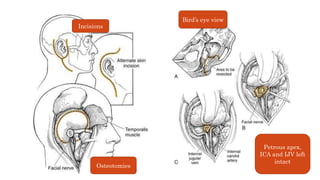
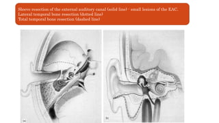
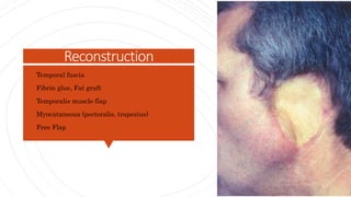
Carcinoma Temporal Bone
Dr Tabeer Arif
Layout
 Introduction
 Relevant Anatomy
 Etiology
 Clinical Presentation
 Differential diagnosis
 Investigations
 Treatment
 Complications
 Outcome
Introduction
 Rare, rapidly progressive disease with limited
therapeutic success
 Female predominance
 Malignant temporal bone tumors < 0.2% of all
head and neck tumors
 Most common (80%) of all primary Temporal bone
tumors – SCC
Anatomy
Spread of tumour
Aetiology
 Chronic Suppurative Otitis Media
 Radiation
 Familial disposition for skin SCC
 HPV – (jin et all – 1997)
 Ultraviolet radiation, chlorinated disinfectants
Clinical
Presentation
 Otorrhea (bloody, offensive)
 Otalgia
 Hearing loss
 Facial palsy
 External Auditory Canal lesion
 Tinnitus
 Headache
 Vertigo
Examination
 Ulcerated lesion or granular
mass or polypoid mass
 Cranial nerve examination
 Neck examination including
parotid gland
Differential
Diagnosis
 Skull base osteomyelitis
 CSOM
 Other tumors of temporal bone
 Melanoma
 Adenocarcinoma
 Adenoid cystic carcinoma
 Lymphoma
 Metastsis
Investigations
 Deep biopsy – CT guided biopsy
 CT
 MRI
 PTA
 Angiography & Balloon Occlusion Test
 Systemic workup (for GA fitness)
Staging
(Pittsburgh
Classification)
Stage Description
T1 Tumour limited to EAC
T2 Tumour with limited erosion of EAC or <0.5cm
involvement of soft tissue on radiological
assessment
T3 Tumour eroding the osseous EAC (full
thickness) with limited (<0.5cm) soft tissue
involvement or middle ear and/or mastoid
T4 Tumour eroding the cochlear, petrous apex,
medial wall of middle ear, carotid canal, jugular
foramen or dura or extensive soft tissue
involvement (>0.5cm). Facial paralysis at
presentation
N Status
N0 No nodal metastasis
N1 Metastasis to regional lymph nodes
M Status
M0 No distant metastasis
M1 Distance metastasis present
Stage
Stage I T1,N0,M0
Stage II T2,N0,M0
Stage III T3,N0,M0 T1,N1,M0
Stage IV T4,N0,M0 T2-4, N1,M0 any T, any N M1
Treatment
 Lateral Temporal bone resection
 T1, T2
 Lesion lateral to Tympanic membrane
 Extended temporal bone resection
 T3, T4
 Adjuvant therapy
Lateral
temporalbone
resection
 Incision
 Pinna excised partially or completely
 Cortical Mastoidectomy
 Posterior Tympanotomy – extended facial recess
 Anterosuperior extension into
Temporomandibular Joint (TMJ)
 Resection of TMJ and Mandible (condyle)
 Superficial Parotidectomy
 Neck dissection
 Reconstruction
Incision
Cortical Mastoidectomy and posterior tympanotomy
Surgical specimen after lateral temporal bone resection. (A) Lateral surface showing large
periauricular tumor. (B) Medial surface of specimen showing intact ear canal and eardrum.
Extended
temporalbone
resection
 Removal of pinna
 Posterior and middle craniotomy
 Sigmoid sinus and jugular bulb delineated
 Labyrinth removed, IAC transected
 Neck dissection continued inferiorly
 Upper 2/3rds of vertical ramus of mandible
excised
 Petrous tip (medial to internal Carotid) excised
separately
Incisions
Petrous apex,
ICA and IJV left
intact
Osteotomies
Bird’s eye view
Sleeve resection of the external auditory canal (solid line) - small lesions of the EAC.
Lateral temporal bone resection (dotted line)
Total temporal bone resection (dashed line)
Managementof
Neck
 Clinically N0 – Supraomohyoid Neck dissections
 Frozen section of suspicious node +ve for tumour
– Modified Radical Neck Dissection
 Clinically palpable lymph nodes at presentation –
Radical Neck dissection
 Parotidectomy
Reconstruction
• Temporal fascia
• Fibrin glue, Fat graft
• Temporalis muscle flap
• Myocutaneous (pectoralis, trapezius)
• Free Flap
Adjuvant
therapy
 Radiotherapy
 Primary treatment – Lesion limited to pinna,
limited EAC tumour by Silva et all
 Post- op radiotherapy (60-70 Gy)
 Chemotherapy
 Debulking and chemotherapy (5FU) by knegt et all
Intracranial
extension
 Poor prognostic factor
 Dura and brain involved – resected
 Frozen section of resection margins
 Dural defect repair with Fascia Lata
Carotid
involvement
 Poor prognosis
 Adventitia involvement – tumor dissected of
artery
 Gross involvement – ligation and resection of
artery
 Neurological consequences
Palliativecare
o Distant metastasis
o ICA involvement, petrous apex involvement and
extensive Dural or brain parenchymal
involvement
 Palliative surgical resection + post op
Radiotherapy
Complications
 Cranial nerve deficit
 Facial paralysis
 Dysphagia
 Aspiration
 Speech problems
 Nutritional deficiency
 Flap failure
 CSF leak
 Meningitis
 Hearing loss
Post Op Care
 Hearing Aids – BAHA
 Facial reanimation
 Feeding tube
 Speech therapy
 Follow UP –
 2 monthly – first 2 years
 6 monthly – 3rd-5th years
 Yearly – after 5 years
 CT & MRI – 6monthly (first 2 years) and then yearly
Recurrence
 Recurrence without distant metastasis – surgical
resection and post op radiotherapy
 Recurrent disease with metastasis – palliative
care
Outcome
 80-100% 5 year survival rates in stage I & II
 50% - stage III
 15-42% - stage IV
Thank You

Carcinoma temporal bone

  • 1.
  • 2.
    Layout  Introduction  RelevantAnatomy  Etiology  Clinical Presentation  Differential diagnosis  Investigations  Treatment  Complications  Outcome
  • 3.
    Introduction  Rare, rapidlyprogressive disease with limited therapeutic success  Female predominance  Malignant temporal bone tumors < 0.2% of all head and neck tumors  Most common (80%) of all primary Temporal bone tumors – SCC
  • 4.
  • 6.
  • 7.
    Aetiology  Chronic SuppurativeOtitis Media  Radiation  Familial disposition for skin SCC  HPV – (jin et all – 1997)  Ultraviolet radiation, chlorinated disinfectants
  • 8.
    Clinical Presentation  Otorrhea (bloody,offensive)  Otalgia  Hearing loss  Facial palsy  External Auditory Canal lesion  Tinnitus  Headache  Vertigo
  • 9.
    Examination  Ulcerated lesionor granular mass or polypoid mass  Cranial nerve examination  Neck examination including parotid gland
  • 10.
    Differential Diagnosis  Skull baseosteomyelitis  CSOM  Other tumors of temporal bone  Melanoma  Adenocarcinoma  Adenoid cystic carcinoma  Lymphoma  Metastsis
  • 11.
    Investigations  Deep biopsy– CT guided biopsy  CT  MRI  PTA  Angiography & Balloon Occlusion Test  Systemic workup (for GA fitness)
  • 14.
    Staging (Pittsburgh Classification) Stage Description T1 Tumourlimited to EAC T2 Tumour with limited erosion of EAC or <0.5cm involvement of soft tissue on radiological assessment T3 Tumour eroding the osseous EAC (full thickness) with limited (<0.5cm) soft tissue involvement or middle ear and/or mastoid T4 Tumour eroding the cochlear, petrous apex, medial wall of middle ear, carotid canal, jugular foramen or dura or extensive soft tissue involvement (>0.5cm). Facial paralysis at presentation
  • 15.
    N Status N0 Nonodal metastasis N1 Metastasis to regional lymph nodes M Status M0 No distant metastasis M1 Distance metastasis present Stage Stage I T1,N0,M0 Stage II T2,N0,M0 Stage III T3,N0,M0 T1,N1,M0 Stage IV T4,N0,M0 T2-4, N1,M0 any T, any N M1
  • 16.
    Treatment  Lateral Temporalbone resection  T1, T2  Lesion lateral to Tympanic membrane  Extended temporal bone resection  T3, T4  Adjuvant therapy
  • 17.
    Lateral temporalbone resection  Incision  Pinnaexcised partially or completely  Cortical Mastoidectomy  Posterior Tympanotomy – extended facial recess  Anterosuperior extension into Temporomandibular Joint (TMJ)  Resection of TMJ and Mandible (condyle)  Superficial Parotidectomy  Neck dissection  Reconstruction
  • 18.
  • 19.
    Cortical Mastoidectomy andposterior tympanotomy
  • 21.
    Surgical specimen afterlateral temporal bone resection. (A) Lateral surface showing large periauricular tumor. (B) Medial surface of specimen showing intact ear canal and eardrum.
  • 22.
    Extended temporalbone resection  Removal ofpinna  Posterior and middle craniotomy  Sigmoid sinus and jugular bulb delineated  Labyrinth removed, IAC transected  Neck dissection continued inferiorly  Upper 2/3rds of vertical ramus of mandible excised  Petrous tip (medial to internal Carotid) excised separately
  • 23.
    Incisions Petrous apex, ICA andIJV left intact Osteotomies Bird’s eye view
  • 24.
    Sleeve resection ofthe external auditory canal (solid line) - small lesions of the EAC. Lateral temporal bone resection (dotted line) Total temporal bone resection (dashed line)
  • 25.
    Managementof Neck  Clinically N0– Supraomohyoid Neck dissections  Frozen section of suspicious node +ve for tumour – Modified Radical Neck Dissection  Clinically palpable lymph nodes at presentation – Radical Neck dissection  Parotidectomy
  • 26.
    Reconstruction • Temporal fascia •Fibrin glue, Fat graft • Temporalis muscle flap • Myocutaneous (pectoralis, trapezius) • Free Flap
  • 27.
    Adjuvant therapy  Radiotherapy  Primarytreatment – Lesion limited to pinna, limited EAC tumour by Silva et all  Post- op radiotherapy (60-70 Gy)  Chemotherapy  Debulking and chemotherapy (5FU) by knegt et all
  • 28.
    Intracranial extension  Poor prognosticfactor  Dura and brain involved – resected  Frozen section of resection margins  Dural defect repair with Fascia Lata
  • 29.
    Carotid involvement  Poor prognosis Adventitia involvement – tumor dissected of artery  Gross involvement – ligation and resection of artery  Neurological consequences
  • 30.
    Palliativecare o Distant metastasis oICA involvement, petrous apex involvement and extensive Dural or brain parenchymal involvement  Palliative surgical resection + post op Radiotherapy
  • 31.
    Complications  Cranial nervedeficit  Facial paralysis  Dysphagia  Aspiration  Speech problems  Nutritional deficiency  Flap failure  CSF leak  Meningitis  Hearing loss
  • 32.
    Post Op Care Hearing Aids – BAHA  Facial reanimation  Feeding tube  Speech therapy  Follow UP –  2 monthly – first 2 years  6 monthly – 3rd-5th years  Yearly – after 5 years  CT & MRI – 6monthly (first 2 years) and then yearly
  • 33.
    Recurrence  Recurrence withoutdistant metastasis – surgical resection and post op radiotherapy  Recurrent disease with metastasis – palliative care
  • 34.
    Outcome  80-100% 5year survival rates in stage I & II  50% - stage III  15-42% - stage IV
  • 35.

Editor's Notes

  • #4 1:0.8 to 3:1
  • #7 Lymphatic and hematologic spread is rare Intraparotid lymph nodes are echelon nodes
  • #8 Chronic inflammation and irritation NPC and other intracranial (7 yrs after radiation, another histologic type than the cancer previously radiated for) UVR (pinna)
  • #9 More than 70%
  • #10 Exophytic mass within left EAC
  • #12 Metastatic workup (in adenocarcinoma, lymphoma etc) Gadolinium DTPA - MRI
  • #18 Incision – post auricular, pre auricular, extended into neck for ND AS – along line of middle fossa plate curving along sup wall of EAC
  • #20 Complete mastoidectomy – exposure of incus long process, facial canal V seg and lat SCC Extended facial recess FN from geniculate ganglion to SM foramen Corda sacrificed, IS joint disarticulated
  • #21  ( a ) intact facial nerve from second genu to periphery, ( b ) sino-dural angle, ( c ) glenoid fossa, ( d ) cut end of the neck of mandible, ( e ) posterior belly of digastric with removal of mastoid tip, and ( f ) remaining deep lobe of parotid
  • #22 Kutz jr et all in 2015 advocated a LTBR with TMJ excision (glenoid fossa and condyle), total parotidectomy and neck dissection for T3 and T4 lesions of Lateral temporal bone
  • #23 Includind condyle, glenoid fossa,coronoid NTTBR petrous apex not included TTBR petrous air cells, sigmoid sinus even petrous ICA
  • #24  Subtotal temporal bone resection. Incisions include a central external auditory canal core, which is sutured closed. Note the tragus can be preserved for better cosmesis. Temporal craniotomy for subtotal temporal bone resection is smaller than for a total temporal bone resection. Parotid gland with main trunk of facial nerve has been elevated from masseter muscle.  Subtotal temporal bone resection. A, Bird’s-eye view of proposed resection. Note the facial nerve can be exposed for clean sectioning in the labyrinthine segment. B,Osteotome at carotid canal first genu directed at internal auditory canal fundus. C, Specimen removed showing remaining petrous apex and otic capsule. Roman numerals denote cranial nerves.
  • #26 Superficial parotidectomy at least in LTBR, total in TTBR First echelon nodes
  • #27 Pedicled flaps (more durable) suitable for smoker, diabetic, vascular disease Free flaps- greater versatility – gold standard
  • #28 Total dose Primary site + neck
  • #29 Separate specimen
  • #32 Lumbar drains (in situ for a few post op days)