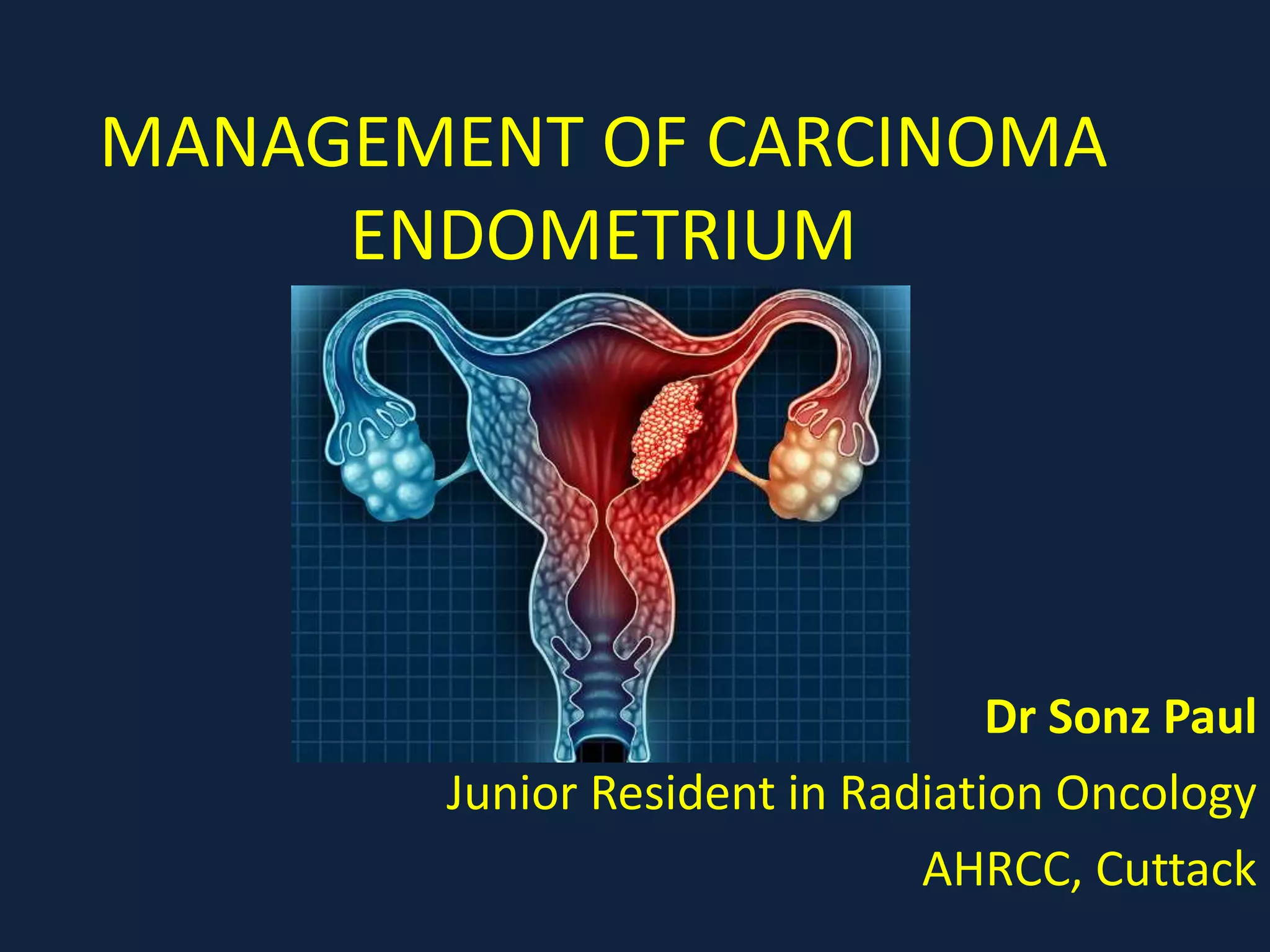
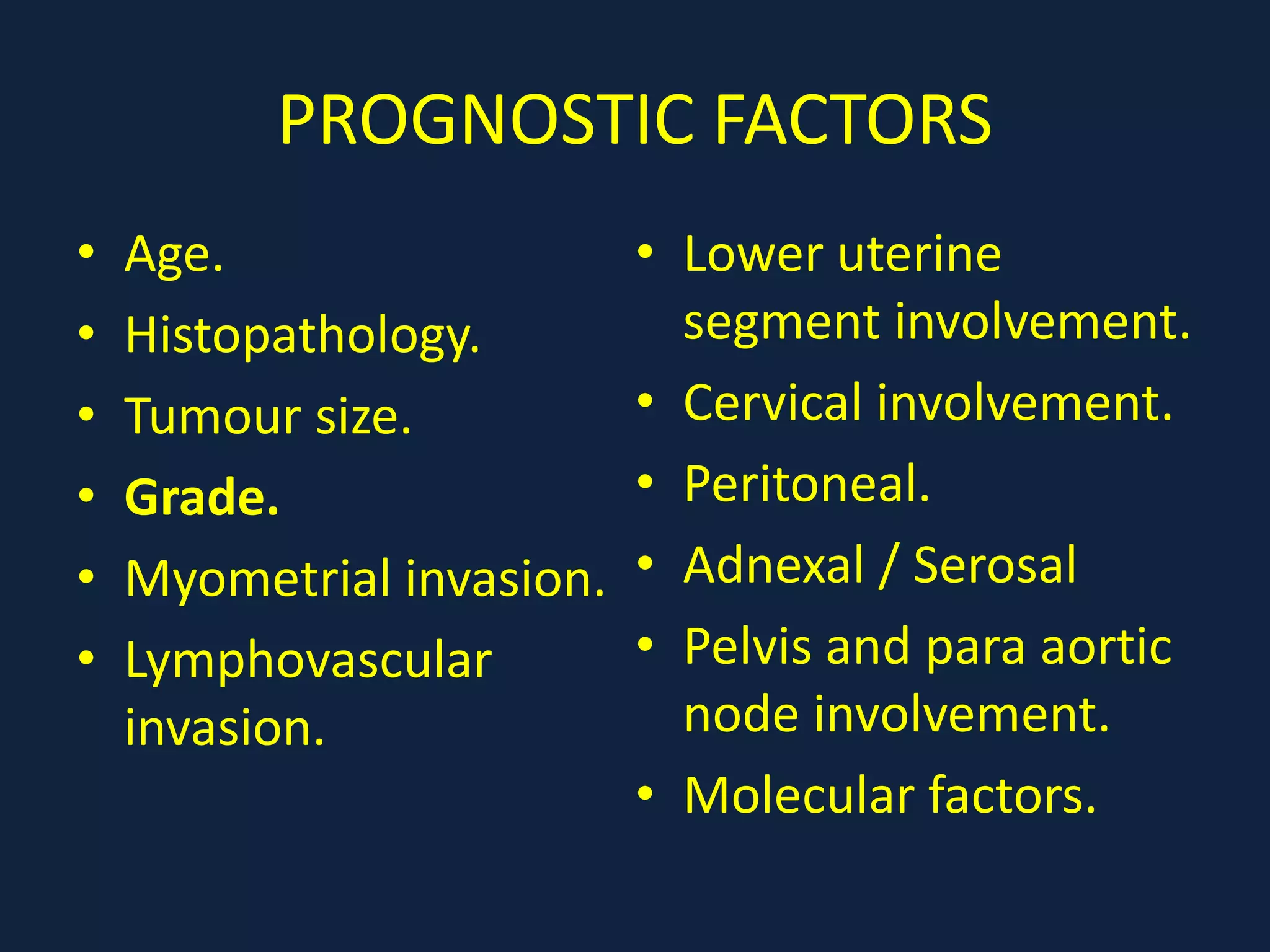
1) Carcinoma endometrium is the most common gynecologic malignancy in Western countries. Risk factors include obesity, early menarche, late menopause, nulliparity, and use of tamoxifen or combined oral contraceptives.
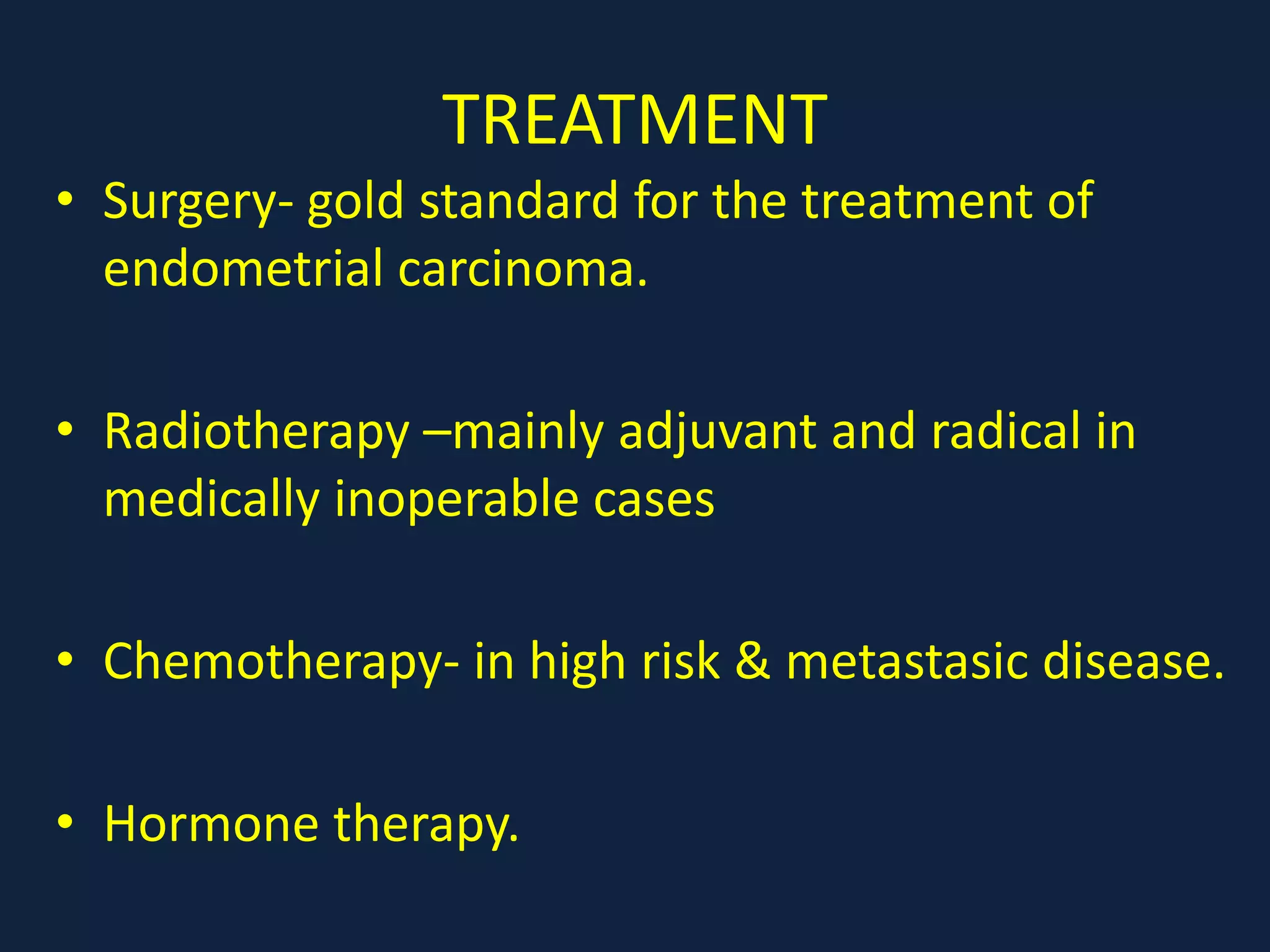
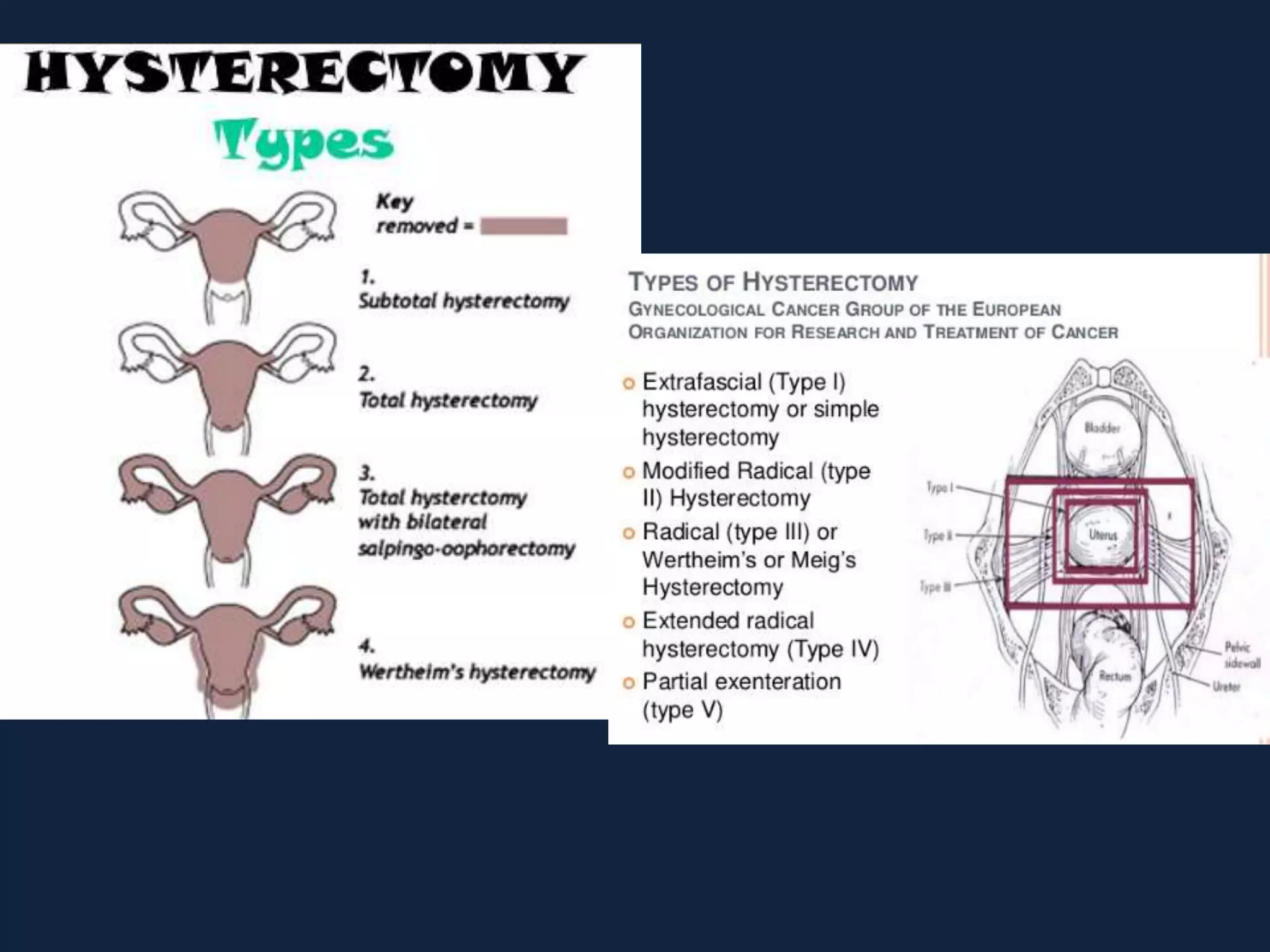
2) Treatment involves surgery (usually a hysterectomy) followed by adjuvant radiation therapy or chemotherapy depending on risk factors and staging. Pelvic radiation therapy techniques have advanced from 4-field box to intensity modulated radiation therapy (IMRT) to reduce toxicity.
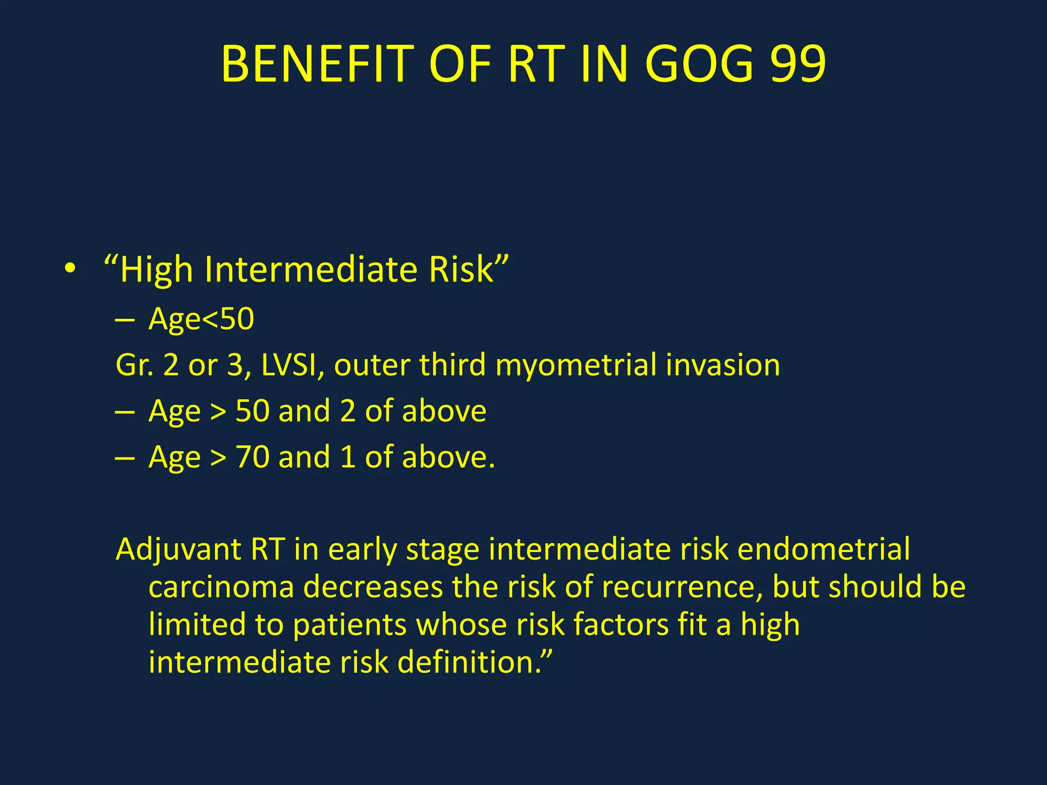
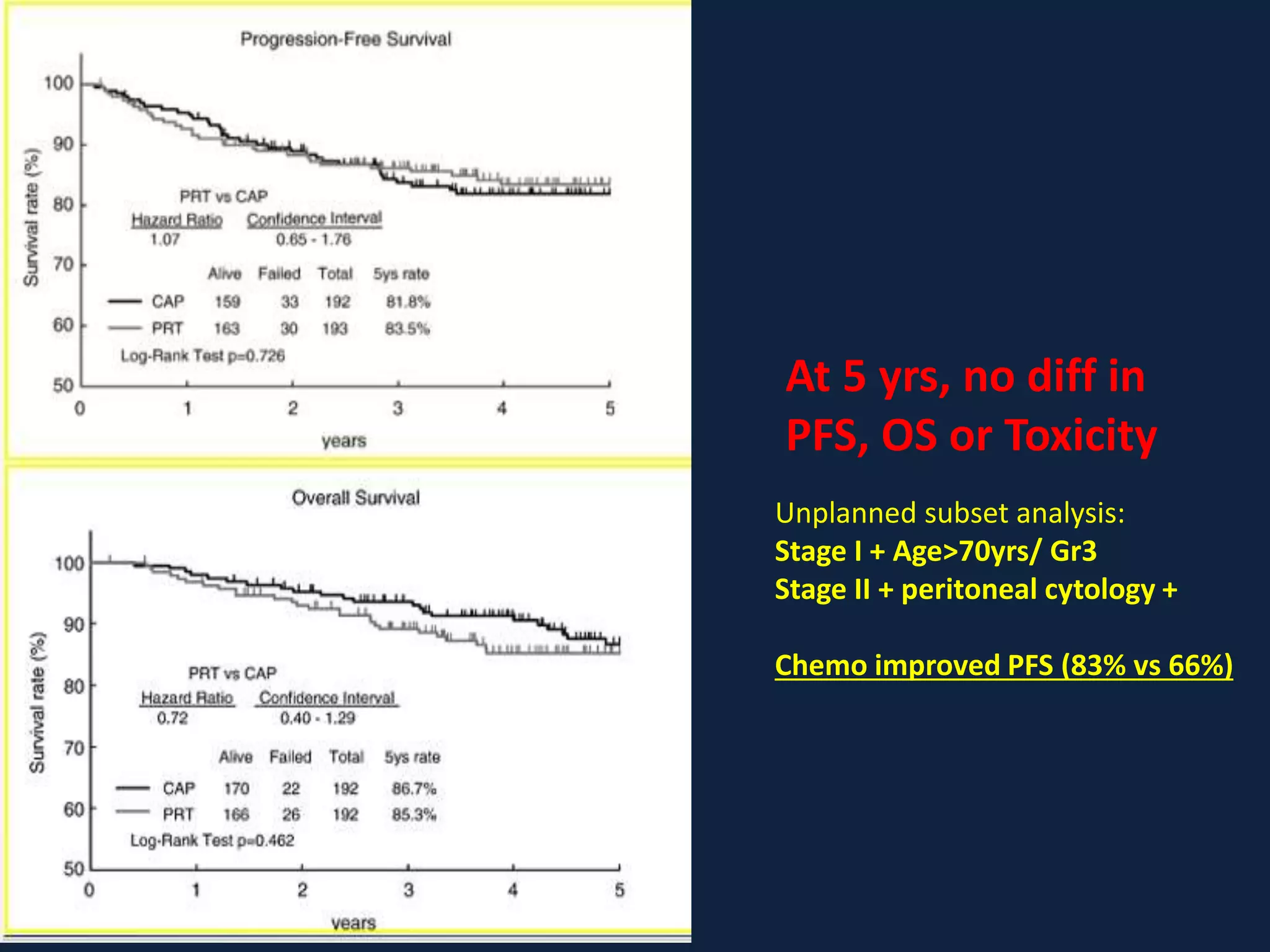
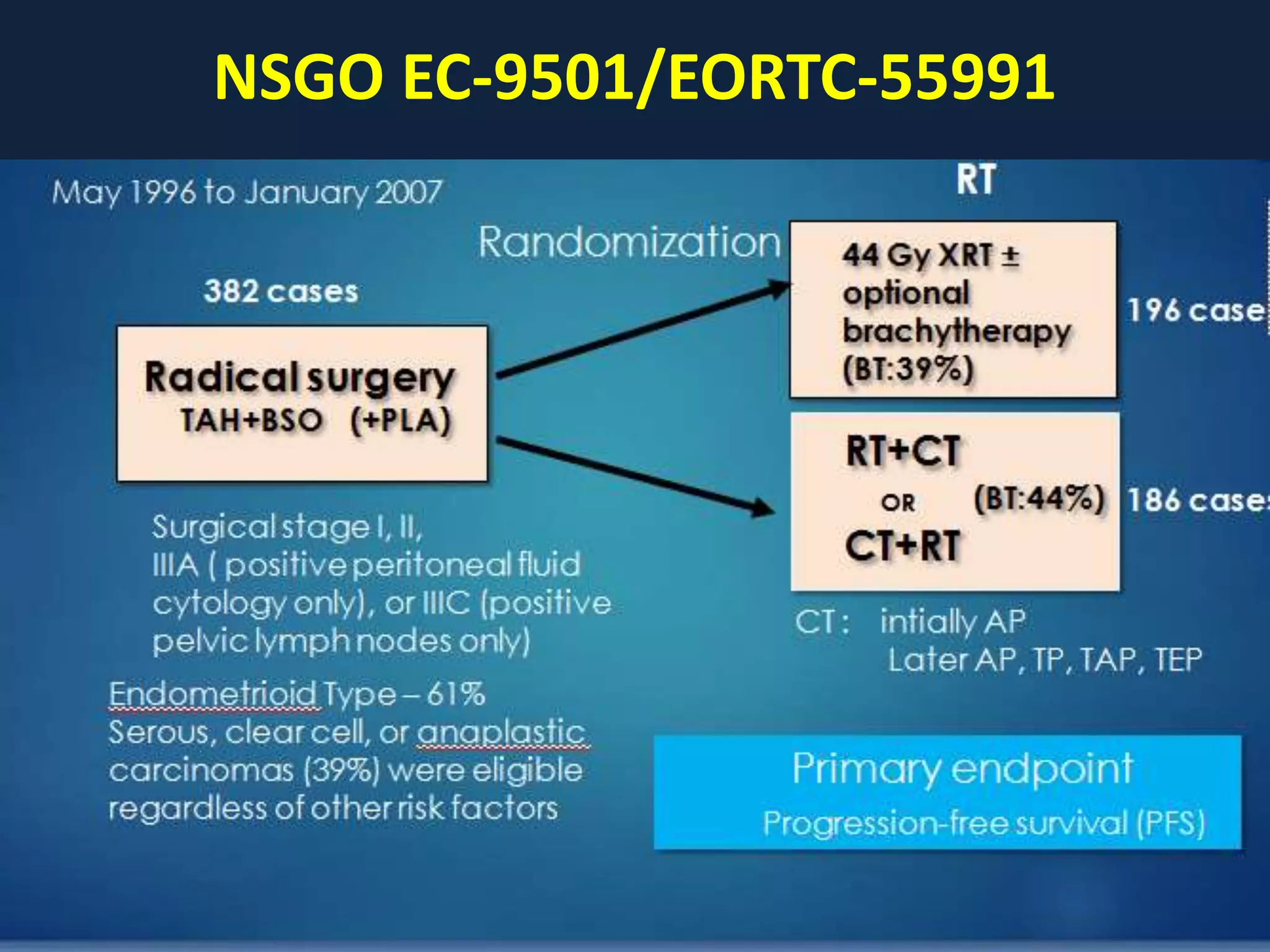
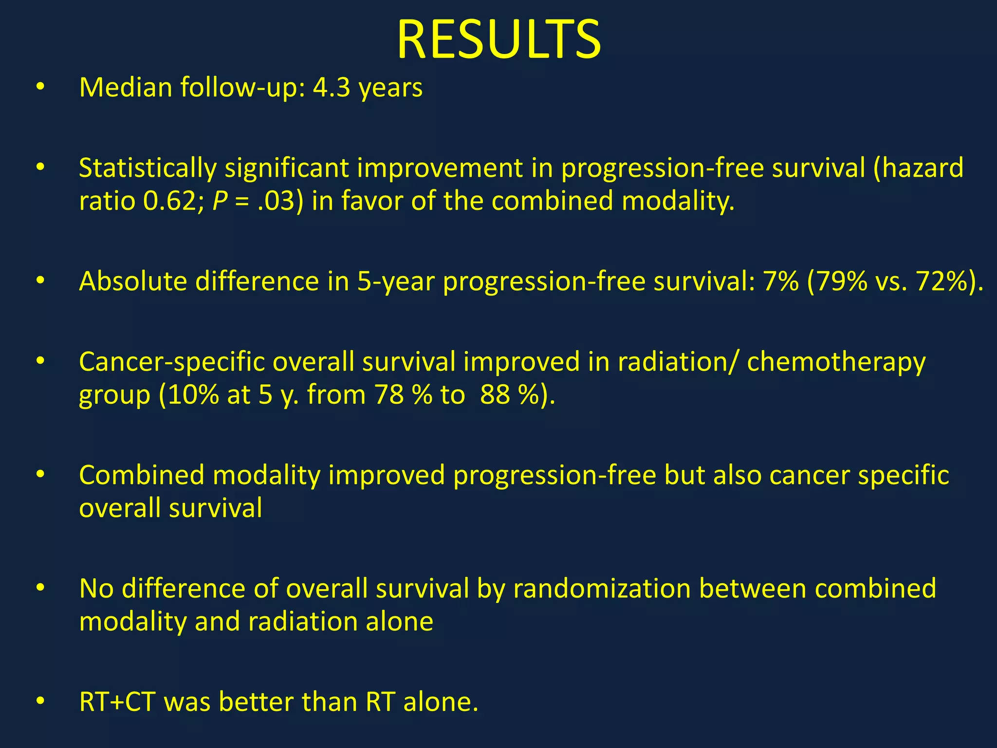
3) Ongoing clinical trials are evaluating the benefits of chemotherapy combined with radiation therapy compared to radiation alone, especially for high risk or advanced stages. Outcomes with current treatments are generally good
2. INTRODUCTION
• Carcinoma endometrium is the most common
gynecologic malignancy in the West, but in India,
the incidence rates are low.
• Most of these cancers present at an early stage
and are associated with a good prognosis.
• However discrepancies still exist with regard to
primary surgical management and post operative
adjuvant therapies directed at reducing
recurrence rates and improving survival.
5. CLINICAL PRESENTATION
• Abnormal Vaginal bleeding in 90% cases.
• Only 5-20% of postmenopausal woman with
abnormal vaginal bleeding will have carcinoma.
• Urinary bleeding.
• Rectal bleeding.
• Constipation.
• Abdominal pain.
• Lower-extremity lymphedema.
• Abdominal distension due to ascites.
• And cough and/or hemoptysis.
6. WORK UP
• Endometrial biopsy.
• Dilatation and curettage (D&C).
• Transvaginal ultrasonography (TVU)
Endometrial Thickness >5mm abnormal
• Saline infusion Sonography.
• Hysteroscopy.
7. WORK UP
• CECT Abdomen and pelvis
• Dynamic contrast MRI of pelvis
• CBC/LFT/RFT/Chest X Ray.
Optional Investigations:
• Cystoscopy, Sigmoidoscopy, S.CA-125 (extra
uterine d/s)
• PET-CT Scan.
8. HISTOPATHOLOGY
Type I Endometrial cancer :
• Endometrioid (75%)
• (secretory, ciliated, papillary or villoglandular)
• Type II Endometrial Cancer :
• Uterine papillary serous
• Clear cell carcinoma
• Mucinous carcinoma
• Squamous cell carcinoma
• Transitional cell carcinoma
• Mixed-cell type
• Undifferentiated carcinoma
• Metastatic carcinoma to the endometrium.
9. TYPE 1 FACTORS TYPE 2
PRE & PERIMENOPAUSAL MENOPAUSE POST MENOPAUSAL
PRIOR ESTROGEN STIMLN+ ESTROGEN STIMULATION NO ESTROGEN STIMLN
MINIMAL MYOMETRIAL INV MORE
LOW GRADE GRADE HIGH GRADE
PI3 K/PTEN/AKT MOL. PATHWAY p53,p16
MUTATION IN PTEN,
MSI, β CATENIN MUTATION
INACTIVATION p16, RED. E
CADHERIN, OVEREXP OF
HER2NEU
11. STAGING: FIGO 2009
• Stage I: Tumour confined to endometrium
IA: No or less than half myometrial invasion
IB: Invasion equal to or more than half of
myometrium
12. STAGING: FIGO 2009
• Stage II: Cervical stromal invasion but not
beyond the uterus
13. STAGING FIGO 2009
• Stage III : Local and/or regional spread of the
tumour
III A: Tumour invades the serosa of the corpus uteri
and/or adnexa
III B: Vaginal and/or parametrial involvement
III C: Metastases to pelvic and /or para aortic
lymph nodes
C1: pelvic node involvement
C2: para aortic lymph node involvement with or
without pelvic lymph node involvement.
14. STAGING FIGO 2009
• Stage IV: Tumour invades bladder and/or
bowel mucosa, and/or distant metastases
IV A: Tumour invasion of bladder and/or bowel
mucosa
IV B: Distant metastases, including abdominal
metastases and/or inguinal lymph nodes
16. TREATMENT
• Surgery- gold standard for the treatment of
endometrial carcinoma.
• Radiotherapy –mainly adjuvant and radical in
medically inoperable cases
• Chemotherapy- in high risk & metastasic disease.
• Hormone therapy.
17. SURGERY
• The mainstay of treatment for endometrial
carcinoma is an total abdominal hysterectomy
and bilateral sapingo-oopherectomy, peritoneal
washing, and ?lymph node dissection.
(TAH+BSO+PW+?LNB ).
• SLN mapping to surgical staging procedures
seems to increase the likelihood of detecting
metastatic cancer cells in regional lymph nodes.
Whether sentinel lymph node mapping will
replace lymphadenectomy needs to be
determined.
18.
19. ESMO GUIDELINES
Stage I IA G1-G2 Hysterectomy + BSO
IA G3 Hysterectomy + BSO+ b/l Pelvic Para Aortic LND
IB G1-3 Hysterectomy + BSO+ b/l Pelvic Para Aortic LND
Stage II Radical Hysterectomy + BSO+ b/l Pelvic Para Aortic LND
Stage III Maximum Cytoreduction with good performance status
Stage IV IV A Anterior & Posterior pelvic exentration
IV B Systemic Therapeutic approach with palliative surgery
20. STAGE III & IV
• Surgery should be performed to determine the
extent of disease and to remove the bulk of
disease if possible.
• Goal of surgery is eradication of macroscopic
disease.
• Postoperative therapy can be tailored according
to the extent of disease.
• Positive impact of cytoreductive surgery on
survival. ( 3 times greater)
27. BENEFIT OF RT IN GOG 99
• “High Intermediate Risk”
– Age<50
Gr. 2 or 3, LVSI, outer third myometrial invasion
– Age > 50 and 2 of above
– Age > 70 and 1 of above.
Adjuvant RT in early stage intermediate risk endometrial
carcinoma decreases the risk of recurrence, but should be
limited to patients whose risk factors fit a high
intermediate risk definition.”
31. PORTEC 2: EBRT vs IVBT
• Nout et al ASCO 2008, Lancet
Eligibility Criteria:
• Age >60 <50% MI Gr. 3
• Age>60 >50%MI Gr. 1-2
• Cervical Glandular Epithelium (+) Gr. 1-2
• Cervical Glandular Epithelium (+) Gr. 3
<50%MI
33. CONCLUSIONS
• IVBT as effective as EBRT for intermediate high
risk EC, despite the slightly but significantly
increased pelvic failure rate in the IVBT arm.
• OS and RFS were similar.
• QOL significantly better with brachytherapy
• IVBT should be the treatment of choice for
patients with High-intermediate risk EC.
34. LOW RISK
PATHOLOGIC
STAGE
AGE <60 Yrs
LVSI(-)
AGE <60 Yrs
LVSI(+)
AGE >/=60 Yrs
LVSI(-)
AGE >/= 60 Yrs
LVSI(+)
Stage IA, Gr 1-2
ESS done NFT NFT/ IVB NFT IVB/ NFT
Stage IA, Gr 1-2
ESS not done NFT NFT/ IVB NFT IVB
35. LOW INTERMEDIATE RISK
PATHOLOGIC
STAGE
AGE <60
LVSI(-)
AGE <60
LVSI(+)
AGE >/=60
LVSI(-)
AGE >/= 60
LVSI(+)
Stage I A, Gr 3
ESS done NFT/ IVB IVB IVB IVB
Stage I A, Gr 3
ESS not done IVB IVB IVB/ EBRT IVB/ EBRT
Stage IB, Gr 2
Stage II(<50% MI), Gr 1-2
ESS done
NFT/ IVB IVB/ NFT IVB IVB/ EBRT
Stage IB, Gr 2
Stage II(<50% MI), Gr 1-2
ESS not done
IVB (IIA)
NFT (IB) IVB IVB IVB/ EBRT
36. HIGH INTERMEDIATE RISK
PATHOLOGIC
STAGE
AGE <60 Yrs
LVSI(-)
AGE <60 Yrs
LVSI(+)
AGE >/=60 Yrs
LVSI(-)
AGE >/= 60 Yrs
LVSI(+)
Stage IB, Gr 3
Stage II (<50%
MI), Gr 3
ESS done
IVB IVB IVB/ EBRT EBRT
Stage IB, Gr 3
Stage II (<50%
MI), Gr 3
ESS not done
IVB/ EBRT EBRT/ IVB EBRT EBRT +/-IVB
Stage II(<50%
MI), Gr 1-2
ESS done
IVB IVB/ EBRT EBRT EBRT +/- IVB
Stage II(<50%
MI), Gr 1-2
ESS not done
EBRT/ IVB EBRT EBRT +/- IVB
boost
EBRT +/- IVB
boost
37. HIGH RISK
PATHOLOGIC
STAGE
AGE <60 Yrs
LVSI(-)
AGE <60 Yrs
LVSI(+)
AGE >/=60 Yrs
LVSI(-)
AGE >/= 60 Yrs
LVSI(+)
Stage II (<50%
MI), Gr 3
ESS done
EBRT/ IVB EBRT EBRT +/- IVB
boost
EBRT +/- IVB
boost IVB
Stage II(<50%
MI), Gr 3
ESS not done
EBRT/ IVB EBRT +/- IVB
boost
EBRT +/- IVB
boost
EBRT +/- IVB
boost
40. At 5 yrs, no diff in
PFS, OS or Toxicity
Unplanned subset analysis:
Stage I + Age>70yrs/ Gr3
Stage II + peritoneal cytology +
Chemo improved PFS (83% vs 66%)
42. RESULTS
• Median follow-up: 4.3 years
• Statistically significant improvement in progression-free survival (hazard
ratio 0.62; P = .03) in favor of the combined modality.
• Absolute difference in 5-year progression-free survival: 7% (79% vs. 72%).
• Cancer-specific overall survival improved in radiation/ chemotherapy
group (10% at 5 y. from 78 % to 88 %).
• Combined modality improved progression-free but also cancer specific
overall survival
• No difference of overall survival by randomization between combined
modality and radiation alone
• RT+CT was better than RT alone.
43. PORTEC 3 TRIAL: CCRTCT vs EBRT
FIGO stage I grade 3 with
deep myometrial invasion
and/or LVSI;
stage II or III;
or serous/clear cell histology
RT (48.6 Gy in 1.8 Gy
fractions)
n-=330
two cycles of cisplatin 50
mg/m² in week 1 and 4 of RT,
followed by four cycles of
carboplatin AUC5 and
paclitaxel 175 mg/m² at 3-
week intervals
n=330
PRIMARY END POINTS
OS
FFS
44. PORTEC 3
CCRT RT p value
OS 81.8% 76.7% 0.18
FFS 75.5% 68.9% 0.08
Patients with stage III EC had greatest benefit
of CTRT: 5-year FFS 69.3% for CTRT vs 58.0%
for RT p=0.032], and 5-year OS was 78.7 % vs
69.8% (p=0.114).
Adjuvant chemotherapy given during and after pelvic radiotherapy for
treatment of HREC did not significantly improve 5-year FFS and OS, compared
with RT alone. For women with stage III EC FFS was however significantly
improved with CTRT by 11% at 5 years.
45. CHEMOTHERAPY
• Chemotherapy can be effective alternative of RT
in high risk endometrial cancer.
• CT should be recommended in high risk and
advanced endometrial cancer as there is high
probability of distant relapse in these group of
patients.
• Combination chemotherapy is better than single
agent chemotherapy .
• Taxane improves locoregional control, PFS and
OS.
• Adjuvant CT-RT has better result than RT alone.
46. HORMONE THERAPY
Hormonal therapy is also an option for advanced stage
disease. Especially if hormone receptor positive
tumours.
MPA 400mg IM weekly or oral 150 mg/ day
or megestrol acetate 160 mg/ day.
If there is an objective response the progestin therapy
can be continued indefinitely.
Complete remission of lung metastasis has also been
seen with progestin alone.
Tamoxifen and progesterone – no added benefit.
48. SIMULATION
• Patients may be simulated in either a supine position or prone position
• Endometrial cancer patients undergoing pelvic IMRT are typically simulated
in the supine position.
• IMRT is preferred over 4 field box technique.
• Immobilization with a customized cradle should be employed to minimize
treatment setup error.
• Patients should be simulated with a comfortably full bladder. At some
centers, two scans are performed (full bladder and empty bladder) and the
two scans are fused to generate an integrated target volume (ITV).
• CT simulation should be done with ≤3 mm slice thickness. Intravenous
contrast is helpful for blood vessel delineation.
• In cases of vaginal involvement, a radio opaque marker should be placed at
the caudal extent of the tumor.
49. FIELDS BORDERS
AP/PA superior : L5-S1
inferior :bottom of the obturator foramina
lateral :2 cm lateral to bony margin of the
pelvic inlet
LATERAL superior and inferior as in AP/PA fields .
anterior :in front of the pubic symphysis
posterior :S2-S3
51. • Ideally, at least 95 % of the PTV should receive 100 % of the
prescription dose, and ≥99 % of the PTV should receive ≥90 % of
the prescription dose.
• The dose maximum should occur within the PTV and dose areas
>100 % of the prescription dose outside of the PTV should be
minimized.
• Patients should undergo at least weekly imaging with megavoltage
(MV) portal, kilovoltage (kV) imaging, or cone beam CT (CBCT) to
verify treatment setup. Patients being treated with IMRT should
undergo image guidance, preferably with daily imaging.
• High-energy linear accelerators (15 MV) are preferred because of
their sparing of the skin and subcutaneous tissue.
• Dose – 45-50.4 Gy(1.8-2Gy/#)
58. IMRT vs CONFORMAL RT
• Pelvic IMRT reduces acute patient reported GI
and GU toxicity compared to standard pelvic RT.
• Pelvic IMRT improves quality of life with regard
to physical functioning and other treatment
effects during treatment .
• Longer term follow up will be needed to
determine if these differences in acute toxicity
result in lower rates of late toxicity.
• Pelvic IMRT reduces need for anti-diarrheal
medications as compared to standard pelvic RT.
59. BRACHYTHERAPY
• After placement of the brachytherapy device, a method of
localization such as a portable x-ray or conventional
radiotherapy CT simulation is indicated.
• If an MRI is obtained, the planning CT should be fused to
the MRI using rigid registration along the brachytherapy
applicator.
• HDR applicators for inoperable primary endometrial
carcinoma are as in cervical carcinoma and include tandem
and ovoids, tandem and ring, and tandem and cylinder.
• The ABS recommends treating the proximal 3–5 cm of the
vagina for endometrial carcinoma and consideration of
treatment of the full length of the vagina for serous and
clear cell histologies.
60. BRACHYTHERAPY
• According to ABS recommendations, intracavitary
brachytherapy should be used only for non-bulky
recurrences with thickness <0.5 cm after completion.
• Interstitial brachytherapy should be offered for
recurrences with thickness >0.5 cm after EBRT.
• At the level of the vagina, the dose distribution should
be optimized to deliver the prescribed dose to a depth
of 0.5 cm unless treating to the surface.
• In adjuvant setting it should be given after 4-6 weeks.
67. SUMMARY
• Endometrial cancer most commonly presents with bleeding
per vagina.
• MRI is the investigation of choice to determine the local
extent of the tumour.
• Stage,myometrial invasion,age ,grade and LVSI are
important prognostic factors.
• Surgery is the treatment of choice.no survival benefit of
simple hysterectomy over radical hysterectomy except in
gross cervical involvement.
68. SUMMARY
• From stage IB with grade tumours adjuvant radiation is mandatory.
• Intravaginal brachytherapy is preferred over EBRT in high intermediate
risk groups
• EBRT must be given from stage II.
• Stage III onwards chemoradiation followed by adjuvant CT should be
given.
• IMRT is the preferred over 3DCRT.
• Dose for EBRT is 45-50.5Gy(1.8-2 Gy).
• Intravaginal Brachytherapy dose is 21Gy/3#.