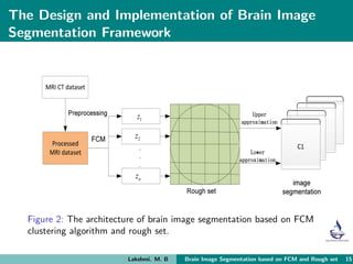
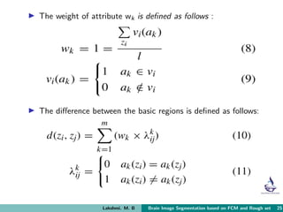
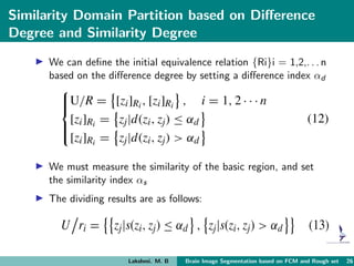
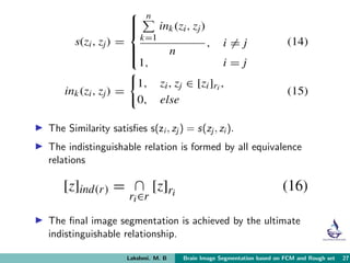
Brain image segmentation based on FCM Clustering Algorithm and Rough Set
1. Brain Image Segmentation based on FCM
Clustering Algorithm and Rough set
Lakshmi. M. B
Sr. No. 9219708
S2, MTech CSE
Sahrdaya College of Engineering and Technology, Kodakara
July 15, 2020
Guided by,
Asst. Prof. Priya. K. V
Lakshmi. M. B Brain Image Segmentation based on FCM and Rough set 1
2. Abstract
New image segmentation method = FCM clustering algorithm
+
Rough set theory
First, the attribute value table is constructed.
The image is divided into several small regions.
Then, the weight values of each attribute are obtained.
Calculate the difference between regions.
The similarity evaluation of each region is realized.
Lakshmi. M. B Brain Image Segmentation based on FCM and Rough set 2
3. Finally, the regions are merged and there is complete image
segmentation.
Validated in the segmentation of artificially generated images,
brain CT images,and MRI images.
The proposed method can reduce the error rate and achieve
better segmentation results for the fuzzy boundary region.
Also the results prove that the algorithm has strong anti-noise
ability.
Lakshmi. M. B Brain Image Segmentation based on FCM and Rough set 3
4. Introduction
Image segmentation refers to dividing an image into regions
with different characteristics and proposing objects of interest.
FCM image segmentation algorithm – divide the sample
points of vector space into C subspaces according to some
distance measure.
Standard FCM algorithm converges too slowly, and results
in inaccurate segmentation of boundary points which makes
the edges blurred.
Lakshmi. M. B Brain Image Segmentation based on FCM and Rough set 4
5. Solution: Many scholars combine rough set theory to solve
this problem and overcome the defects of FCM clustering.
Proposed method:
1. The attribute value table is constructed.
2. The image is divided into several small regions.
3. The weight values of each attribute are obtained.
4. Calculate the difference between regions.
5. The similarity evaluation of each region is realized.
6. Regions are merged and there is complete image segmentation.
Lakshmi. M. B Brain Image Segmentation based on FCM and Rough set 5
6. Related Works
Threshold-based segmentation is simple and has
advantages in processing speed, but it is not suitable for the
segmentation of blurred boundary areas in images.
For the segmentation of blurred boundary areas,
unsupervised clustering method is usually used.
FCM method is very effective for the segmentation of fuzzy
boundary areas and has been widely used.
Histogram-based threshold segmentation is the most
widely used method for image segmentation.
Lakshmi. M. B Brain Image Segmentation based on FCM and Rough set 6
7. It’s advantage is that they do not need any prior information
of the image.
They are only based on the gray level and do not consider the
spatial correlation of the same or similar elements.
In order to overcome this shortcoming, the rough
homogeneity method was adopted which only considers the
spatial correlation of adjacent pixels in the same plane.
Lakshmi. M. B Brain Image Segmentation based on FCM and Rough set 7
8. Fuzzy C-means (FCM) Clustering Algorithm
Fuzzy clustering algorithm uses fuzzy method to cluster.
It can reflect the real world more objectively.
It searches for the optimal extremes through repeated
iterations.
Euclidean distance is used as the distance measure in the
objective function of FCM algorithm.
Clustering analysis largely depends on the distribution of data
sets.
Lakshmi. M. B Brain Image Segmentation based on FCM and Rough set 8
9. Figure 1: The mapping principle of FCM from input space to
characteristic space
Lakshmi. M. B Brain Image Segmentation based on FCM and Rough set 9
10. Linear sample space – FCM can achieve good clustering
effect;
Non-linear sample space – the clustering effect will be
unsatisfactory.
Kernel-fuzzy C-means clustering algorithm uses kernel
function, and then carries out FCM clustering.
This overcomes the disadvantage that FCM is not suitable for
the non-linear data distribution to a certain extent.
Lakshmi. M. B Brain Image Segmentation based on FCM and Rough set 10
11. Rough Set Theory
First proposed by Z. Pawlak.
It is a powerful tool for dealing with uncertain, incomplete and
inaccurate knowledge.
Rough set granulates knowledge based on the concepts of
upper and lower approximation, and reduction and so on.
It directly simulates the logical thinking ability of human
beings.
Lakshmi. M. B Brain Image Segmentation based on FCM and Rough set 11
12. It also provides an effective processing technology for the
processing of intelligent information.
Rough set data analysis – attribute reduction, rule extraction,
value reduction, minimization of decision rules, etc.
At present, rough set has been applied to image enhancement,
image segmentation, and image filtering and so on.
Image processing based on rough sets can achieve better
results.
Lakshmi. M. B Brain Image Segmentation based on FCM and Rough set 12
13. Brain Image Segmentation
Brain imaging plays an important role in today’s medical
diagnosis.
Image segmentation aiming at acquiring tissues and organs
and related biological structures is one of the most important
steps in medical imaging.
Traditional manual segmentation cannot deal with a large
number of medical images effectively.
Lakshmi. M. B Brain Image Segmentation based on FCM and Rough set 13
14. Automatic brain image segmentation faces two major
challenges:-
1. the diversity of biological structure itself, and
2. the low contrast and noise caused by the defects of brain
image technology itself.
Goal of brain image segmentation – describe the different
anatomical structures on brain images according to the
discontinuity in feature space.
Combining spatial information with pixel classification will be
a meaningful segmentation method.
Lakshmi. M. B Brain Image Segmentation based on FCM and Rough set 14
15. The Design and Implementation of Brain Image
Segmentation Framework
Figure 2: The architecture of brain image segmentation based on FCM
clustering algorithm and rough set.
Lakshmi. M. B Brain Image Segmentation based on FCM and Rough set 15
16. Image Preprocessing
The main purpose is to eliminate the noise and to improve the
classification effect.
A binary masking image is established to detect large targets,
which is given by the formula:
Bm = I> Bth
where, Bm is binary masked image, I is MR image,
Bth is the threshold of image binarization.
Lakshmi. M. B Brain Image Segmentation based on FCM and Rough set 16
17. The background image can be calculated by the formula:
IB = Bm × I where, IB is a black background image.
In order to avoid the omission of image edge vexes, here w/2
background vexes are added to each edge.
Next, the source image is divided into Ne sub-images
Lakshmi. M. B Brain Image Segmentation based on FCM and Rough set 17
18. Figure 3: The implementation of FCM.
Lakshmi. M. B Brain Image Segmentation based on FCM and Rough set 18
19. The size of the sub-image corresponds to the extraction
window.
After graying the color image, it is binarized.
The most commonly used method of gray image binarization
is threshold method.
All pixels whose gray value is greater than or equal to the
threshold value belong to a specific object, and their gray
value is 255.
Or, the pixels are excluded from the object area, and the gray
value is 0.
Lakshmi. M. B Brain Image Segmentation based on FCM and Rough set 19
20. The gray value of the brain region and the background region
is 1 and 0 respectively.
The gray value of the brain region is restored by mask
operation.
Masking Operation – use of selected images or objects to
block all or part of the processed image to control image
processing.
The image value in the region of interest remains unchanged,
while the pixel value of the image outside the region is set to
0.
Lakshmi. M. B Brain Image Segmentation based on FCM and Rough set 20
21. Information Table Construction and Region Division based
on FCM
Construct an information table that can be used for image
segmentation.
Different clustering numbers are used as attributes of the
pixels to be segmented, and clustering segmentation results
are used as attributes.
The clustering criterion function is defined as a membership
function:
Lakshmi. M. B Brain Image Segmentation based on FCM and Rough set 21
22. In the method of fuzzy mean clustering, the sum of
membership degrees of samples to each cluster is required to
be 1.
The necessary condition for obtaining the minimum is:
Lakshmi. M. B Brain Image Segmentation based on FCM and Rough set 22
23. All kinds of clustering centers and samples are obtained as the
algorithm converges.
For each kind of membership degree, the fuzzy clustering
division is completed.
Finally, the results of the fuzzy clustering are de-fuzzified.
The fuzzy clustering is transformed into deterministic
classification.
Lakshmi. M. B Brain Image Segmentation based on FCM and Rough set 23
24. Calculation of Attribute Weight and Basic Regional
Difference based on Value Reduction
Rough set describes knowledge from the point of view of
pattern classification.
Knowledge space is divided into different pattern equivalence
classes, thus it is represented as granular structure.
Consider different attributes and give them different weights
in order to make reasonable decisions.
In reduction, the more frequent the attributes appear, the
more important the classification is.
Lakshmi. M. B Brain Image Segmentation based on FCM and Rough set 24
25. The weight of attribute wk is defined as follows :
The difference between the basic regions is defined as follows:
Lakshmi. M. B Brain Image Segmentation based on FCM and Rough set 25
26. Similarity Domain Partition based on Difference
Degree and Similarity Degree
We can define the initial equivalence relation {Ri}i = 1,2,. . . n
based on the difference degree by setting a difference index αd
We must measure the similarity of the basic region, and set
the similarity index αs
The dividing results are as follows:
Lakshmi. M. B Brain Image Segmentation based on FCM and Rough set 26
27. The Similarity satisfies s(zi , zj ) = s(zj , zi ).
The indistinguishable relation is formed by all equivalence
relations
The final image segmentation is achieved by the ultimate
indistinguishable relationship.
Lakshmi. M. B Brain Image Segmentation based on FCM and Rough set 27
28. The Image Segment Results Compared With Other
Algorithms
Two groups of weighted images with different resolutions were
selected for simulation experiments:
1. The simulation Image dataset is from IBSR brain image
database, the size of images was 256∗256∗63 individual
elements.
2. The clinical case images were selected, and the size of images
was 512∗512∗512 voxels.
All simulation experiments were implemented on MATLAB
platform.
Lakshmi. M. B Brain Image Segmentation based on FCM and Rough set 28
29. Comparison of Segmentation Effects of Different
Optimization Methods
Sensitivity (Se), specificity (Sq), positive likelihood (PL) and
negative likelihood (NL) are selected to evaluate the
superiority of different algorithms.
The larger Se, Sq, PL, the better the clustering effect and the
more accurate the organizational segmentation is.
The smaller NL, the better the segmentation effect is.
Lakshmi. M. B Brain Image Segmentation based on FCM and Rough set 29
30. Figure: The comparison simulation under different image segment
algorithm.
Lakshmi. M. B Brain Image Segmentation based on FCM and Rough set 30
31. Table: The index comparison under different algorithms
Lakshmi. M. B Brain Image Segmentation based on FCM and Rough set 31
32. Comparison of Segmentation Effect with Different
Clustering Algorithms
The clustering process uses DBI and DUNN index to evaluate
the clustering algorithms.
The smaller the DBI index is, the bigger the DUNN index is,
the better the clustering effect is.
Lakshmi. M. B Brain Image Segmentation based on FCM and Rough set 32
33. Table: The index comparison of K-means and FCM clustering
algorithms
The statistical significance test between DBI and DUNN obtained
by the two algorithms is shown below:
Lakshmi. M. B Brain Image Segmentation based on FCM and Rough set 33
34. Comparison of the Results of Different
Segmentation Algorithms
Tanimoto coefficients are used to evaluate the results of
different segmentation algorithms, and the similarities between
the segmentation results of different segmentation algorithms
and the real results are measured.
Tanimoto coefficient is given by the formula:
The bigger the Tanimoto coefficient is, the more accurate the
image segmentation is.
Lakshmi. M. B Brain Image Segmentation based on FCM and Rough set 34
35. Tanimoto coefficients obtained by different segmentation
algorithms.
Lakshmi. M. B Brain Image Segmentation based on FCM and Rough set 35
36. Comparing the results of this algorithm with different algorithms
Lakshmi. M. B Brain Image Segmentation based on FCM and Rough set 36
37. The average Tanimoto coefficients based on this algorithm are
higher than other clustering algorithm.
The average Tanimoto coefficients obtained by the algorithms
are different.
The proposed algorithm is superior to other clustering in white
matter and cerebrospinal fluid segmentation.
In gray matter segmentation, there is no significant difference
between the proposed algorithm and other clustering
algorithm.
Lakshmi. M. B Brain Image Segmentation based on FCM and Rough set 37
38. Clinical Image Segmentation Results
The tissue density of the imaging area is expressed by different
gray levels from black to white.
The low-density represents area such as soft tissue-white area
and the high-density area, such as skeleton.
Clinical images include white matter, gray matter,
cerebrospinal fluid, etc., which increase the difficulty of brain
image segmentation to a certain extent.
Lakshmi. M. B Brain Image Segmentation based on FCM and Rough set 38
39. The segment results of MRI medical image.
Lakshmi. M. B Brain Image Segmentation based on FCM and Rough set 39
40. Brain CT images are often used in clinical diagnosis of
craniocerebral radiotherapy and craniocerebral surgery.
With this algorithm, white matter can be divided into a
connected whole region, so that white matter and gray matter
have a clearer segmentation boundary.
In FCM algorithm, the misadvised soft tissues are segmented
accurately, and the skeleton and soft tissues are clearly
segmented from the background.
Lakshmi. M. B Brain Image Segmentation based on FCM and Rough set 40
41. Clinical Image Segmentation Results Compared with
System Tools
Brain tissue includes white matter, gray matter, cerebrospinal
fluid, lesions and so on.
The cluster number C=5 was selected.
The value of the fuzzy factor is chosen as P=2
Three different iteration stop thresholds em are selected, and
the maximum iteration times are set to 300.
Lakshmi. M. B Brain Image Segmentation based on FCM and Rough set 41
42. The segmentation effect of brain image based on this algorithm.
Lakshmi. M. B Brain Image Segmentation based on FCM and Rough set 42
43. The Segment Results Compared with FCM
There are many difficulties in the imaging process that are
difficult to distinguish.
It is necessary to combine with actual needs to produce
practical image processing methods for different diseases.
Here, we are combining the rough set theory with image
processing technology.
The larger the Vpc of the experimental results, the better the
clustering effect of the results is
Lakshmi. M. B Brain Image Segmentation based on FCM and Rough set 43
44. The relationship between iterative threshold and iterative time
Lakshmi. M. B Brain Image Segmentation based on FCM and Rough set 44
45. The index Vxb mainly reflects the quotient between the
degree of compactness within a class and the degree of
separation between different classes.
The smaller the final value of Vxb, the better the clustering
effect of this method.
Compared with FCM, our algorithm is slightly better in
clustering, but has a great improvement in time.
Lakshmi. M. B Brain Image Segmentation based on FCM and Rough set 45
46. The segment results of MRI medical image compared with FCM
Lakshmi. M. B Brain Image Segmentation based on FCM and Rough set 46
47. The Anti-Jamming Performance of Image
Segmentation Algorithms
The main objective of medical image processing is to achieve
the doctor’s visual effect and distinguish the focus, which
requires a robust segmentation algorithm.
To verify the anti-jamming performance, we setup the image
to receive different degrees of noise interference.
We observed the segmentation effect in the presence of noise
interference.
Lakshmi. M. B Brain Image Segmentation based on FCM and Rough set 47
48. Comparison of robustness between two methods
Lakshmi. M. B Brain Image Segmentation based on FCM and Rough set 48
49. Conclusion
This method can reduce the error rate and has better
segmentation effect on the blurred boundary areas.
This has strong stability and high accuracy.
Also shows superiority in both simulated images and clinical
examples.
The results shows that it has better robustness than the FCM
algorithm and has strong clinical application ability.
Lakshmi. M. B Brain Image Segmentation based on FCM and Rough set 49
50. Reference
Hong Huang, Fanzhi Meng, Shaohua Zhou, Feng Jiang, and
Gunasekaran Manogaran, “Brain Image Segmentation Based on
FCM Clustering Algorithm and Rough Set,” IEEE Access, vol. 7,
pp. 12386 - 12396, 2019.
Available: https://ieeexplore.ieee.org/document/8612906
Lakshmi. M. B Brain Image Segmentation based on FCM and Rough set 50
51. Lakshmi. M. B Brain Image Segmentation based on FCM and Rough set 51