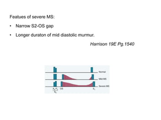
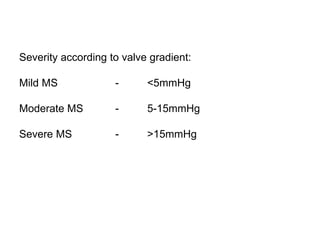
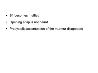
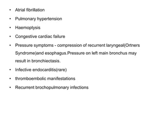
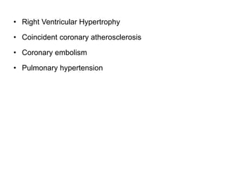
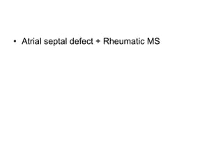
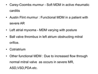
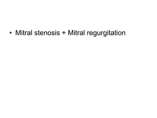
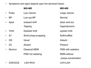
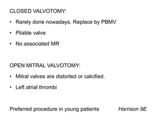
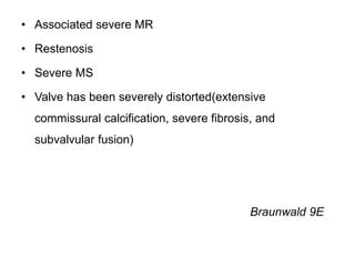
SlideShare a Scribd company logo
1 of 112
MITRAL STENOSIS
Dr.Monica.K
INTRODUCTION
• Mitral stenosis (MS) is characterized by obstruction to left ventricular
inflow at the level of mitral valve due to structural abnormality of the
mitral valve apparatus.
• Rheumatic fever is the leading cause of mitral stenosis (MS).
• Pure or predominant MS occurs in approximately 40% of all patients
with rheumatic heart disease and a history of rheumatic fever.
• With reductions in the incidence of acute rheumatic fever, the
incidence of MS has declined considerably over the past several
decades
• Two third of the cases are females.
PATHOPHYSIOLOGY
NATURAL HISTORY
• Mitral stenosis is a progressive,lifelong disease.
• Usually slow and stable in early years
• Progressive acceleration in later years
• 20-40 years latency from rheumatic fever to symptom
onset.
CLINICAL DISCUSSION
• Mitral valve apparatus is composed of?
• valve leaflets with
commissures
• valve annulus
• chordae tendinae
• papillary muscles
• myocardium(papillary muscles
are attached here)
• What are the causes of Mitral stenosis
• Rheumatic Heart disease (most common)
• Congenital
• Severe mitral annular calcification
• Rheumatoid arthritis
• Systemic lupus erythematosus
• Amyloidosis
• Drugs - methylsergide
• Carcinoid syndrome
Other causes of LV inflow obstruction
• Left atrial myxoma
• Cotriatrium
• Ball valve thrombus in left atrium Harrison 19E Pg.1539
• Why mitral valve is most commonly
involved in rheumatic heart disease?
• Mitral valve surface area is more than aortic area
• The pressure gradient across mitral valve is greater than
that across aortic valve.
Hence more prone for injury.
• What are the pathological processes that
occur in rheumatic mitral valve?
• In rheumatic MS, chronic inflammation leads to diffuse
thickening of the valve leaflets with formation of fibrous
tissue and/or calcific deposits. The mitral commissures
fuse, the chordae tendineae fuse and shorten, the
valvular cusps become rigid, and these changes, in
turn,lead to narrowing at the apex of the funnel-shaped
(“fish-mouth”)valve.
• MS is usually the result of repeated episodes of carditis
alternating with healing.
Hurst's 11E
• What is Selzer and Cohn's hypothesis
• The initial mitral valvulitis leads to abnormal flow patterns
across the mitral valv that promote thickening,fibrosis
and calcification of leaflets.The thickened leaflets fuse
along their edges i.e commissures, resulting in a
stenosed mitral valve which has a 'fish mouth' or 'button
hole' appearance.
» API 2015
• Classical symptoms in MS?
• fatiguability (d/t low cardiac output)
• dyspnoea,orthopnoea,PND,cough,haemoptysis,recurrent
bronchitis (d/t left atrial failure- pulmonary congestion)
• swelling of legs,right hypochondrial pain (d/t RVF)
• palpitation (d/t atrial fibrillation)
• What are the classical physical findings in
a case of mitral stenosis?
• Low volume pulse
• Apex Beat - Normal in position. Tapping in character.
• Diastolic thrill may be palpable in mitral area.
• Left Parasternal heave
Auscultation
• Loud S1
• Opening Snap just after S2
• A low pitched rough rumbling mid diastolic murmur with presystolic
accentuation over the apex best heard with the bell of stethescope
in left lateral position during expiration.
• With the development of pulmonary hypertension, Palpable P2,loud
P2, Ejection systolic murmur in pulmonary area and a Pansystolic
murmur may be heard along left sternal border(Functional TR)
• Causes for acute deterioration in MS?
• severe exertion
• excitement
• fever
• severe anemia
• paroxysmal AF
• pregnancy
• thyrotoxicosis
• infection
• Describe classical mitral facies
• It is the pinkish purple patches
on cheeks. As cardiac output is
low in MS, vasoconstriction
produces peripheral cyanosis
in lips,tip of nose and cheeks.
Malar flush is seen in malar
area due to vasodilation. All
these constitute mitral facies
• What is the normal mitral valve area and
severity grading of MS according to valve
area?
• Normal mitral valve area is 4-6 sq.cm
• Mild MS - 1.5-2.5 sq.cm
• Moderate MS - 1-1.5 sq.cm
• Severe/critical MS - <1 sq.cm
• What is the reason for loud S1 in MS?
• Due to persistent diastolic gradient across the
mitral valve, the valve cusps remain wide open
throughout the diastole and as soon as the
ventricular systole starts, the widely opened
mitral valve cusps close rapidly, giving rise to
loud S1.
• M1T1 summation
• Flow across deformed valve
• What are the causes of loud S1?
• Mitral stenosis
• Tricuspid stenosis
• Sinus tachycardia
• Short PR interval
• Hyperkinetic circulation (severe anemia,pyrexia,exercise
etc)
• What are the causes of muffled S1 in MS?
• Associated with mitral or aortic regurgitaion
• Mitral valve calcification
• Active rheumatic carditis(due to prolonged PR interval)
• Digitalis overdose(due to prolonged PR interval)
• Left heart failure
• Atrial fibrillation(varying S1)
• Rotation of the heart due to gross right ventricular
hypertrophy.
• Obesity,emphysema,pericardial effusion,thick chest wall.
• JVP IN MS
• Prominent a waves - In presence of PHT
• Prominent v wave - In presence of TR (functional)
• Prominent y descent - In presence of TR (functional)
• Absent a waves - Atrial fibrillation
• How can the severity of MS assessed
clinically?
Featues of severe MS:
• Narrow S2-OS gap
• Longer duraton of mid diastolic murmur.
Harrison 19E Pg.1540
• Severity according to valve gradient?
Severity according to valve gradient:
Mild MS - <5mmHg
Moderate MS - 5-15mmHg
Severe MS - >15mmHg
• Why is the murmur mid diastolic in MS?
• The interval between closing of aortic valve and opening
of mitral valve is known as 'isovolumetric relaxation
period'. Diastolic murmur is heard as soon as the blood
flows from left atrium to left ventricle after the opening of
mitral valve. There is no blood flow in the isovolumetric
relaxation period and this is why the murmur of MS is
mid-diastolic,not early diastolic.
• What is the cause for presystolic
accentuation?
• The last part of ventricular diastole actually coincides
with atrial systole. atrial systole increases the blood flow
across the stenotic valve and thus there is presystolic
accentuation.
• What is an opening snap?
• Opening snap is produced due to bellowing down of the
closed mitral valve cusps at the onset of ventricular
diastole.
It has the following characteristics:
• Sharp and high pitched
• Best heard with diaphragm of stethescope
• Best heard after expiration with the patient in standing
position
• Loudest in between the apex and left sternal border
• Present in early diastole (at the end of isovolumetric
relaxation period)
• What does opening snap in MS indicate?
• MS is organic
• Valve cusps are pliable
• Significant MS
• High atrioventricular pressure gradient
• Severe AR,MR,atrial fibrillation,left heart failure absent
• MS is readily amenable for surgery
• Diminishing S2-OS gap indicates increasing severity.
To differentiate OS from splitting of S2
• Ask the patient to stand.As standing reduces the venous return, S2-
OS gap increases and A2-P2 gap decreases (dynamic auscultation).
• When is Opening snap absent in MS?
• Mild MS
• Severe MS
• Calcific MS
• Congenital MS
• MS with associated significant MR/AR/AS
• What is the cause of Atrial fibrillation in
MS?
• Left atrial enlargement results in disorganisation of atrial
muscle bundles coupled with inhomogenous refratory
periods
• What are the changes noticed when Atrial
fibrillation develops in MS?
• Irregularly irregular pulse with a pulse deficit >10
• Varying intensity of S1
• Presystolic accentuation of diastolic murmur disappears
as atria contracts in an uncordinated and ineffective
manner. But presystolic accentuation may be present in
short cycles in AF.(Crile's hypothesis)
• Opening snap may not heard
• No a waves in JVP
• What is the mechanism behind acute
pulmonary edema in presence of AF in
MS?
• Decrease in the diastolic flow period caused by
increased heart rate
• Loss of atrial contribution to cardiac output.
• What are the changes that occur after left
atrial failure in MS?
• S1 becomes muffled
• Opening snap is not heard
• Presystolic accentuation of the murmur disappears
• What are the complications of MS?
• Atrial fibrillation
• Pulmonary hypertension
• Haemoptysis
• Congestive cardiac failure
• Pressure symptoms - compression of recurrent laryngeal(Ortners
Syndrome)and esophagus.Pressure on left main bronchus may
result in bronchiectasis.
• Infective endocarditis(rare)
• thromboembolic manifestations
• Recurrent brochopulmonary infections
• What are the possible explanations for
haemoptysis?
• Due to rupture of thin walled bronchial or pulmonary
veins resulting from sudden rise in LA pressure.
• Chronic bronchitis
• Acute pulmonary edema (Pink frothy sputum)
• Pulmonary infarction
• Overdose of anticoagulant therapy
• Pulmonary haemosiderosis
• Causes of pulmonary hypertension in MS
(1) passive backward transmission of the elevated LA
pressure;
(2) pulmonary arteriolar constriction (the so called“second
stenosis”), which presumably is triggered by LA and
pulmonary venous hypertension (reactive pulmonary
hypertension);
(3) interstitial edema in the walls of the small pulmonary
vessels; and
(4) at end stage, organic obliterative changes in the
pulmonary vascular bed(active PHT)
• What is Jordan's hypothesis?
• When the pulmonary venous pressure increases fluid
passes into alveolar walls and promotes thickening and
fibrosis of the wall resulting in hypoxemia. This
hypoxemia is sensed by chemoreceptors that lead to
reflex pulmonary vasoconstriction.This triad of passive
pressure transmission,reflex vasoconstriction and
obliterative changes in the vascular bed causes
Pulmonary Hypertension.
» API 2015
• What are the possible explanations for
syncope in MS?
• Cerebral embolism from atrial fibrillation
• Due to low cardiac output ( PHT,AF with RVR)
• Right Ventricular Hypertrophy
• Coincident coronary atherosclerosis
• Coronary embolism
• Pulmonary hypertension
• What is Lutembacher's syndrome?
• Atrial septal defect + Rheumatic MS
• Differential diagnosis for MDM in apex?
• Carey-Coombs murmur - Soft MDM in active rheumatic
carditis
• Austin Flint murmur : Functional MDM in a patient with
severe AR
• Left atrial myxoma - MDM varying with posture
• Ball valve thrombus in left atrium obstructing mitral
orifice.
• Cotriatrium
• Other functional MDM : Due to increased flow through
normal mitral valve as occurs in severe MR,
ASD,VSD,PDA etc.
COMBINED VALVULAR LESIONS
• Mitral stenosis + Mitral regurgitation
• Symptoms and signs depend upon the dominant lesion
MS>MR MR>MS
• Pulse Low volume Large volume
• BP Low sys BP Normal
• Apex outward shift down and out
Tapping Hyperdynamic
• Thrill Diastolic thrill systolic thrill
• S1 Short,sharp,snapping Soft/muffled
• OS Usual Absent
• S3 Absent Present
• Murmur Classical MDM PSM with radiation
PSM not radiating MDM without
presys accentuation
• CXR,ECG LAH+RVH LVH+LAH
• In presence of significant MR Mitral valve
replacement is the treatment of choice.
• Mitral stenosis+aortic stenosis
• Aortic involvement present in one third of cases of MS.
• Combined MS and AS is usually of rheumatic origin.
• Other cause - Congenital SHONE COMPLEX - MS+AS+COA
• Decreased cardiac output more pronounced in combined lesion.
• Signs of AS are masked in presence of severe MS. Ejection systolic
murmur may decrease in intensity, Apical impulse is less than
heaving
• In presence of significant AS,Signs of MS i.e. S1 may not be loud
and opening snap and classical MDM may not be heard.
• If the degree of AS appears to be mild and the mitral valve is
acceptable for balloon valvotomy, this should be attempted first. If
mitral balloon valvotomy is successful, the aortic valve should then
be reevaluated.
• Mitral stenosis+Aortic regurgitation
• When both AR and MS coexist, severe MS usually coexists with
mild AR.However, the coexistent AR is occasionally severe.
• The combination of coexistent severe MS and severe AR may
present confusing pathophysiology and often leads to misdiagnosis.
MS restricts LV filling, blunting the impact of AR on LV volume.Thus,
even severe AR may fail to cause a hyperdynamic circulation, so
that typical signs of AR are absent during physical examination.
• Likewise, echocardiographic LV cavitary dimensions may be only
mildly enlarged.
• In most cases, it is advisable to perform mitral valvotomy first and
then follow the patient for symptomatic improvement. If symptoms
disappear, correction of AR can be delayed.
• Mitral stenosis+Tricuspid regurgitation
• It is important to establish whether tricuspid regurgitation
is primary or secondary to pulmonary hypertension.
• In general, if pulmonary hypertension is severe and the
tricuspid valve anatomy is not grossly distorted,
improvement in TR can be expected after correction of
MS.
• On the other hand, if there is severe rheumatic deformity
of the tricuspid valve, competence is likely to be restored
only by surgery.(tricuspid annuloplasty)
• Specific investigations in MS?
• ECG
• Echocardiogram - TTE and TEE
• Chest X-ray - PA view and RAO view
• Cardiac catheterisation
• ECG findings in MS
• P mitrale (left atrial enlargement)
• P pulmonale (right atrial enlargement)
• RVH
• Features of atrial fibrillation
• Chest X ray features of MS
• Mitralisation of heart - Straightening of left heart border d/t small
aortic knuckle, dilated pulmonary artery and prominent left atrial
appendage.
• Double contour of right heart border (shadow within shadow) - LA
enlargement.
• Evidence of pulmonary hypertension - Dilated pulmonary arteries at
hilum with peripheral pruning.
• Elevation of left upper lobe bronchus and splaying of carina - LA
enlargement.
• Kerley Blines - dense,short, horizontal lines in costophrenic region
when pulmonary venous pressure is between 20-30mmHg.
• Kerley A lines - Straight,dense lines running towards hilum when
pulmonary venous pressure is >30mmHg.
• Upturned apex - inverted moustache sign
• RAO view - Barium swallow
may demonstrate sickling of
barium filled esophagus due to
compression by enlarged LA.
• Lateral view - Obliteration of
retrosternal air space.
Chamber enlargement can be
looked for.
• Echocardiography to assess?
• Chamber enlargement
• valve pathology and movement
• thrombus
• vegetation
• LV and RV function
• Pulmonary arterial and other pressures
• Gold standard method for assessing
severity of MS?
• Simultaneous measurement of cardiac output and the
gradient between the LA and the LV and calculation of
valve area by cardiac catheterisation remains the "gold
standard" method for assessing the severity of MS
Hurst's 11E
• What is the role of cardiac catheterisation
in MS?
• To measure arterial pressures and valvular pressure
gradient
• Valve area
• cardiac output
• other valvular lesions
• assess the coronaries
• LV function
• What are the treatment options?
• Medical management
• Percutaneous mitral balloon valvuloplasty
• Valvotomy - closed and open
• Valve replacement - Mechanical/bioprosthesis
• Role of medical management
• Restrict strenous physical activity
• Heart failure measures
• Rheumatic fever prophylaxis
• Anticoagulation in AF
• Patients with mild MS and moderate asymptomatic MS
usually managed conservatively. Interventional
procedures done usually in symptomatic MS.
• Indications for Percutaneous mitral balloon
valvuloplasty
• Significant symptoms
• Isolated MS
• No/mild MR
• Mobile non-calcified valve
• LA free of thrombus
• Asymptomatic- Severe PHT - Pulmonary systolic pressure greater
than 50 mm Hg at rest or 60 mm Hg with exercise.
Pregnant women - PBMV is the procedure of choice
Harrison 19E
• Complications of PBMV?
• Cardiac perforation
• Embolic complications
• Iatrogenic ASD
• Cardiac tamponade
• Postprocedural MR
• Indications of PBMV in mild MS
• If patients wishes to get pregnant
• Prior to major surgery
• Indications for mitral valvotomy
CLOSED VALVOTOMY:
• Rarely done nowadays. Replace by PBMV
• Pliable valve
• No associated MR
OPEN MITRAL VALVOTOMY:
• Mitral valves are distorted or calcified.
• Left atrial thrombi
Preferred procedure in young patients Harrison 9E
• Indications for mitral valve replacement
• Associated severe MR
• Restenosis
• Severe MS
• Valve has been severely distorted(extensive
commissural calcification, severe fibrosis, and
subvalvular fusion)
Braunwald 9E
• What are the types of prosthetic valves?
Mechanical prosthesis :
• Caged ball valve (Starr-Edwards prosthesis)
• Tilting disc valve (St.Jude,Bjork-Shiley valves)
A mechanical valve is preferred when MV replacement for MS is
necessary when AF is present because of the need for chronic
anticoagulation and in patients younger than 65 years in sinus
rhythm.
Bioprosthesis:
• Porcine prosthesis
• Pericardial xenograft prosthesis
A bioprosthetic valve is appropriate in patients who cannot take
warfarin and is reasonable in all patients older than 65 years
Braunwald 9E
THANK YOU

More Related Content

What's hot (20)

Aortic valve stenosis
Aortic valve stenosisAortic valve stenosis
Aortic valve stenosis
 
Aortic regurgitation
Aortic regurgitationAortic regurgitation
Aortic regurgitation
 
Mitral valve prolapse
Mitral valve prolapseMitral valve prolapse
Mitral valve prolapse
 
Echo Mitral Stenosis
Echo Mitral StenosisEcho Mitral Stenosis
Echo Mitral Stenosis
 
LBBB
LBBBLBBB
LBBB
 
Mitral Regurgitation
Mitral RegurgitationMitral Regurgitation
Mitral Regurgitation
 
Anatomy and function of the coronary arteries
Anatomy and function of the coronary arteriesAnatomy and function of the coronary arteries
Anatomy and function of the coronary arteries
 
Mitral regurgitation
Mitral regurgitationMitral regurgitation
Mitral regurgitation
 
Aortic valve disease
Aortic valve diseaseAortic valve disease
Aortic valve disease
 
Bundle branch blocks by Dr Sujith Chadala
Bundle branch blocks by Dr Sujith ChadalaBundle branch blocks by Dr Sujith Chadala
Bundle branch blocks by Dr Sujith Chadala
 
Mitral valve
Mitral valveMitral valve
Mitral valve
 
Echo assessment of cardiomyopathy pdf
Echo assessment of cardiomyopathy pdfEcho assessment of cardiomyopathy pdf
Echo assessment of cardiomyopathy pdf
 
Aortic Stenosis
Aortic StenosisAortic Stenosis
Aortic Stenosis
 
Mitral valve anatomy - ppt by kunwar sidharth
Mitral valve    anatomy - ppt by kunwar sidharthMitral valve    anatomy - ppt by kunwar sidharth
Mitral valve anatomy - ppt by kunwar sidharth
 
S3 and s4 heart sounds
S3 and s4 heart soundsS3 and s4 heart sounds
S3 and s4 heart sounds
 
JVP
JVPJVP
JVP
 
heart auscultation
heart auscultationheart auscultation
heart auscultation
 
Mitral regurgitation
Mitral regurgitationMitral regurgitation
Mitral regurgitation
 
Ventricular septal defect (vsd)
Ventricular septal defect (vsd)Ventricular septal defect (vsd)
Ventricular septal defect (vsd)
 
Atrial septal defect
Atrial septal defectAtrial septal defect
Atrial septal defect
 

Similar to MITRAL STENOSIS: CAUSES, SYMPTOMS, SIGNS AND COMPLICATIONS

mitral s.pdf
mitral s.pdfmitral s.pdf
mitral s.pdfHibaP5
 
Valvular Heart Disease.pptx
Valvular Heart Disease.pptxValvular Heart Disease.pptx
Valvular Heart Disease.pptxShilpasree Saha
 
Mitral stenosis.pptx
Mitral stenosis.pptxMitral stenosis.pptx
Mitral stenosis.pptxManoj Aryal
 
Valvular Heart Disease-Fifth year students-27-7-22 . Samir Rafla.pptx
Valvular Heart Disease-Fifth year students-27-7-22 . Samir Rafla.pptxValvular Heart Disease-Fifth year students-27-7-22 . Samir Rafla.pptx
Valvular Heart Disease-Fifth year students-27-7-22 . Samir Rafla.pptxSamirRafla1
 
Mitral regurgitation
Mitral regurgitationMitral regurgitation
Mitral regurgitationUphar Gupta
 
Valvular Heart Disease, Esther
Valvular Heart Disease, EstherValvular Heart Disease, Esther
Valvular Heart Disease, EstherEsther Mary Mathew
 
Pathophysiology aortic valve disease
Pathophysiology aortic valve diseasePathophysiology aortic valve disease
Pathophysiology aortic valve diseaseIndia CTVS
 
Valvular heart diseases
Valvular heart diseasesValvular heart diseases
Valvular heart diseasesabelfelege
 
VALVULAR DISEASES & DEFORMITIES.pptx
VALVULAR DISEASES & DEFORMITIES.pptxVALVULAR DISEASES & DEFORMITIES.pptx
VALVULAR DISEASES & DEFORMITIES.pptxSAMOEINESH
 
14- Acute Myocardial Infarction.pdf
14- Acute Myocardial Infarction.pdf14- Acute Myocardial Infarction.pdf
14- Acute Myocardial Infarction.pdfMarwanSweity
 
Acquired valvular heart disease
Acquired valvular heart diseaseAcquired valvular heart disease
Acquired valvular heart diseaseKamalAdhikari13
 
Mitral stenosis.pdf
Mitral stenosis.pdfMitral stenosis.pdf
Mitral stenosis.pdfNikhiraj1
 
Pathology of the heart.. Disorders of heart
Pathology of the heart.. Disorders of heartPathology of the heart.. Disorders of heart
Pathology of the heart.. Disorders of heartVijitaPriya
 
MITRAL STENOSIS PATHOLOGY.pptx
MITRAL STENOSIS PATHOLOGY.pptxMITRAL STENOSIS PATHOLOGY.pptx
MITRAL STENOSIS PATHOLOGY.pptxsandhiyaraja5
 

Similar to MITRAL STENOSIS: CAUSES, SYMPTOMS, SIGNS AND COMPLICATIONS (20)

mitral s.pdf
mitral s.pdfmitral s.pdf
mitral s.pdf
 
Mitral stenosis
Mitral stenosis Mitral stenosis
Mitral stenosis
 
Valvular Heart Disease.pptx
Valvular Heart Disease.pptxValvular Heart Disease.pptx
Valvular Heart Disease.pptx
 
Mitral stenosis.pptx
Mitral stenosis.pptxMitral stenosis.pptx
Mitral stenosis.pptx
 
Valvular Heart Disease-Fifth year students-27-7-22 . Samir Rafla.pptx
Valvular Heart Disease-Fifth year students-27-7-22 . Samir Rafla.pptxValvular Heart Disease-Fifth year students-27-7-22 . Samir Rafla.pptx
Valvular Heart Disease-Fifth year students-27-7-22 . Samir Rafla.pptx
 
Valvular heart disease2 . samir rafla
Valvular heart disease2 . samir raflaValvular heart disease2 . samir rafla
Valvular heart disease2 . samir rafla
 
Mitral regurgitation
Mitral regurgitationMitral regurgitation
Mitral regurgitation
 
Valvular Heart Disease, Esther
Valvular Heart Disease, EstherValvular Heart Disease, Esther
Valvular Heart Disease, Esther
 
Pathophysiology aortic valve disease
Pathophysiology aortic valve diseasePathophysiology aortic valve disease
Pathophysiology aortic valve disease
 
Valvular heart diseases
Valvular heart diseasesValvular heart diseases
Valvular heart diseases
 
VALVULAR DISEASES & DEFORMITIES.pptx
VALVULAR DISEASES & DEFORMITIES.pptxVALVULAR DISEASES & DEFORMITIES.pptx
VALVULAR DISEASES & DEFORMITIES.pptx
 
Aortic stenosis.pptx
Aortic stenosis.pptxAortic stenosis.pptx
Aortic stenosis.pptx
 
14- Acute Myocardial Infarction.pdf
14- Acute Myocardial Infarction.pdf14- Acute Myocardial Infarction.pdf
14- Acute Myocardial Infarction.pdf
 
Valvular Heart Diseases
Valvular Heart DiseasesValvular Heart Diseases
Valvular Heart Diseases
 
Aortic stenosis- Dr Shaz Pamangadan
Aortic stenosis- Dr Shaz PamangadanAortic stenosis- Dr Shaz Pamangadan
Aortic stenosis- Dr Shaz Pamangadan
 
Acquired valvular heart disease
Acquired valvular heart diseaseAcquired valvular heart disease
Acquired valvular heart disease
 
Mitral stenosis.pdf
Mitral stenosis.pdfMitral stenosis.pdf
Mitral stenosis.pdf
 
Atherosclerosis
AtherosclerosisAtherosclerosis
Atherosclerosis
 
Pathology of the heart.. Disorders of heart
Pathology of the heart.. Disorders of heartPathology of the heart.. Disorders of heart
Pathology of the heart.. Disorders of heart
 
MITRAL STENOSIS PATHOLOGY.pptx
MITRAL STENOSIS PATHOLOGY.pptxMITRAL STENOSIS PATHOLOGY.pptx
MITRAL STENOSIS PATHOLOGY.pptx
 

Recently uploaded

18-04-UA_REPORT_MEDIALITERAСY_INDEX-DM_23-1-final-eng.pdf
18-04-UA_REPORT_MEDIALITERAСY_INDEX-DM_23-1-final-eng.pdf18-04-UA_REPORT_MEDIALITERAСY_INDEX-DM_23-1-final-eng.pdf
18-04-UA_REPORT_MEDIALITERAСY_INDEX-DM_23-1-final-eng.pdfssuser54595a
 
Introduction to ArtificiaI Intelligence in Higher Education
Introduction to ArtificiaI Intelligence in Higher EducationIntroduction to ArtificiaI Intelligence in Higher Education
Introduction to ArtificiaI Intelligence in Higher Educationpboyjonauth
 
Presiding Officer Training module 2024 lok sabha elections
Presiding Officer Training module 2024 lok sabha electionsPresiding Officer Training module 2024 lok sabha elections
Presiding Officer Training module 2024 lok sabha electionsanshu789521
 
Accessible design: Minimum effort, maximum impact
Accessible design: Minimum effort, maximum impactAccessible design: Minimum effort, maximum impact
Accessible design: Minimum effort, maximum impactdawncurless
 
Enzyme, Pharmaceutical Aids, Miscellaneous Last Part of Chapter no 5th.pdf
Enzyme, Pharmaceutical Aids, Miscellaneous Last Part of Chapter no 5th.pdfEnzyme, Pharmaceutical Aids, Miscellaneous Last Part of Chapter no 5th.pdf
Enzyme, Pharmaceutical Aids, Miscellaneous Last Part of Chapter no 5th.pdfSumit Tiwari
 
“Oh GOSH! Reflecting on Hackteria's Collaborative Practices in a Global Do-It...
“Oh GOSH! Reflecting on Hackteria's Collaborative Practices in a Global Do-It...“Oh GOSH! Reflecting on Hackteria's Collaborative Practices in a Global Do-It...
“Oh GOSH! Reflecting on Hackteria's Collaborative Practices in a Global Do-It...Marc Dusseiller Dusjagr
 
call girls in Kamla Market (DELHI) 🔝 >༒9953330565🔝 genuine Escort Service 🔝✔️✔️
call girls in Kamla Market (DELHI) 🔝 >༒9953330565🔝 genuine Escort Service 🔝✔️✔️call girls in Kamla Market (DELHI) 🔝 >༒9953330565🔝 genuine Escort Service 🔝✔️✔️
call girls in Kamla Market (DELHI) 🔝 >༒9953330565🔝 genuine Escort Service 🔝✔️✔️9953056974 Low Rate Call Girls In Saket, Delhi NCR
 
CARE OF CHILD IN INCUBATOR..........pptx
CARE OF CHILD IN INCUBATOR..........pptxCARE OF CHILD IN INCUBATOR..........pptx
CARE OF CHILD IN INCUBATOR..........pptxGaneshChakor2
 
Separation of Lanthanides/ Lanthanides and Actinides
Separation of Lanthanides/ Lanthanides and ActinidesSeparation of Lanthanides/ Lanthanides and Actinides
Separation of Lanthanides/ Lanthanides and ActinidesFatimaKhan178732
 
Mastering the Unannounced Regulatory Inspection
Mastering the Unannounced Regulatory InspectionMastering the Unannounced Regulatory Inspection
Mastering the Unannounced Regulatory InspectionSafetyChain Software
 
SOCIAL AND HISTORICAL CONTEXT - LFTVD.pptx
SOCIAL AND HISTORICAL CONTEXT - LFTVD.pptxSOCIAL AND HISTORICAL CONTEXT - LFTVD.pptx
SOCIAL AND HISTORICAL CONTEXT - LFTVD.pptxiammrhaywood
 
Hybridoma Technology ( Production , Purification , and Application )
Hybridoma Technology  ( Production , Purification , and Application  ) Hybridoma Technology  ( Production , Purification , and Application  )
Hybridoma Technology ( Production , Purification , and Application ) Sakshi Ghasle
 
How to Make a Pirate ship Primary Education.pptx
How to Make a Pirate ship Primary Education.pptxHow to Make a Pirate ship Primary Education.pptx
How to Make a Pirate ship Primary Education.pptxmanuelaromero2013
 
Kisan Call Centre - To harness potential of ICT in Agriculture by answer farm...
Kisan Call Centre - To harness potential of ICT in Agriculture by answer farm...Kisan Call Centre - To harness potential of ICT in Agriculture by answer farm...
Kisan Call Centre - To harness potential of ICT in Agriculture by answer farm...Krashi Coaching
 
Organic Name Reactions for the students and aspirants of Chemistry12th.pptx
Organic Name Reactions  for the students and aspirants of Chemistry12th.pptxOrganic Name Reactions  for the students and aspirants of Chemistry12th.pptx
Organic Name Reactions for the students and aspirants of Chemistry12th.pptxVS Mahajan Coaching Centre
 
Concept of Vouching. B.Com(Hons) /B.Compdf
Concept of Vouching. B.Com(Hons) /B.CompdfConcept of Vouching. B.Com(Hons) /B.Compdf
Concept of Vouching. B.Com(Hons) /B.CompdfUmakantAnnand
 
mini mental status format.docx
mini    mental       status     format.docxmini    mental       status     format.docx
mini mental status format.docxPoojaSen20
 

Recently uploaded (20)

18-04-UA_REPORT_MEDIALITERAСY_INDEX-DM_23-1-final-eng.pdf
18-04-UA_REPORT_MEDIALITERAСY_INDEX-DM_23-1-final-eng.pdf18-04-UA_REPORT_MEDIALITERAСY_INDEX-DM_23-1-final-eng.pdf
18-04-UA_REPORT_MEDIALITERAСY_INDEX-DM_23-1-final-eng.pdf
 
Introduction to ArtificiaI Intelligence in Higher Education
Introduction to ArtificiaI Intelligence in Higher EducationIntroduction to ArtificiaI Intelligence in Higher Education
Introduction to ArtificiaI Intelligence in Higher Education
 
Presiding Officer Training module 2024 lok sabha elections
Presiding Officer Training module 2024 lok sabha electionsPresiding Officer Training module 2024 lok sabha elections
Presiding Officer Training module 2024 lok sabha elections
 
Accessible design: Minimum effort, maximum impact
Accessible design: Minimum effort, maximum impactAccessible design: Minimum effort, maximum impact
Accessible design: Minimum effort, maximum impact
 
Enzyme, Pharmaceutical Aids, Miscellaneous Last Part of Chapter no 5th.pdf
Enzyme, Pharmaceutical Aids, Miscellaneous Last Part of Chapter no 5th.pdfEnzyme, Pharmaceutical Aids, Miscellaneous Last Part of Chapter no 5th.pdf
Enzyme, Pharmaceutical Aids, Miscellaneous Last Part of Chapter no 5th.pdf
 
Model Call Girl in Tilak Nagar Delhi reach out to us at 🔝9953056974🔝
Model Call Girl in Tilak Nagar Delhi reach out to us at 🔝9953056974🔝Model Call Girl in Tilak Nagar Delhi reach out to us at 🔝9953056974🔝
Model Call Girl in Tilak Nagar Delhi reach out to us at 🔝9953056974🔝
 
“Oh GOSH! Reflecting on Hackteria's Collaborative Practices in a Global Do-It...
“Oh GOSH! Reflecting on Hackteria's Collaborative Practices in a Global Do-It...“Oh GOSH! Reflecting on Hackteria's Collaborative Practices in a Global Do-It...
“Oh GOSH! Reflecting on Hackteria's Collaborative Practices in a Global Do-It...
 
call girls in Kamla Market (DELHI) 🔝 >༒9953330565🔝 genuine Escort Service 🔝✔️✔️
call girls in Kamla Market (DELHI) 🔝 >༒9953330565🔝 genuine Escort Service 🔝✔️✔️call girls in Kamla Market (DELHI) 🔝 >༒9953330565🔝 genuine Escort Service 🔝✔️✔️
call girls in Kamla Market (DELHI) 🔝 >༒9953330565🔝 genuine Escort Service 🔝✔️✔️
 
CARE OF CHILD IN INCUBATOR..........pptx
CARE OF CHILD IN INCUBATOR..........pptxCARE OF CHILD IN INCUBATOR..........pptx
CARE OF CHILD IN INCUBATOR..........pptx
 
Separation of Lanthanides/ Lanthanides and Actinides
Separation of Lanthanides/ Lanthanides and ActinidesSeparation of Lanthanides/ Lanthanides and Actinides
Separation of Lanthanides/ Lanthanides and Actinides
 
Código Creativo y Arte de Software | Unidad 1
Código Creativo y Arte de Software | Unidad 1Código Creativo y Arte de Software | Unidad 1
Código Creativo y Arte de Software | Unidad 1
 
Mastering the Unannounced Regulatory Inspection
Mastering the Unannounced Regulatory InspectionMastering the Unannounced Regulatory Inspection
Mastering the Unannounced Regulatory Inspection
 
SOCIAL AND HISTORICAL CONTEXT - LFTVD.pptx
SOCIAL AND HISTORICAL CONTEXT - LFTVD.pptxSOCIAL AND HISTORICAL CONTEXT - LFTVD.pptx
SOCIAL AND HISTORICAL CONTEXT - LFTVD.pptx
 
Hybridoma Technology ( Production , Purification , and Application )
Hybridoma Technology  ( Production , Purification , and Application  ) Hybridoma Technology  ( Production , Purification , and Application  )
Hybridoma Technology ( Production , Purification , and Application )
 
How to Make a Pirate ship Primary Education.pptx
How to Make a Pirate ship Primary Education.pptxHow to Make a Pirate ship Primary Education.pptx
How to Make a Pirate ship Primary Education.pptx
 
Kisan Call Centre - To harness potential of ICT in Agriculture by answer farm...
Kisan Call Centre - To harness potential of ICT in Agriculture by answer farm...Kisan Call Centre - To harness potential of ICT in Agriculture by answer farm...
Kisan Call Centre - To harness potential of ICT in Agriculture by answer farm...
 
Organic Name Reactions for the students and aspirants of Chemistry12th.pptx
Organic Name Reactions  for the students and aspirants of Chemistry12th.pptxOrganic Name Reactions  for the students and aspirants of Chemistry12th.pptx
Organic Name Reactions for the students and aspirants of Chemistry12th.pptx
 
Concept of Vouching. B.Com(Hons) /B.Compdf
Concept of Vouching. B.Com(Hons) /B.CompdfConcept of Vouching. B.Com(Hons) /B.Compdf
Concept of Vouching. B.Com(Hons) /B.Compdf
 
mini mental status format.docx
mini    mental       status     format.docxmini    mental       status     format.docx
mini mental status format.docx
 
TataKelola dan KamSiber Kecerdasan Buatan v022.pdf
TataKelola dan KamSiber Kecerdasan Buatan v022.pdfTataKelola dan KamSiber Kecerdasan Buatan v022.pdf
TataKelola dan KamSiber Kecerdasan Buatan v022.pdf
 

MITRAL STENOSIS: CAUSES, SYMPTOMS, SIGNS AND COMPLICATIONS

  • 2. INTRODUCTION • Mitral stenosis (MS) is characterized by obstruction to left ventricular inflow at the level of mitral valve due to structural abnormality of the mitral valve apparatus. • Rheumatic fever is the leading cause of mitral stenosis (MS). • Pure or predominant MS occurs in approximately 40% of all patients with rheumatic heart disease and a history of rheumatic fever. • With reductions in the incidence of acute rheumatic fever, the incidence of MS has declined considerably over the past several decades • Two third of the cases are females.
  • 4. NATURAL HISTORY • Mitral stenosis is a progressive,lifelong disease. • Usually slow and stable in early years • Progressive acceleration in later years • 20-40 years latency from rheumatic fever to symptom onset.
  • 6. • Mitral valve apparatus is composed of?
  • 7. • valve leaflets with commissures • valve annulus • chordae tendinae • papillary muscles • myocardium(papillary muscles are attached here)
  • 8. • What are the causes of Mitral stenosis
  • 9. • Rheumatic Heart disease (most common) • Congenital • Severe mitral annular calcification • Rheumatoid arthritis • Systemic lupus erythematosus • Amyloidosis • Drugs - methylsergide • Carcinoid syndrome Other causes of LV inflow obstruction • Left atrial myxoma • Cotriatrium • Ball valve thrombus in left atrium Harrison 19E Pg.1539
  • 10. • Why mitral valve is most commonly involved in rheumatic heart disease?
  • 11. • Mitral valve surface area is more than aortic area • The pressure gradient across mitral valve is greater than that across aortic valve. Hence more prone for injury.
  • 12. • What are the pathological processes that occur in rheumatic mitral valve?
  • 13. • In rheumatic MS, chronic inflammation leads to diffuse thickening of the valve leaflets with formation of fibrous tissue and/or calcific deposits. The mitral commissures fuse, the chordae tendineae fuse and shorten, the valvular cusps become rigid, and these changes, in turn,lead to narrowing at the apex of the funnel-shaped (“fish-mouth”)valve. • MS is usually the result of repeated episodes of carditis alternating with healing. Hurst's 11E
  • 14. • What is Selzer and Cohn's hypothesis
  • 15. • The initial mitral valvulitis leads to abnormal flow patterns across the mitral valv that promote thickening,fibrosis and calcification of leaflets.The thickened leaflets fuse along their edges i.e commissures, resulting in a stenosed mitral valve which has a 'fish mouth' or 'button hole' appearance. » API 2015
  • 17. • fatiguability (d/t low cardiac output) • dyspnoea,orthopnoea,PND,cough,haemoptysis,recurrent bronchitis (d/t left atrial failure- pulmonary congestion) • swelling of legs,right hypochondrial pain (d/t RVF) • palpitation (d/t atrial fibrillation)
  • 18. • What are the classical physical findings in a case of mitral stenosis?
  • 19. • Low volume pulse • Apex Beat - Normal in position. Tapping in character. • Diastolic thrill may be palpable in mitral area. • Left Parasternal heave Auscultation • Loud S1 • Opening Snap just after S2 • A low pitched rough rumbling mid diastolic murmur with presystolic accentuation over the apex best heard with the bell of stethescope in left lateral position during expiration. • With the development of pulmonary hypertension, Palpable P2,loud P2, Ejection systolic murmur in pulmonary area and a Pansystolic murmur may be heard along left sternal border(Functional TR)
  • 20. • Causes for acute deterioration in MS?
  • 21. • severe exertion • excitement • fever • severe anemia • paroxysmal AF • pregnancy • thyrotoxicosis • infection
  • 22. • Describe classical mitral facies
  • 23. • It is the pinkish purple patches on cheeks. As cardiac output is low in MS, vasoconstriction produces peripheral cyanosis in lips,tip of nose and cheeks. Malar flush is seen in malar area due to vasodilation. All these constitute mitral facies
  • 24. • What is the normal mitral valve area and severity grading of MS according to valve area?
  • 25. • Normal mitral valve area is 4-6 sq.cm • Mild MS - 1.5-2.5 sq.cm • Moderate MS - 1-1.5 sq.cm • Severe/critical MS - <1 sq.cm
  • 26. • What is the reason for loud S1 in MS?
  • 27. • Due to persistent diastolic gradient across the mitral valve, the valve cusps remain wide open throughout the diastole and as soon as the ventricular systole starts, the widely opened mitral valve cusps close rapidly, giving rise to loud S1. • M1T1 summation • Flow across deformed valve
  • 28. • What are the causes of loud S1?
  • 29. • Mitral stenosis • Tricuspid stenosis • Sinus tachycardia • Short PR interval • Hyperkinetic circulation (severe anemia,pyrexia,exercise etc)
  • 30. • What are the causes of muffled S1 in MS?
  • 31. • Associated with mitral or aortic regurgitaion • Mitral valve calcification • Active rheumatic carditis(due to prolonged PR interval) • Digitalis overdose(due to prolonged PR interval) • Left heart failure • Atrial fibrillation(varying S1) • Rotation of the heart due to gross right ventricular hypertrophy. • Obesity,emphysema,pericardial effusion,thick chest wall.
  • 33. • Prominent a waves - In presence of PHT • Prominent v wave - In presence of TR (functional) • Prominent y descent - In presence of TR (functional) • Absent a waves - Atrial fibrillation
  • 34. • How can the severity of MS assessed clinically?
  • 35. Featues of severe MS: • Narrow S2-OS gap • Longer duraton of mid diastolic murmur. Harrison 19E Pg.1540
  • 36. • Severity according to valve gradient?
  • 37. Severity according to valve gradient: Mild MS - <5mmHg Moderate MS - 5-15mmHg Severe MS - >15mmHg
  • 38. • Why is the murmur mid diastolic in MS?
  • 39. • The interval between closing of aortic valve and opening of mitral valve is known as 'isovolumetric relaxation period'. Diastolic murmur is heard as soon as the blood flows from left atrium to left ventricle after the opening of mitral valve. There is no blood flow in the isovolumetric relaxation period and this is why the murmur of MS is mid-diastolic,not early diastolic.
  • 40. • What is the cause for presystolic accentuation?
  • 41. • The last part of ventricular diastole actually coincides with atrial systole. atrial systole increases the blood flow across the stenotic valve and thus there is presystolic accentuation.
  • 42. • What is an opening snap?
  • 43. • Opening snap is produced due to bellowing down of the closed mitral valve cusps at the onset of ventricular diastole. It has the following characteristics: • Sharp and high pitched • Best heard with diaphragm of stethescope • Best heard after expiration with the patient in standing position • Loudest in between the apex and left sternal border • Present in early diastole (at the end of isovolumetric relaxation period)
  • 44. • What does opening snap in MS indicate?
  • 45. • MS is organic • Valve cusps are pliable • Significant MS • High atrioventricular pressure gradient • Severe AR,MR,atrial fibrillation,left heart failure absent • MS is readily amenable for surgery • Diminishing S2-OS gap indicates increasing severity. To differentiate OS from splitting of S2 • Ask the patient to stand.As standing reduces the venous return, S2- OS gap increases and A2-P2 gap decreases (dynamic auscultation).
  • 46. • When is Opening snap absent in MS?
  • 47. • Mild MS • Severe MS • Calcific MS • Congenital MS • MS with associated significant MR/AR/AS
  • 48. • What is the cause of Atrial fibrillation in MS?
  • 49. • Left atrial enlargement results in disorganisation of atrial muscle bundles coupled with inhomogenous refratory periods
  • 50. • What are the changes noticed when Atrial fibrillation develops in MS?
  • 51. • Irregularly irregular pulse with a pulse deficit >10 • Varying intensity of S1 • Presystolic accentuation of diastolic murmur disappears as atria contracts in an uncordinated and ineffective manner. But presystolic accentuation may be present in short cycles in AF.(Crile's hypothesis) • Opening snap may not heard • No a waves in JVP
  • 52. • What is the mechanism behind acute pulmonary edema in presence of AF in MS?
  • 53. • Decrease in the diastolic flow period caused by increased heart rate • Loss of atrial contribution to cardiac output.
  • 54. • What are the changes that occur after left atrial failure in MS?
  • 55. • S1 becomes muffled • Opening snap is not heard • Presystolic accentuation of the murmur disappears
  • 56. • What are the complications of MS?
  • 57. • Atrial fibrillation • Pulmonary hypertension • Haemoptysis • Congestive cardiac failure • Pressure symptoms - compression of recurrent laryngeal(Ortners Syndrome)and esophagus.Pressure on left main bronchus may result in bronchiectasis. • Infective endocarditis(rare) • thromboembolic manifestations • Recurrent brochopulmonary infections
  • 58. • What are the possible explanations for haemoptysis?
  • 59. • Due to rupture of thin walled bronchial or pulmonary veins resulting from sudden rise in LA pressure. • Chronic bronchitis • Acute pulmonary edema (Pink frothy sputum) • Pulmonary infarction • Overdose of anticoagulant therapy • Pulmonary haemosiderosis
  • 60. • Causes of pulmonary hypertension in MS
  • 61. (1) passive backward transmission of the elevated LA pressure; (2) pulmonary arteriolar constriction (the so called“second stenosis”), which presumably is triggered by LA and pulmonary venous hypertension (reactive pulmonary hypertension); (3) interstitial edema in the walls of the small pulmonary vessels; and (4) at end stage, organic obliterative changes in the pulmonary vascular bed(active PHT)
  • 62. • What is Jordan's hypothesis?
  • 63. • When the pulmonary venous pressure increases fluid passes into alveolar walls and promotes thickening and fibrosis of the wall resulting in hypoxemia. This hypoxemia is sensed by chemoreceptors that lead to reflex pulmonary vasoconstriction.This triad of passive pressure transmission,reflex vasoconstriction and obliterative changes in the vascular bed causes Pulmonary Hypertension. » API 2015
  • 64. • What are the possible explanations for syncope in MS?
  • 65. • Cerebral embolism from atrial fibrillation • Due to low cardiac output ( PHT,AF with RVR)
  • 66. • Right Ventricular Hypertrophy • Coincident coronary atherosclerosis • Coronary embolism • Pulmonary hypertension
  • 67. • What is Lutembacher's syndrome?
  • 68. • Atrial septal defect + Rheumatic MS
  • 69. • Differential diagnosis for MDM in apex?
  • 70. • Carey-Coombs murmur - Soft MDM in active rheumatic carditis • Austin Flint murmur : Functional MDM in a patient with severe AR • Left atrial myxoma - MDM varying with posture • Ball valve thrombus in left atrium obstructing mitral orifice. • Cotriatrium • Other functional MDM : Due to increased flow through normal mitral valve as occurs in severe MR, ASD,VSD,PDA etc.
  • 72. • Mitral stenosis + Mitral regurgitation
  • 73. • Symptoms and signs depend upon the dominant lesion MS>MR MR>MS • Pulse Low volume Large volume • BP Low sys BP Normal • Apex outward shift down and out Tapping Hyperdynamic • Thrill Diastolic thrill systolic thrill • S1 Short,sharp,snapping Soft/muffled • OS Usual Absent • S3 Absent Present • Murmur Classical MDM PSM with radiation PSM not radiating MDM without presys accentuation • CXR,ECG LAH+RVH LVH+LAH
  • 74. • In presence of significant MR Mitral valve replacement is the treatment of choice.
  • 76. • Aortic involvement present in one third of cases of MS. • Combined MS and AS is usually of rheumatic origin. • Other cause - Congenital SHONE COMPLEX - MS+AS+COA • Decreased cardiac output more pronounced in combined lesion. • Signs of AS are masked in presence of severe MS. Ejection systolic murmur may decrease in intensity, Apical impulse is less than heaving • In presence of significant AS,Signs of MS i.e. S1 may not be loud and opening snap and classical MDM may not be heard. • If the degree of AS appears to be mild and the mitral valve is acceptable for balloon valvotomy, this should be attempted first. If mitral balloon valvotomy is successful, the aortic valve should then be reevaluated.
  • 77. • Mitral stenosis+Aortic regurgitation
  • 78. • When both AR and MS coexist, severe MS usually coexists with mild AR.However, the coexistent AR is occasionally severe. • The combination of coexistent severe MS and severe AR may present confusing pathophysiology and often leads to misdiagnosis. MS restricts LV filling, blunting the impact of AR on LV volume.Thus, even severe AR may fail to cause a hyperdynamic circulation, so that typical signs of AR are absent during physical examination. • Likewise, echocardiographic LV cavitary dimensions may be only mildly enlarged. • In most cases, it is advisable to perform mitral valvotomy first and then follow the patient for symptomatic improvement. If symptoms disappear, correction of AR can be delayed.
  • 80. • It is important to establish whether tricuspid regurgitation is primary or secondary to pulmonary hypertension. • In general, if pulmonary hypertension is severe and the tricuspid valve anatomy is not grossly distorted, improvement in TR can be expected after correction of MS. • On the other hand, if there is severe rheumatic deformity of the tricuspid valve, competence is likely to be restored only by surgery.(tricuspid annuloplasty)
  • 82. • ECG • Echocardiogram - TTE and TEE • Chest X-ray - PA view and RAO view • Cardiac catheterisation
  • 84. • P mitrale (left atrial enlargement) • P pulmonale (right atrial enlargement) • RVH • Features of atrial fibrillation
  • 85. • Chest X ray features of MS
  • 86. • Mitralisation of heart - Straightening of left heart border d/t small aortic knuckle, dilated pulmonary artery and prominent left atrial appendage. • Double contour of right heart border (shadow within shadow) - LA enlargement. • Evidence of pulmonary hypertension - Dilated pulmonary arteries at hilum with peripheral pruning. • Elevation of left upper lobe bronchus and splaying of carina - LA enlargement. • Kerley Blines - dense,short, horizontal lines in costophrenic region when pulmonary venous pressure is between 20-30mmHg. • Kerley A lines - Straight,dense lines running towards hilum when pulmonary venous pressure is >30mmHg. • Upturned apex - inverted moustache sign
  • 87.
  • 88. • RAO view - Barium swallow may demonstrate sickling of barium filled esophagus due to compression by enlarged LA. • Lateral view - Obliteration of retrosternal air space. Chamber enlargement can be looked for.
  • 90. • Chamber enlargement • valve pathology and movement • thrombus • vegetation • LV and RV function • Pulmonary arterial and other pressures
  • 91. • Gold standard method for assessing severity of MS?
  • 92. • Simultaneous measurement of cardiac output and the gradient between the LA and the LV and calculation of valve area by cardiac catheterisation remains the "gold standard" method for assessing the severity of MS Hurst's 11E
  • 93. • What is the role of cardiac catheterisation in MS?
  • 94. • To measure arterial pressures and valvular pressure gradient • Valve area • cardiac output • other valvular lesions • assess the coronaries • LV function
  • 95. • What are the treatment options?
  • 96. • Medical management • Percutaneous mitral balloon valvuloplasty • Valvotomy - closed and open • Valve replacement - Mechanical/bioprosthesis
  • 97. • Role of medical management
  • 98. • Restrict strenous physical activity • Heart failure measures • Rheumatic fever prophylaxis • Anticoagulation in AF • Patients with mild MS and moderate asymptomatic MS usually managed conservatively. Interventional procedures done usually in symptomatic MS.
  • 99. • Indications for Percutaneous mitral balloon valvuloplasty
  • 100. • Significant symptoms • Isolated MS • No/mild MR • Mobile non-calcified valve • LA free of thrombus • Asymptomatic- Severe PHT - Pulmonary systolic pressure greater than 50 mm Hg at rest or 60 mm Hg with exercise. Pregnant women - PBMV is the procedure of choice Harrison 19E
  • 101.
  • 103. • Cardiac perforation • Embolic complications • Iatrogenic ASD • Cardiac tamponade • Postprocedural MR
  • 104. • Indications of PBMV in mild MS
  • 105. • If patients wishes to get pregnant • Prior to major surgery
  • 106. • Indications for mitral valvotomy
  • 107. CLOSED VALVOTOMY: • Rarely done nowadays. Replace by PBMV • Pliable valve • No associated MR OPEN MITRAL VALVOTOMY: • Mitral valves are distorted or calcified. • Left atrial thrombi Preferred procedure in young patients Harrison 9E
  • 108. • Indications for mitral valve replacement
  • 109. • Associated severe MR • Restenosis • Severe MS • Valve has been severely distorted(extensive commissural calcification, severe fibrosis, and subvalvular fusion) Braunwald 9E
  • 110. • What are the types of prosthetic valves?
  • 111. Mechanical prosthesis : • Caged ball valve (Starr-Edwards prosthesis) • Tilting disc valve (St.Jude,Bjork-Shiley valves) A mechanical valve is preferred when MV replacement for MS is necessary when AF is present because of the need for chronic anticoagulation and in patients younger than 65 years in sinus rhythm. Bioprosthesis: • Porcine prosthesis • Pericardial xenograft prosthesis A bioprosthetic valve is appropriate in patients who cannot take warfarin and is reasonable in all patients older than 65 years Braunwald 9E