SlideShare a Scribd company logo
1 of 40
ANATOMY OF ORBIT
REFERENCES
1. A K Khurana & Indu Khurana – Anatomy and
physiology of eye.
2. Parsons diseases of the EYE - by Ramanjit
sihota and Radhika tandon.
3. Comprehensive Ophthalmology by A K
Khurana.
> Bony orbit is formed from the
mesenchyme surrounding the
optic vesicle beginning at 06
weeks of embryonic age.
FORMED BY SEVEN BONES
• 1. Frontal
• 2. Ethamoid
• 3. Lacrimal
• 4. Palatine
• 5. Maxilla
• 6. Zygomatic
• 7. Sphenoid
SHAPE
• The two bony orbits are quadrangular truncated
pyramids b/w anterior cranial fossa above and
maxillary sinuses below.
• The medial walls of two orbits are parallel to each
other.
• Lateral wall of each orbit lies at an angle
> 45 ’ with medial wall
> 90 ‘ with lateral wall of other orbit.
DIMENSIONS
• Depth – 42 mm along medial wall
50 mm along lateral wall.
. Base – 40 mm in width
35 mm in height.
. Inraorbital width – 25mm(medial margins)
. Extraorbital width – 100mm(lateral margins)
MEDIAL WALL
• Quadrilateral in shape.
• Formed by, from front to back-
1. frontal process of maxilla
2. the lacrimal bone
3. orbital plate of the ethamoid
4. body of sphenoid
Cont…
• The anterior part of medial wall bears the lacrimal
sac fossa. Medial to lacrimal fossa lie the anterior
ethamoidal sinus(upper part) and middle meatus of
nose(lower part).
• Attachments – posteriorly are
1. Horner’s muscle(lacrimal fibres of orbicularis)
2. Septum orbitale
3. Check ligament of the medial rectus muscle
RELATIONS
• Medial to the medial wall lie anterior ethamoidal
sinuses, middle meatus of nose, middle and
posterior ethamoidal sinuses and sphenoidal sinus.
• Orbital surface of medial wall is related to superior
oblique muscle in the upper part near the roof and
medial rectus muscle in the middle part. In
between these two muscles- anterior and
posterior ethamoidal nerve, infratrochlear nerve
and terminal branch of ophthalmic artery.
CLINICAL APPLICATIONS
• Medial wall is the thinnest wall of orbit(0.2-0.4 mm)
and also called as lamina papyracea.
• Ethamoiditis being the commonest cause of orbital
cellulitis.
• Medila wall is frequently eroded by inflammatory
lesions, cysts and neoplasms that originate from
adjacent air sinuses.
• Easily fractured during injuries as well as orbitotomy
operations.
• During surgery along this wall, heamorrhage is most
troublesome due to injury to ethamoidal vessels.
• The medial wall can be easily visualised with routine
PA radiographs of orbit.
INFERIOR WALL (FLOOR)
• Triangular in shape, shortest of all walls and
extending about 35 to 40 mm from inf margin.
• Formed by 3 bones-
1. Maxillary bone medially
2. Zygomatic bone laterally and
3. Palatine bone posteriorly.
. It slopes downwards from posterior to anterior by
~ 20’.
. Posterior part of floor is separated from lateral
wall by inferior orbital fissure.
RELATIONS
• Below it is related to maxillary sinus and
palatine air cells.
• Above it is related to inferior rectus muscle,
inferior oblique muscle and nerve to inferior
oblique.
• Inferior oblique muscle originates just lateral
to opening of nasolacrimal duct.
CLINICAL APPLICATIONS
• Orbital floor being thin is commonly involved
in ‘ blow-out fractures’ and also is easily
invaded by tumors of maxillary antrum.
• Floor is best visualised with standard
posteroanterior radiographs.
LATERAL WALL
• It is thickest , strongest, triangular in shape.
• It is formed by –
1. Zygomatic bone anteriorly
2. Greater wing of sphenoid bone posteriorly.
. On the posterior part of lateral wall – a bony projection –
spina recti lateralis.
. On anterior part – a bony projection – lateral orbital
tubercle of Whitnall.( att to check ligament of lateral
rectus muscle, and to suspensary ligament of eyeball,
lateral palpebral ligament and aponeurosis of levator
muscle.
. Posteriorly lateral wall is separated from the roof by
superior orbital fissure and from the floor by the inferior
orbital fissure.
RELATIONS
• Laterally , the lateral wall separates the orbit
from temporal fossa anteriorly and from the
middle cranial fossa posteriorly.
• Medially it is related to lateral rectus, lacrimal
nerve and vessels and zygomatic nerve.
CLINICAL APPLICATIONS
• Lateral wall covers only posterior half of
eyeball. Anterior half is not covered by bone.
Hence ,palpation of retrobulbar tumors is easier
from lateral.
• Lateral orbital surgical approach is common.
• No foramina in lateral wall, so anterior part can
be breached without serious haemorrhgae.
ROOF (VAULT)
• Triangular in shape and formed by mainly
orbital plate of frontal bone and posteriorly by
the lesser wing of sphenoid bone at apex.
• Anterolateral part has a depression – fossa for
the lacrimal gland.
• Roof slopes backwards and downwards towards
apex.
• Trochlear fossa- small depression at the junction
of roof and medial wall- for the pulley of
superior oblique.
RELATIONS
• Above its related to frontal lobe of cerebrum,
meninges and frontal sinuses.
• Below are periorbita, frontal nerve, levator
palpebrae superioris, superior rectus, superior
oblique, trochlear nerve and lacrimal gland.
• At junction of roof and lateral wall is a gap
posteriorly- superior orbital fissure.
CLINICAL APPLICATIONS
> Roof is thin throughout its extent and the
periorbita easily peels away from its
undersurface
> On the cranial side , the dura can be easily
lifted, because the roof is not perforated by
nerves or vessels
> Sharp objects injury to roof can penetrate
and damage the frontal lobe.
SCHEMATIC VIEW OF ORBITAL CAVITY
BASE OF ORBIT
• Anterior open end of orbit is base, bounded by
orbital margins.
• Margins are formed by a ring of compact bone,
gives attachment to the septum orbitale.
> Superior orbital margin – formed by orbital arch
of the frontal bone – lateral 2/3 is sharp and
medial 1/3 is rounded – at the junction is
supraorbital notch, which transmits supraorbital
nerve and artery.
10 mm medial to notch is supratrochlear groove
which transmits supratrochlear nerve and artery.
Cont….
> Lateral margin – its strongest , formed by
zygomatic process of frontal bone and zygomatic
bone. It is deficient anteriorly.
> Inferior orbital margin - formed by zygomatic
bone laterally and maxilla medially. 4-5mm
below margin is infraorbital foramen which
transmits infraorbital nerve and vessels.
> Medial orbital margin - formed by ant lacrimal
crest on frontal process of maxilla below and
frontal bone above. Its upper part becomes
continuous with the posterior lacrimal crest.
APEX OF ORBIT
• It is the posterior end of orbit. Where 4 walls
converge
• Apex has two orifices optic canal and sup
orbital fissure situated in sphenoid bone.
• At the apex jus below optic canal inf orbital
fissure joins the sup orbital fissure and is
contiguous with foramen rotundum .
OPTIC CANAL
• It connects orbit to middle cranial fossa.
• Transmits optic nerve and ophthalmic artery.
• Normal adult dimensions are attained by the age
of 4-5 years.
• Average length is 6-11mm.
• Orbital end is vertically oval, cranial end is
horizontally oval, and center is circular.
• Tumors like optic nerve glioma and meningiomas
may lead to enlargement of optic canal(detected
on X-ray films.
SUPERIOR ORBITAL FISSURE
• Comma shaped aperture in orbital cavity, bounded
by lesser and greater wing of sphenoid bone. It is
situated lateral to optic foramen.
• Fissure is divided into upper, middle and lower parts
by common tendinous ring(for origin of recti).
• Structures passing through - are
1. Lacrimal, frontal and trochlear nerve.
2. Sup ophthalmic vein and recurrent branch of
ophthalmic artery.
3. Sup and inf divisions of 3rd C N, nasociliary branch
of ophthalmic division of 5th and 6th C N.
4. Inf ophthalmic vein.
INFERIOR ORBITAL FISSURE
• Inf orbital fissure lies just below the sup orbital
fissure b/w lateral wall and floor of orbit, giving
access to the pterygopalatine and
inferotemporal fossae.
• Structures passing through – are
1. Infraorbital and zygomatic branches of the
maxillary div of 5th C N.
2. Orbital branch of pterygopalatine ganglion.
3. Branch of inf ophthalmic vein(communicates
with pterygoid plexus).
PERIORBITA
• It is the periosteum lining the orbital surface of
the bones of orbit.
• In the optic canal , the dural sheath of optic nerve
is closely adherent to periorbita.
• At the orbital margin , periorbia is thickened to
form the arcus marginale to which the septum
orbitale is attached.
• At the post lacrimal crest , periorbita splits into
two layers and encloses lacrimal sac and reunite
at ant lacrimal crest.
• At the apex the periorbita is thickened to form
the common tendinous ring of zinn.
FASCIA BULBI
• Fascia bulbi or tenon’s capsule is a dense,
ealastic, and vascular connective tissue that
envelops the globe from the limbus to the optic
disc.
• The lower part of fascia bulbi is thickened and
take part in the formation of a sling or hammock
on which the globe rests( suspensary ligament of
lockwood).
• Around the distal end of the optic nerve. The
fascia is fused with the dural sheath of the optic
nerve.
SURGICAL SPACES IN THE ORBIT
1. SUBPERIOSTEAL SPACE
It is a potential space b/w orbital bones and
periorbita. Dermoid cyst, epidermoid cyst,
mucocele, subperiosteal abscess, myeloma,
osteomatous tumors, haematoma and fibrous
dysplasia are commonly seen in this space.
Plain X-ray are most useful in diagnosing the
tumors of this space.
2. PERIPHERAL ORBITAL SPACE
• It is bounded peripherally by periorbita, internally
by four extraocular muscles with their
intermuscular septa and anteriorly bythe septum
orbitale.
• Tumors in this space produce eccentric proptosis
and can usually be palpated.
• Common tumors are – malignant lympoma,
capillary haemangioma of childhood, intrensic
neoplasms, of lacrimal gland and pseudotumors.
3. CENTRAL SPACE
• Also called muscular cone or retrobulbar space.
• Bounded anteriorly by tenon’s capsule and peripherally
by recti muscles and their intermuscular septa.
• Contents – optic nerve and its meninges, divisions of 3rd
C N, 6th CN, nasociliary nerve, ciliary ganglion,
ophthalmic artery and sup ophthalmic vein.
• Tumors of this space includes - cavernous
haemangioma of adults, solitary neurofibroma,
neurilemomas, nodular orbital haemangiomas and optic
nerve gliomas . And usually produce axial proptosis.
4. SUB-TENON’S SPACE
• It is a potential space around the eyeball b/w
the sclera and tenon’s capsule.
• Pus collected in this space is drained by
incision of tenon’s capsule through the
conjunctiva.
APERTURES AT THE BASE OF ORBIT
• The base of orbit is partially closed by the
globe and extraocular muscles with their facial
expansions . These expansions and two
oblique muscles bound about 5 orifices b/w
orbital margin and globe.
• Through these orifices fat may herniate from
orbit to come into contact with septum
orbitale.
• 1. Superior aperture
• 2. Superomedial aperture
• 3. Inferomedial aperture
• 4. Inferior aperture
• 5. Inferolateral aperture.
THANK YOU

More Related Content

What's hot (20)

Anatomy of the orbit
Anatomy of the orbit Anatomy of the orbit
Anatomy of the orbit
 
eyelid anatomy slideshare
eyelid anatomy slideshareeyelid anatomy slideshare
eyelid anatomy slideshare
 
Anatomy of lacrimal apparatus
Anatomy of lacrimal apparatusAnatomy of lacrimal apparatus
Anatomy of lacrimal apparatus
 
Orbital blood supply
Orbital blood supplyOrbital blood supply
Orbital blood supply
 
The orbit of eye
The orbit of eyeThe orbit of eye
The orbit of eye
 
Anatomy of orbit
Anatomy of orbitAnatomy of orbit
Anatomy of orbit
 
Anatomy of the eyelids
Anatomy of the eyelidsAnatomy of the eyelids
Anatomy of the eyelids
 
Approach to orbital surgery.
Approach to orbital surgery.Approach to orbital surgery.
Approach to orbital surgery.
 
Orbit Anatomy and Surgical Spaces
Orbit Anatomy and Surgical Spaces Orbit Anatomy and Surgical Spaces
Orbit Anatomy and Surgical Spaces
 
Accommodation: Theories and Mechanism
Accommodation: Theories and MechanismAccommodation: Theories and Mechanism
Accommodation: Theories and Mechanism
 
Anatomy of Retina
Anatomy of RetinaAnatomy of Retina
Anatomy of Retina
 
Orbit Anatomy
Orbit AnatomyOrbit Anatomy
Orbit Anatomy
 
anatomy of orbital
anatomy of orbital anatomy of orbital
anatomy of orbital
 
Anatomy of orbit
Anatomy of orbitAnatomy of orbit
Anatomy of orbit
 
Anatomy of Eyelid
Anatomy of EyelidAnatomy of Eyelid
Anatomy of Eyelid
 
Anatomy of lens
Anatomy of lensAnatomy of lens
Anatomy of lens
 
Anatomy and embryology of anterior chamber angle ppt new
Anatomy and embryology of anterior chamber angle ppt newAnatomy and embryology of anterior chamber angle ppt new
Anatomy and embryology of anterior chamber angle ppt new
 
Tear film Dr Ferdous
Tear film Dr Ferdous  Tear film Dr Ferdous
Tear film Dr Ferdous
 
Anatomy and physiology of eyelids
Anatomy and physiology of eyelidsAnatomy and physiology of eyelids
Anatomy and physiology of eyelids
 
Anatomy of the retina
Anatomy of the retinaAnatomy of the retina
Anatomy of the retina
 

Similar to Anatomy of orbit

Similar to Anatomy of orbit (20)

Anatomy of orbit ophthalm
Anatomy of orbit ophthalmAnatomy of orbit ophthalm
Anatomy of orbit ophthalm
 
The Orbit
The OrbitThe Orbit
The Orbit
 
Anatomy of orbit
Anatomy of orbitAnatomy of orbit
Anatomy of orbit
 
orbit malavika NEW.pptx
orbit malavika NEW.pptxorbit malavika NEW.pptx
orbit malavika NEW.pptx
 
The Orbit
The OrbitThe Orbit
The Orbit
 
Orbit
OrbitOrbit
Orbit
 
Antomy of orbit 25 4-19
Antomy of orbit 25 4-19Antomy of orbit 25 4-19
Antomy of orbit 25 4-19
 
By pd anatomy of the orbit
By pd anatomy of the orbitBy pd anatomy of the orbit
By pd anatomy of the orbit
 
Anatomy of orbit and its clinical applications.
Anatomy of orbit and its clinical applications.Anatomy of orbit and its clinical applications.
Anatomy of orbit and its clinical applications.
 
Surgical anatomy of Noe complex in context of trauma
Surgical anatomy of Noe complex in context of traumaSurgical anatomy of Noe complex in context of trauma
Surgical anatomy of Noe complex in context of trauma
 
Orbital anatomy and orbital frcture
Orbital anatomy and orbital frctureOrbital anatomy and orbital frcture
Orbital anatomy and orbital frcture
 
Anatomy of orbit by pushkar dhir
Anatomy of orbit by pushkar dhirAnatomy of orbit by pushkar dhir
Anatomy of orbit by pushkar dhir
 
Orbital anatomy
Orbital anatomy Orbital anatomy
Orbital anatomy
 
THE ORBIT.pptx
THE ORBIT.pptxTHE ORBIT.pptx
THE ORBIT.pptx
 
ORBIT.pptx
ORBIT.pptxORBIT.pptx
ORBIT.pptx
 
orbit anatomy.pptx
orbit anatomy.pptxorbit anatomy.pptx
orbit anatomy.pptx
 
Anatomy and congenital anomalies of orbit
Anatomy and congenital anomalies of orbitAnatomy and congenital anomalies of orbit
Anatomy and congenital anomalies of orbit
 
Orbit final
Orbit finalOrbit final
Orbit final
 
Anatomy of orbit
Anatomy of orbitAnatomy of orbit
Anatomy of orbit
 
ANAT PPT.pptx
ANAT PPT.pptxANAT PPT.pptx
ANAT PPT.pptx
 

Recently uploaded

Chandigarh Call Girls Service ❤️🍑 9809698092 👄🫦Independent Escort Service Cha...
Chandigarh Call Girls Service ❤️🍑 9809698092 👄🫦Independent Escort Service Cha...Chandigarh Call Girls Service ❤️🍑 9809698092 👄🫦Independent Escort Service Cha...
Chandigarh Call Girls Service ❤️🍑 9809698092 👄🫦Independent Escort Service Cha...Sheetaleventcompany
 
Bandra East [ best call girls in Mumbai Get 50% Off On VIP Escorts Service 90...
Bandra East [ best call girls in Mumbai Get 50% Off On VIP Escorts Service 90...Bandra East [ best call girls in Mumbai Get 50% Off On VIP Escorts Service 90...
Bandra East [ best call girls in Mumbai Get 50% Off On VIP Escorts Service 90...Angel
 
Ahmedabad Call Girls Book Now 9630942363 Top Class Ahmedabad Escort Service A...
Ahmedabad Call Girls Book Now 9630942363 Top Class Ahmedabad Escort Service A...Ahmedabad Call Girls Book Now 9630942363 Top Class Ahmedabad Escort Service A...
Ahmedabad Call Girls Book Now 9630942363 Top Class Ahmedabad Escort Service A...GENUINE ESCORT AGENCY
 
Race Course Road } Book Call Girls in Bangalore | Whatsapp No 6378878445 VIP ...
Race Course Road } Book Call Girls in Bangalore | Whatsapp No 6378878445 VIP ...Race Course Road } Book Call Girls in Bangalore | Whatsapp No 6378878445 VIP ...
Race Course Road } Book Call Girls in Bangalore | Whatsapp No 6378878445 VIP ...dishamehta3332
 
❤️Amritsar Escorts Service☎️9815674956☎️ Call Girl service in Amritsar☎️ Amri...
❤️Amritsar Escorts Service☎️9815674956☎️ Call Girl service in Amritsar☎️ Amri...❤️Amritsar Escorts Service☎️9815674956☎️ Call Girl service in Amritsar☎️ Amri...
❤️Amritsar Escorts Service☎️9815674956☎️ Call Girl service in Amritsar☎️ Amri...Sheetaleventcompany
 
Call Girl In Indore 📞9235973566📞 Just📲 Call Inaaya Indore Call Girls Service ...
Call Girl In Indore 📞9235973566📞 Just📲 Call Inaaya Indore Call Girls Service ...Call Girl In Indore 📞9235973566📞 Just📲 Call Inaaya Indore Call Girls Service ...
Call Girl In Indore 📞9235973566📞 Just📲 Call Inaaya Indore Call Girls Service ...Sheetaleventcompany
 
❤️Call Girl Service In Chandigarh☎️9814379184☎️ Call Girl in Chandigarh☎️ Cha...
❤️Call Girl Service In Chandigarh☎️9814379184☎️ Call Girl in Chandigarh☎️ Cha...❤️Call Girl Service In Chandigarh☎️9814379184☎️ Call Girl in Chandigarh☎️ Cha...
❤️Call Girl Service In Chandigarh☎️9814379184☎️ Call Girl in Chandigarh☎️ Cha...Sheetaleventcompany
 
Chandigarh Call Girls Service ❤️🍑 9809698092 👄🫦Independent Escort Service Cha...
Chandigarh Call Girls Service ❤️🍑 9809698092 👄🫦Independent Escort Service Cha...Chandigarh Call Girls Service ❤️🍑 9809698092 👄🫦Independent Escort Service Cha...
Chandigarh Call Girls Service ❤️🍑 9809698092 👄🫦Independent Escort Service Cha...Sheetaleventcompany
 
Control of Local Blood Flow: acute and chronic
Control of Local Blood Flow: acute and chronicControl of Local Blood Flow: acute and chronic
Control of Local Blood Flow: acute and chronicMedicoseAcademics
 
Pune Call Girl Service 📞9xx000xx09📞Just Call Divya📲 Call Girl In Pune No💰Adva...
Pune Call Girl Service 📞9xx000xx09📞Just Call Divya📲 Call Girl In Pune No💰Adva...Pune Call Girl Service 📞9xx000xx09📞Just Call Divya📲 Call Girl In Pune No💰Adva...
Pune Call Girl Service 📞9xx000xx09📞Just Call Divya📲 Call Girl In Pune No💰Adva...Sheetaleventcompany
 
Jaipur Call Girl Service 📞9xx000xx09📞Just Call Divya📲 Call Girl In Jaipur No💰...
Jaipur Call Girl Service 📞9xx000xx09📞Just Call Divya📲 Call Girl In Jaipur No💰...Jaipur Call Girl Service 📞9xx000xx09📞Just Call Divya📲 Call Girl In Jaipur No💰...
Jaipur Call Girl Service 📞9xx000xx09📞Just Call Divya📲 Call Girl In Jaipur No💰...Sheetaleventcompany
 
Call 8250092165 Patna Call Girls ₹4.5k Cash Payment With Room Delivery
Call 8250092165 Patna Call Girls ₹4.5k Cash Payment With Room DeliveryCall 8250092165 Patna Call Girls ₹4.5k Cash Payment With Room Delivery
Call 8250092165 Patna Call Girls ₹4.5k Cash Payment With Room DeliveryJyoti singh
 
👉Chandigarh Call Girl Service📲Niamh 8868886958 📲Book 24hours Now📲👉Sexy Call G...
👉Chandigarh Call Girl Service📲Niamh 8868886958 📲Book 24hours Now📲👉Sexy Call G...👉Chandigarh Call Girl Service📲Niamh 8868886958 📲Book 24hours Now📲👉Sexy Call G...
👉Chandigarh Call Girl Service📲Niamh 8868886958 📲Book 24hours Now📲👉Sexy Call G...Sheetaleventcompany
 
Gorgeous Call Girls Dehradun {8854095900} ❤️VVIP ROCKY Call Girls in Dehradun...
Gorgeous Call Girls Dehradun {8854095900} ❤️VVIP ROCKY Call Girls in Dehradun...Gorgeous Call Girls Dehradun {8854095900} ❤️VVIP ROCKY Call Girls in Dehradun...
Gorgeous Call Girls Dehradun {8854095900} ❤️VVIP ROCKY Call Girls in Dehradun...Sheetaleventcompany
 
ANATOMY AND PHYSIOLOGY OF RESPIRATORY SYSTEM.pptx
ANATOMY AND PHYSIOLOGY OF RESPIRATORY SYSTEM.pptxANATOMY AND PHYSIOLOGY OF RESPIRATORY SYSTEM.pptx
ANATOMY AND PHYSIOLOGY OF RESPIRATORY SYSTEM.pptxSwetaba Besh
 
7 steps How to prevent Thalassemia : Dr Sharda Jain & Vandana Gupta
7 steps How to prevent Thalassemia : Dr Sharda Jain & Vandana Gupta7 steps How to prevent Thalassemia : Dr Sharda Jain & Vandana Gupta
7 steps How to prevent Thalassemia : Dr Sharda Jain & Vandana GuptaLifecare Centre
 
ANATOMY AND PHYSIOLOGY OF REPRODUCTIVE SYSTEM.pptx
ANATOMY AND PHYSIOLOGY OF REPRODUCTIVE SYSTEM.pptxANATOMY AND PHYSIOLOGY OF REPRODUCTIVE SYSTEM.pptx
ANATOMY AND PHYSIOLOGY OF REPRODUCTIVE SYSTEM.pptxSwetaba Besh
 
Jual Obat Aborsi Di Dubai UAE Wa 0838-4800-7379 Obat Penggugur Kandungan Cytotec
Jual Obat Aborsi Di Dubai UAE Wa 0838-4800-7379 Obat Penggugur Kandungan CytotecJual Obat Aborsi Di Dubai UAE Wa 0838-4800-7379 Obat Penggugur Kandungan Cytotec
Jual Obat Aborsi Di Dubai UAE Wa 0838-4800-7379 Obat Penggugur Kandungan Cytotecjualobat34
 
Premium Call Girls Nagpur {9xx000xx09} ❤️VVIP POOJA Call Girls in Nagpur Maha...
Premium Call Girls Nagpur {9xx000xx09} ❤️VVIP POOJA Call Girls in Nagpur Maha...Premium Call Girls Nagpur {9xx000xx09} ❤️VVIP POOJA Call Girls in Nagpur Maha...
Premium Call Girls Nagpur {9xx000xx09} ❤️VVIP POOJA Call Girls in Nagpur Maha...Sheetaleventcompany
 
Goa Call Girl Service 📞9xx000xx09📞Just Call Divya📲 Call Girl In Goa No💰Advanc...
Goa Call Girl Service 📞9xx000xx09📞Just Call Divya📲 Call Girl In Goa No💰Advanc...Goa Call Girl Service 📞9xx000xx09📞Just Call Divya📲 Call Girl In Goa No💰Advanc...
Goa Call Girl Service 📞9xx000xx09📞Just Call Divya📲 Call Girl In Goa No💰Advanc...Sheetaleventcompany
 

Recently uploaded (20)
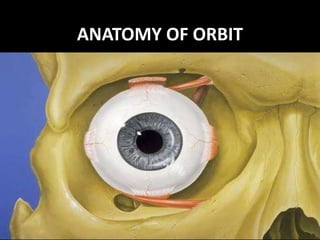
Chandigarh Call Girls Service ❤️🍑 9809698092 👄🫦Independent Escort Service Cha...
Chandigarh Call Girls Service ❤️🍑 9809698092 👄🫦Independent Escort Service Cha...Chandigarh Call Girls Service ❤️🍑 9809698092 👄🫦Independent Escort Service Cha...
Chandigarh Call Girls Service ❤️🍑 9809698092 👄🫦Independent Escort Service Cha...
 
Bandra East [ best call girls in Mumbai Get 50% Off On VIP Escorts Service 90...
Bandra East [ best call girls in Mumbai Get 50% Off On VIP Escorts Service 90...Bandra East [ best call girls in Mumbai Get 50% Off On VIP Escorts Service 90...
Bandra East [ best call girls in Mumbai Get 50% Off On VIP Escorts Service 90...
 
Ahmedabad Call Girls Book Now 9630942363 Top Class Ahmedabad Escort Service A...
Ahmedabad Call Girls Book Now 9630942363 Top Class Ahmedabad Escort Service A...Ahmedabad Call Girls Book Now 9630942363 Top Class Ahmedabad Escort Service A...
Ahmedabad Call Girls Book Now 9630942363 Top Class Ahmedabad Escort Service A...
 
Race Course Road } Book Call Girls in Bangalore | Whatsapp No 6378878445 VIP ...
Race Course Road } Book Call Girls in Bangalore | Whatsapp No 6378878445 VIP ...Race Course Road } Book Call Girls in Bangalore | Whatsapp No 6378878445 VIP ...
Race Course Road } Book Call Girls in Bangalore | Whatsapp No 6378878445 VIP ...
 
❤️Amritsar Escorts Service☎️9815674956☎️ Call Girl service in Amritsar☎️ Amri...
❤️Amritsar Escorts Service☎️9815674956☎️ Call Girl service in Amritsar☎️ Amri...❤️Amritsar Escorts Service☎️9815674956☎️ Call Girl service in Amritsar☎️ Amri...
❤️Amritsar Escorts Service☎️9815674956☎️ Call Girl service in Amritsar☎️ Amri...
 
Call Girl In Indore 📞9235973566📞 Just📲 Call Inaaya Indore Call Girls Service ...
Call Girl In Indore 📞9235973566📞 Just📲 Call Inaaya Indore Call Girls Service ...Call Girl In Indore 📞9235973566📞 Just📲 Call Inaaya Indore Call Girls Service ...
Call Girl In Indore 📞9235973566📞 Just📲 Call Inaaya Indore Call Girls Service ...
 
❤️Call Girl Service In Chandigarh☎️9814379184☎️ Call Girl in Chandigarh☎️ Cha...
❤️Call Girl Service In Chandigarh☎️9814379184☎️ Call Girl in Chandigarh☎️ Cha...❤️Call Girl Service In Chandigarh☎️9814379184☎️ Call Girl in Chandigarh☎️ Cha...
❤️Call Girl Service In Chandigarh☎️9814379184☎️ Call Girl in Chandigarh☎️ Cha...
 
Chandigarh Call Girls Service ❤️🍑 9809698092 👄🫦Independent Escort Service Cha...
Chandigarh Call Girls Service ❤️🍑 9809698092 👄🫦Independent Escort Service Cha...Chandigarh Call Girls Service ❤️🍑 9809698092 👄🫦Independent Escort Service Cha...
Chandigarh Call Girls Service ❤️🍑 9809698092 👄🫦Independent Escort Service Cha...
 
Control of Local Blood Flow: acute and chronic
Control of Local Blood Flow: acute and chronicControl of Local Blood Flow: acute and chronic
Control of Local Blood Flow: acute and chronic
 
Pune Call Girl Service 📞9xx000xx09📞Just Call Divya📲 Call Girl In Pune No💰Adva...
Pune Call Girl Service 📞9xx000xx09📞Just Call Divya📲 Call Girl In Pune No💰Adva...Pune Call Girl Service 📞9xx000xx09📞Just Call Divya📲 Call Girl In Pune No💰Adva...
Pune Call Girl Service 📞9xx000xx09📞Just Call Divya📲 Call Girl In Pune No💰Adva...
 
Jaipur Call Girl Service 📞9xx000xx09📞Just Call Divya📲 Call Girl In Jaipur No💰...
Jaipur Call Girl Service 📞9xx000xx09📞Just Call Divya📲 Call Girl In Jaipur No💰...Jaipur Call Girl Service 📞9xx000xx09📞Just Call Divya📲 Call Girl In Jaipur No💰...
Jaipur Call Girl Service 📞9xx000xx09📞Just Call Divya📲 Call Girl In Jaipur No💰...
 
Call 8250092165 Patna Call Girls ₹4.5k Cash Payment With Room Delivery
Call 8250092165 Patna Call Girls ₹4.5k Cash Payment With Room DeliveryCall 8250092165 Patna Call Girls ₹4.5k Cash Payment With Room Delivery
Call 8250092165 Patna Call Girls ₹4.5k Cash Payment With Room Delivery
 
👉Chandigarh Call Girl Service📲Niamh 8868886958 📲Book 24hours Now📲👉Sexy Call G...
👉Chandigarh Call Girl Service📲Niamh 8868886958 📲Book 24hours Now📲👉Sexy Call G...👉Chandigarh Call Girl Service📲Niamh 8868886958 📲Book 24hours Now📲👉Sexy Call G...
👉Chandigarh Call Girl Service📲Niamh 8868886958 📲Book 24hours Now📲👉Sexy Call G...
 
Gorgeous Call Girls Dehradun {8854095900} ❤️VVIP ROCKY Call Girls in Dehradun...
Gorgeous Call Girls Dehradun {8854095900} ❤️VVIP ROCKY Call Girls in Dehradun...Gorgeous Call Girls Dehradun {8854095900} ❤️VVIP ROCKY Call Girls in Dehradun...
Gorgeous Call Girls Dehradun {8854095900} ❤️VVIP ROCKY Call Girls in Dehradun...
 
ANATOMY AND PHYSIOLOGY OF RESPIRATORY SYSTEM.pptx
ANATOMY AND PHYSIOLOGY OF RESPIRATORY SYSTEM.pptxANATOMY AND PHYSIOLOGY OF RESPIRATORY SYSTEM.pptx
ANATOMY AND PHYSIOLOGY OF RESPIRATORY SYSTEM.pptx
 
7 steps How to prevent Thalassemia : Dr Sharda Jain & Vandana Gupta
7 steps How to prevent Thalassemia : Dr Sharda Jain & Vandana Gupta7 steps How to prevent Thalassemia : Dr Sharda Jain & Vandana Gupta
7 steps How to prevent Thalassemia : Dr Sharda Jain & Vandana Gupta
 
ANATOMY AND PHYSIOLOGY OF REPRODUCTIVE SYSTEM.pptx
ANATOMY AND PHYSIOLOGY OF REPRODUCTIVE SYSTEM.pptxANATOMY AND PHYSIOLOGY OF REPRODUCTIVE SYSTEM.pptx
ANATOMY AND PHYSIOLOGY OF REPRODUCTIVE SYSTEM.pptx
 
Jual Obat Aborsi Di Dubai UAE Wa 0838-4800-7379 Obat Penggugur Kandungan Cytotec
Jual Obat Aborsi Di Dubai UAE Wa 0838-4800-7379 Obat Penggugur Kandungan CytotecJual Obat Aborsi Di Dubai UAE Wa 0838-4800-7379 Obat Penggugur Kandungan Cytotec
Jual Obat Aborsi Di Dubai UAE Wa 0838-4800-7379 Obat Penggugur Kandungan Cytotec
 
Premium Call Girls Nagpur {9xx000xx09} ❤️VVIP POOJA Call Girls in Nagpur Maha...
Premium Call Girls Nagpur {9xx000xx09} ❤️VVIP POOJA Call Girls in Nagpur Maha...Premium Call Girls Nagpur {9xx000xx09} ❤️VVIP POOJA Call Girls in Nagpur Maha...
Premium Call Girls Nagpur {9xx000xx09} ❤️VVIP POOJA Call Girls in Nagpur Maha...
 
Goa Call Girl Service 📞9xx000xx09📞Just Call Divya📲 Call Girl In Goa No💰Advanc...
Goa Call Girl Service 📞9xx000xx09📞Just Call Divya📲 Call Girl In Goa No💰Advanc...Goa Call Girl Service 📞9xx000xx09📞Just Call Divya📲 Call Girl In Goa No💰Advanc...
Goa Call Girl Service 📞9xx000xx09📞Just Call Divya📲 Call Girl In Goa No💰Advanc...
 

Anatomy of orbit

  • 2. REFERENCES 1. A K Khurana & Indu Khurana – Anatomy and physiology of eye. 2. Parsons diseases of the EYE - by Ramanjit sihota and Radhika tandon. 3. Comprehensive Ophthalmology by A K Khurana.
  • 3. > Bony orbit is formed from the mesenchyme surrounding the optic vesicle beginning at 06 weeks of embryonic age.
  • 4. FORMED BY SEVEN BONES • 1. Frontal • 2. Ethamoid • 3. Lacrimal • 4. Palatine • 5. Maxilla • 6. Zygomatic • 7. Sphenoid
  • 5. SHAPE • The two bony orbits are quadrangular truncated pyramids b/w anterior cranial fossa above and maxillary sinuses below. • The medial walls of two orbits are parallel to each other. • Lateral wall of each orbit lies at an angle > 45 ’ with medial wall > 90 ‘ with lateral wall of other orbit.
  • 6. DIMENSIONS • Depth – 42 mm along medial wall 50 mm along lateral wall. . Base – 40 mm in width 35 mm in height. . Inraorbital width – 25mm(medial margins) . Extraorbital width – 100mm(lateral margins)
  • 7. MEDIAL WALL • Quadrilateral in shape. • Formed by, from front to back- 1. frontal process of maxilla 2. the lacrimal bone 3. orbital plate of the ethamoid 4. body of sphenoid
  • 8. Cont… • The anterior part of medial wall bears the lacrimal sac fossa. Medial to lacrimal fossa lie the anterior ethamoidal sinus(upper part) and middle meatus of nose(lower part). • Attachments – posteriorly are 1. Horner’s muscle(lacrimal fibres of orbicularis) 2. Septum orbitale 3. Check ligament of the medial rectus muscle
  • 9. RELATIONS • Medial to the medial wall lie anterior ethamoidal sinuses, middle meatus of nose, middle and posterior ethamoidal sinuses and sphenoidal sinus. • Orbital surface of medial wall is related to superior oblique muscle in the upper part near the roof and medial rectus muscle in the middle part. In between these two muscles- anterior and posterior ethamoidal nerve, infratrochlear nerve and terminal branch of ophthalmic artery.
  • 10.
  • 11. CLINICAL APPLICATIONS • Medial wall is the thinnest wall of orbit(0.2-0.4 mm) and also called as lamina papyracea. • Ethamoiditis being the commonest cause of orbital cellulitis. • Medila wall is frequently eroded by inflammatory lesions, cysts and neoplasms that originate from adjacent air sinuses. • Easily fractured during injuries as well as orbitotomy operations. • During surgery along this wall, heamorrhage is most troublesome due to injury to ethamoidal vessels. • The medial wall can be easily visualised with routine PA radiographs of orbit.
  • 12. INFERIOR WALL (FLOOR) • Triangular in shape, shortest of all walls and extending about 35 to 40 mm from inf margin. • Formed by 3 bones- 1. Maxillary bone medially 2. Zygomatic bone laterally and 3. Palatine bone posteriorly. . It slopes downwards from posterior to anterior by ~ 20’. . Posterior part of floor is separated from lateral wall by inferior orbital fissure.
  • 13. RELATIONS • Below it is related to maxillary sinus and palatine air cells. • Above it is related to inferior rectus muscle, inferior oblique muscle and nerve to inferior oblique. • Inferior oblique muscle originates just lateral to opening of nasolacrimal duct.
  • 14.
  • 15. CLINICAL APPLICATIONS • Orbital floor being thin is commonly involved in ‘ blow-out fractures’ and also is easily invaded by tumors of maxillary antrum. • Floor is best visualised with standard posteroanterior radiographs.
  • 16. LATERAL WALL • It is thickest , strongest, triangular in shape. • It is formed by – 1. Zygomatic bone anteriorly 2. Greater wing of sphenoid bone posteriorly. . On the posterior part of lateral wall – a bony projection – spina recti lateralis. . On anterior part – a bony projection – lateral orbital tubercle of Whitnall.( att to check ligament of lateral rectus muscle, and to suspensary ligament of eyeball, lateral palpebral ligament and aponeurosis of levator muscle. . Posteriorly lateral wall is separated from the roof by superior orbital fissure and from the floor by the inferior orbital fissure.
  • 17. RELATIONS • Laterally , the lateral wall separates the orbit from temporal fossa anteriorly and from the middle cranial fossa posteriorly. • Medially it is related to lateral rectus, lacrimal nerve and vessels and zygomatic nerve.
  • 18.
  • 19. CLINICAL APPLICATIONS • Lateral wall covers only posterior half of eyeball. Anterior half is not covered by bone. Hence ,palpation of retrobulbar tumors is easier from lateral. • Lateral orbital surgical approach is common. • No foramina in lateral wall, so anterior part can be breached without serious haemorrhgae.
  • 20. ROOF (VAULT) • Triangular in shape and formed by mainly orbital plate of frontal bone and posteriorly by the lesser wing of sphenoid bone at apex. • Anterolateral part has a depression – fossa for the lacrimal gland. • Roof slopes backwards and downwards towards apex. • Trochlear fossa- small depression at the junction of roof and medial wall- for the pulley of superior oblique.
  • 21. RELATIONS • Above its related to frontal lobe of cerebrum, meninges and frontal sinuses. • Below are periorbita, frontal nerve, levator palpebrae superioris, superior rectus, superior oblique, trochlear nerve and lacrimal gland. • At junction of roof and lateral wall is a gap posteriorly- superior orbital fissure.
  • 22.
  • 23. CLINICAL APPLICATIONS > Roof is thin throughout its extent and the periorbita easily peels away from its undersurface > On the cranial side , the dura can be easily lifted, because the roof is not perforated by nerves or vessels > Sharp objects injury to roof can penetrate and damage the frontal lobe.
  • 24. SCHEMATIC VIEW OF ORBITAL CAVITY
  • 25. BASE OF ORBIT • Anterior open end of orbit is base, bounded by orbital margins. • Margins are formed by a ring of compact bone, gives attachment to the septum orbitale. > Superior orbital margin – formed by orbital arch of the frontal bone – lateral 2/3 is sharp and medial 1/3 is rounded – at the junction is supraorbital notch, which transmits supraorbital nerve and artery. 10 mm medial to notch is supratrochlear groove which transmits supratrochlear nerve and artery.
  • 26. Cont…. > Lateral margin – its strongest , formed by zygomatic process of frontal bone and zygomatic bone. It is deficient anteriorly. > Inferior orbital margin - formed by zygomatic bone laterally and maxilla medially. 4-5mm below margin is infraorbital foramen which transmits infraorbital nerve and vessels. > Medial orbital margin - formed by ant lacrimal crest on frontal process of maxilla below and frontal bone above. Its upper part becomes continuous with the posterior lacrimal crest.
  • 27. APEX OF ORBIT • It is the posterior end of orbit. Where 4 walls converge • Apex has two orifices optic canal and sup orbital fissure situated in sphenoid bone. • At the apex jus below optic canal inf orbital fissure joins the sup orbital fissure and is contiguous with foramen rotundum .
  • 28. OPTIC CANAL • It connects orbit to middle cranial fossa. • Transmits optic nerve and ophthalmic artery. • Normal adult dimensions are attained by the age of 4-5 years. • Average length is 6-11mm. • Orbital end is vertically oval, cranial end is horizontally oval, and center is circular. • Tumors like optic nerve glioma and meningiomas may lead to enlargement of optic canal(detected on X-ray films.
  • 29. SUPERIOR ORBITAL FISSURE • Comma shaped aperture in orbital cavity, bounded by lesser and greater wing of sphenoid bone. It is situated lateral to optic foramen. • Fissure is divided into upper, middle and lower parts by common tendinous ring(for origin of recti). • Structures passing through - are 1. Lacrimal, frontal and trochlear nerve. 2. Sup ophthalmic vein and recurrent branch of ophthalmic artery. 3. Sup and inf divisions of 3rd C N, nasociliary branch of ophthalmic division of 5th and 6th C N. 4. Inf ophthalmic vein.
  • 30. INFERIOR ORBITAL FISSURE • Inf orbital fissure lies just below the sup orbital fissure b/w lateral wall and floor of orbit, giving access to the pterygopalatine and inferotemporal fossae. • Structures passing through – are 1. Infraorbital and zygomatic branches of the maxillary div of 5th C N. 2. Orbital branch of pterygopalatine ganglion. 3. Branch of inf ophthalmic vein(communicates with pterygoid plexus).
  • 31. PERIORBITA • It is the periosteum lining the orbital surface of the bones of orbit. • In the optic canal , the dural sheath of optic nerve is closely adherent to periorbita. • At the orbital margin , periorbia is thickened to form the arcus marginale to which the septum orbitale is attached. • At the post lacrimal crest , periorbita splits into two layers and encloses lacrimal sac and reunite at ant lacrimal crest. • At the apex the periorbita is thickened to form the common tendinous ring of zinn.
  • 32. FASCIA BULBI • Fascia bulbi or tenon’s capsule is a dense, ealastic, and vascular connective tissue that envelops the globe from the limbus to the optic disc. • The lower part of fascia bulbi is thickened and take part in the formation of a sling or hammock on which the globe rests( suspensary ligament of lockwood). • Around the distal end of the optic nerve. The fascia is fused with the dural sheath of the optic nerve.
  • 33. SURGICAL SPACES IN THE ORBIT
  • 34. 1. SUBPERIOSTEAL SPACE It is a potential space b/w orbital bones and periorbita. Dermoid cyst, epidermoid cyst, mucocele, subperiosteal abscess, myeloma, osteomatous tumors, haematoma and fibrous dysplasia are commonly seen in this space. Plain X-ray are most useful in diagnosing the tumors of this space.
  • 35. 2. PERIPHERAL ORBITAL SPACE • It is bounded peripherally by periorbita, internally by four extraocular muscles with their intermuscular septa and anteriorly bythe septum orbitale. • Tumors in this space produce eccentric proptosis and can usually be palpated. • Common tumors are – malignant lympoma, capillary haemangioma of childhood, intrensic neoplasms, of lacrimal gland and pseudotumors.
  • 36. 3. CENTRAL SPACE • Also called muscular cone or retrobulbar space. • Bounded anteriorly by tenon’s capsule and peripherally by recti muscles and their intermuscular septa. • Contents – optic nerve and its meninges, divisions of 3rd C N, 6th CN, nasociliary nerve, ciliary ganglion, ophthalmic artery and sup ophthalmic vein. • Tumors of this space includes - cavernous haemangioma of adults, solitary neurofibroma, neurilemomas, nodular orbital haemangiomas and optic nerve gliomas . And usually produce axial proptosis.
  • 37. 4. SUB-TENON’S SPACE • It is a potential space around the eyeball b/w the sclera and tenon’s capsule. • Pus collected in this space is drained by incision of tenon’s capsule through the conjunctiva.
  • 38. APERTURES AT THE BASE OF ORBIT • The base of orbit is partially closed by the globe and extraocular muscles with their facial expansions . These expansions and two oblique muscles bound about 5 orifices b/w orbital margin and globe. • Through these orifices fat may herniate from orbit to come into contact with septum orbitale.
  • 39. • 1. Superior aperture • 2. Superomedial aperture • 3. Inferomedial aperture • 4. Inferior aperture • 5. Inferolateral aperture.