Orbital blowout fracture
•
20 likes•4,048 views

Orbital trauma and surgical management

Recommended
More Related Content
What's hot
What's hot (20)
Similar to Orbital blowout fracture
Similar to Orbital blowout fracture (20)
More from Cairo university
More from Cairo university (20)
Recently uploaded
Recently uploaded (20)
Orbital blowout fracture
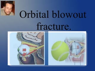
- 2. An orbital blowout fracture is a traumatic deformity of the orbital floor or medial wall, typically resulting from impact of a blunt object larger than the orbital aperture, or eye socket. Etiology:
- 3. Anatomical considerations: 3-Optic nerve foramen III = Oculomotor nerve (Motor) IV = Trochlear nerve (Motor) VI = Abducens nerve V1 = Ophthalmic (Sensory) 1-Superior orbital fissure 2-Inferior orbital fissure V2 = Infra-orbital nerve. II = Optic nerve. Intra-orbital neurovascular structures. 1 2 3
- 4. Fronta l Sphenoi d Zygomatic Maxillary Ethmoid Lacrimal Anatomical considerations: The medial wall is slightly thinner (0.25 mm vs 0.50 mm) of floor thickness
- 5. Anatomical considerations: Lateral rectus muscle Medial rectus muscle Superior rectus muscle Inferior rectus muscle Superior oblique muscle Inferior oblique muscle Ocular muscles:
- 6. Extra-ocular muscles Eye movement of lateral rectus muscle,
- 7. Extra-ocular muscles Eye movement of superior oblique muscle,
- 8. II- Optic (vision) III = Oculomotor nerve (Motor) double vision (diplopia) eyelid drooping (ptosis) pupil dilation (mydriasis) IV = Trochlear nerve (Motor)”Sup. Obliq. M double vision (diplopia) Squint.lateral sup VI = Abducens nerve. double vision (diplopia)”Lat. Rec. M Squint.-medial V1 = Ophthalmic (Sensory) Corneal anesthesia. V2 = Infra-Orbital (Sensory) Skin of lower eye lid-nose-upper lip. Clinical significance of intra-orbital structures:
- 9. Orbital trauma: Edema or hematoma within the internal orbit. Orbital blowout fracture. Clinical significance of intra-orbital structures: Increase the intra-orbital pressure, compromising the intra-orbital neurovascular structures.
- 10. Signs and symptoms: Loss of sensation due to infraorbital nerve injury Diplopia and enophthalmos. Orbital pain Limitation of eye movement. Orbital and lid subcutaneous ecchymosis or emphysema. Diagnosis:
- 11. Diagnosis: Clinical examination: Ocular motility: Test eye movements in all possible directions. Presence of squint or improper eye movement denoting muscle entrapment or neural damage. Forced duction test used for differentiation.
- 12. Pupil function: Pupillary light reflex provides a useful diagnostic tool for testing the integrity of the sensory (CN II) and motor (CN III) functions of the eye. Direct reflex. Consensual reflex Diagnosis: Clinical examination:
- 13. Diagnosis: Imaging: Plain radiographs do not sensitively capture blowout fractures. C.T: (coronal cut) teardrop sign, polypoid mass consists of herniated orbital contents, periorbital fat and inferior rectus muscle. The affected sinus is partially opacified on radiograph. Air-fluid level in maxillary sinus due to presence of blood.
- 14. Diagnosis: Imaging: Medial wall fracture is less common than floor fracture due to presence of honey comb ethmoid bone.
- 15. Inflammatory myopathy Vitreous hemorrhage Diagnosis: Imaging: MRI : Detection of Inflammatory myopathy Optic nerve condition Vitrous hemorrhage. Optic nerve damage
- 16. All patients should follow-up with an ophthalmologist within one week of the fracture (Retinal examination, Intra-ocular pressure). To prevent orbital emphysema, patients are advised to avoid blowing of the nose. Nasal decongestants are commonly used. It is also common practice to administer prophylactic antibiotics when the fracture enters a sinus.(Amoxicillin-clavulanate and azithromycin) Corticosteroids are used to decrease swelling. Surgical repair of a "blowout" is safely postponed for up to two weeks, if necessary, to let the swelling subside. Pre-operative preparation:
- 17. Surgery is indicated if there is : Enophthalmos greater than 2 mm. Double vision on primary or inferior gaze. Entrapment of extraocular muscles. Fracture involves greater than 50% of the orbital floor. Most blowout fractures heal spontaneously without significant consequence. Surgical management It can be safely postponed for up to two weeks
- 18. Reconstruction is usually performed through a Transconjunctival incision. Surgical management Transconjunctival
- 19. Reconstruction through a Subciliary incision. Surgical management
- 20. Reconstruction is usually performed with Titanium mesh or porous polyethylene or polydioxanone . Surgical management polydioxanone polyethylene
- 21. Complications: More recently, there has been success with endoscopic, or minimally invasive, approaches. partial relief from double vision or a sunken eye. Ectropion of lower eye lid. Graft morbidity. Sunken eye Ectropion Graft