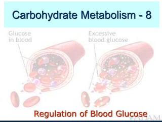
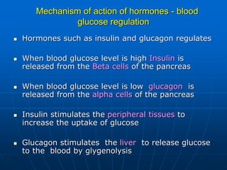
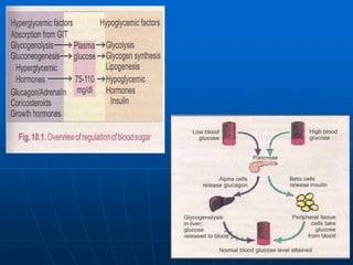
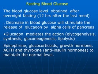
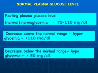
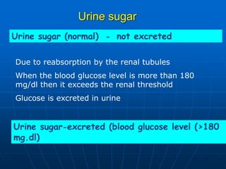
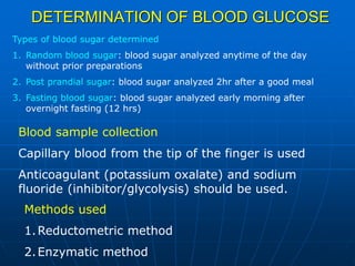
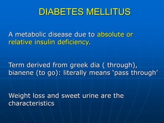
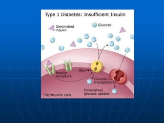
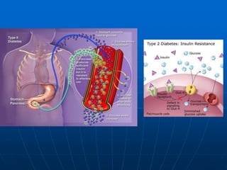
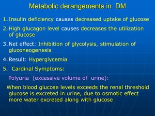
Regulation of Blood Glucose
Carbohydrate Metabolism - 8
Learning Objectives
• Objectives:
• Regulation of blood glucose levels
• Metabolic derangements in diabetes
Introduction
Regulation of blood glucose
• An effectively regulated system
•Hormones such as Insulin, glucagon, adrenaline
mediates this action by controlling the various
metabolic pathways of carbohydrates (glucose,
glycogen)
•Important process for the functioning of brains,
kidney and RBC’S
•Glucose is the major source of energy or fuel
Factors maintaining the blood sugar
Basically depends on the balance between glucose
entering and leaving the ECF
I. Factors controlling the entry of glucose into blood
a. Absorption from intestines
b. Glycogenolysis
c. Gluconeogenesis
II. Factors controlling the depletion of glucose in blood
a. Utilisation by tissues for energy
b. Glycogen synthesis
c. Conversion into fat (lipogenesis)
Mechanism of action of hormones - blood
glucose regulation
 Hormones such as insulin and glucagon regulates
 When blood glucose level is high Insulin is
released from the Beta cells of the pancreas
 When blood glucose level is low glucagon is
released from the alpha cells of the pancreas
 Insulin stimulates the peripheral tissues to
increase the uptake of glucose
 Glucagon stimulates the liver to release glucose
to the blood by glygenolysis
Post prandial glucose
The blood glucose level obtained following
(2 hrs) after a good meal is referred as post
prandial glucose After meal, glucose is
absorbed form the intestine and enters blood.
Increased in blood glucose will stimulate the
release of insulin by beta cells.
Insulin mediates the action (peripheral tissue
uptake, glycogen synthesis, lipogenesis)
Fasting Blood Glucose
The blood glucose level obtained after
overnight fasting (12 hrs after the last meal)
. Decrease in blood glucose will stimulate the
release of glucagon by alpha cells of pancreas
•Glucagon mediates the action (glycogenolysis,
synthesis, gluconeogenesis, lipolysis)
Epinephrine, glucocorticoids, growth hormone,
ACTH and thyroxine (anti-insulin hormones) to
maintain the normal level.
NORMAL PLASMA GLUCOSE LEVEL
Fasting plasma glucose level
(normal) normoglycemia - 75-110 mg/dl
Increase above the normal range - hyper
glycemia ~ >110 mg/dl
Decrease below the normal range- hypo
glycemia ~ > 50 mg/dl
Urine sugar
Urine sugar (normal) - not excreted
Urine sugar-excreted (blood glucose level (>180
mg.dl)
Due to reabsorption by the renal tubules
When the blood glucose level is more than 180
mg/dl then it exceeds the renal threshold
Glucose is excreted in urine
DETERMINATION OF BLOOD GLUCOSE
Blood sample collection
Capillary blood from the tip of the finger is used
Anticoagulant (potassium oxalate) and sodium
fluoride (inhibitor/glycolysis) should be used.
Methods used
1.Reductometric method
2.Enzymatic method
Types of blood sugar determined
1. Random blood sugar: blood sugar analyzed anytime of the day
without prior preparations
2. Post prandial sugar: blood sugar analyzed 2hr after a good meal
3. Fasting blood sugar: blood sugar analyzed early morning after
overnight fasting (12 hrs)
Reductometric methods
Based on the reducing property of glucose
Alkaline copper reagent is reduced by glucose (cupric
to cuprous) and react with phosphomolybdate to
form a colored compound, measured colorimetrically
e.g: Folin-wu method, Nelson Somayagi method.
Enzymatic method
Autoanalysers are used
The extent of oxidation of glucose by the addition of
enzymes is measured
e.g: Glucose oxidase method
Glucometer (quick monitoring kit)
Reagent (enzyme) is immobilized and in dry form
in a strip
Blood is placed on the strip
Color intensity is measured by photometry
GLUCOSE TOLERANCE TEST
Ability of a person to metabolize a given load of
glucose is referred to as glucose tolerance
Done to assess the status of the carbohydrate
metabolism
A known quantity of glucose is administered
and the blood level of glucose is determined
GTT is a standardized test and highly useful for
the diagnosis of Diabetes mellitus
Method
1. Preparation of the patient – 12 hrs fasting,
good carbohydrate meal (prior 3 days),
avoid drugs.
2. Administration of glucose – Oral, dose -
75g/250-300 ml of water)
3. Sample collection : blood and urine samples
( 0hr, 1, 11/2,2,21/2)
4. Blood sugar analysis
5. Interpretation : GTT curve :
Plasma glucose VS time
DIABETES MELLITUS
A metabolic disease due to absolute or
relative insulin deficiency.
Term derived from greek dia ( through),
bianene (to go): literally means ‘pass through’
Weight loss and sweet urine are the
characteristics
Classification of Diabetes mellitus
1.Type 1 DM: Insulin dependent DM(IDDM), 5%
total cases, circulating insulin is deficient.
Sub classes: Immune mediated, idiopathic
2. Type 2 DM: Non Insulin dependent DM (NIDDM),
most common type, circulating insulin is normal or
slightly altered depending on the stage
Sub classes: obese, non-obese and maturity
onset diabetes of young (MODY)
3. Diabetic prone states: Gestational diabetes (GDM)
Impaired glucose tolerance (IGT), Impaired
fasting glycemia
4. Secondary causes: a. Endocrinopathies (cushing’s disease<
thyrotoxicosis, acromegaly) b. Drug-induced (steroids, beta
blockers) c. pancreatic diseases (chronic pancretitis)
TYPE1:
•Decreased insulin production
•Low level of circulating insulin, increased blood and urine glucose
•Patient is dependent on insulin injections
•Onset below 30 yrs common in adolescent
•Clinical features: polyuria, polydipsia, loss of body weight, ketosis
•Auto immune basis is seen in most cases- circulating antibodies
against insulin is seen in 50%
•Circulating antibodies against islet cell cytoplasmic protiens are
seen in 80% cases.
•Genetically inherited, viral infection
Without insulin, patients with type 1 diabetes develop
severely elevated blood sugar levels. This leads to increased
urine glucose, which in turn leads to excessive loss of fluid
and electrolytes in the urine.
Lack of insulin also causes the inability to store fat and protein
along with breakdown of existing fat and protein stores.
This dysregulation, results in the process of ketosis and the
release of ketones into the blood. Ketones turn the blood
acidic, a condition called diabetic ketoacidosis (DKA).
Symptoms of diabetic ketoacidosis include nausea, vomiting,
and abdominal pain.
Without prompt medical treatment, patients with diabetic
ketoacidosis can rapidly go into shock, coma, and even death.
TYPE 11
•Decreased biological response to insulin or insulin
resistance
•Relative insulin deficiency
• beta cell failure
•Commonly seen above 40 yrs, 60% cases are
obese, 40% non-obese
•Some show mutation in insulin-receptor gene, ob
gene in adipose tissue and plasma leptin levels are
high
•MODY due to defective glucokinase, gene
mutation.
In patients with type 2 diabetes, stress, infection, and medications (such as
corticosteroids) can also lead to severely elevated blood sugar levels.
Accompanied by dehydration, severe blood sugar elevation in patients with
type 2 diabetes can lead to an increase in blood osmolality (hyperosmolar
state). This condition can lead to coma (hyperosmolar coma). A hyperosmolar
coma usually occurs in elderly patients with type 2 diabetes. Like diabetic
ketoacidosis, a hyperosmolar coma is a medical emergency. Immediate
treatment with intravenous fluid and insulin is important in reversing the
hyperosmolar state.
Metabolic derangements in DM
1.Insulin deficiency causes decreased uptake of glucose
2.High glucagon level causes decreases the utilization
of glucose
3.Net effect: Inhibition of glycolysis, stimulation of
gluconeogenesis
4.Result: Hyperglycemia
5. Cardinal Symptoms:
Polyuria (excessive volume of urine):
When blood glucose levels exceeds the renal threshold
glucose is excreted in urine, due to osmotic effect
more water excreted along with glucose
Polydipsia (excessive thirst):
To compensate the loss of water thirst centre is
activated, more water is taken.
Polyphagia (more food intake):
Ineffective glucose utilization will result in increased
breakdown of fat and protein – loss of weight
Finally lead to increased uptake of food
Laboratory investigations in DM
1.Random blood sugar estimation
2.Oral GTT
3.Complete lipid profile
4.Microalbuminuria
Clinical monitoring of DM
HYPERGLYCEMIA
•Persistent hyperglycemia is present can lead to chronic complications.
•Periodic check-up of blood glucose level (fasting and postprandial) is
done every 3 months
•Blood glucose levels to be maintained within normal limits
•Treatment regime should be continued to maintain the normal level
Glycated Hemoglobin
Measurement of Glycated hemoglobin is the best index for long term
control of blood glucose.
Enzymatic addition of protein to sugar is glycosylation; non-enzymatic
process is called glycation.
In hyperglycemic condition, proteins undergo glucation; glucose
forms schiff base with the N-terminal amino group of proteins.
Determination of glycated-Hb is not for diagnosis but for monitoring the
DM.
Normal level of HbA1 - 4-8%
In diabetic- 10-15%
HbA1 reveals the mean glucose values for previous 8-10 weeks
Un effected by recent food intake or food changes
HbA1 indicates the average glucose concentration In plasma over a period
of time
An elevated level indicates poor control of DM
Estimation should be done every 3 months in all insulin –treated patient
Advanced Glycation end products (AGE’s)
Found in tissue proteins such as collagen in diabetic patients
AGE formation is proportional to the square of glucose concentration
AGE- Hb is seen in the blood.
•Learning outcomes:
•At the end of the lecture, students should be able to:
•Explain how glucose homeostasis is maintained in the body
•Discuss the metabolic derangements associated with diabetes.

blood glucose + DM.ppt

  • 1.
    Regulation of BloodGlucose Carbohydrate Metabolism - 8
  • 2.
    Learning Objectives • Objectives: •Regulation of blood glucose levels • Metabolic derangements in diabetes
  • 3.
    Introduction Regulation of bloodglucose • An effectively regulated system •Hormones such as Insulin, glucagon, adrenaline mediates this action by controlling the various metabolic pathways of carbohydrates (glucose, glycogen) •Important process for the functioning of brains, kidney and RBC’S •Glucose is the major source of energy or fuel
  • 4.
    Factors maintaining theblood sugar Basically depends on the balance between glucose entering and leaving the ECF I. Factors controlling the entry of glucose into blood a. Absorption from intestines b. Glycogenolysis c. Gluconeogenesis II. Factors controlling the depletion of glucose in blood a. Utilisation by tissues for energy b. Glycogen synthesis c. Conversion into fat (lipogenesis)
  • 5.
    Mechanism of actionof hormones - blood glucose regulation  Hormones such as insulin and glucagon regulates  When blood glucose level is high Insulin is released from the Beta cells of the pancreas  When blood glucose level is low glucagon is released from the alpha cells of the pancreas  Insulin stimulates the peripheral tissues to increase the uptake of glucose  Glucagon stimulates the liver to release glucose to the blood by glygenolysis
  • 7.
    Post prandial glucose Theblood glucose level obtained following (2 hrs) after a good meal is referred as post prandial glucose After meal, glucose is absorbed form the intestine and enters blood. Increased in blood glucose will stimulate the release of insulin by beta cells. Insulin mediates the action (peripheral tissue uptake, glycogen synthesis, lipogenesis)
  • 8.
    Fasting Blood Glucose Theblood glucose level obtained after overnight fasting (12 hrs after the last meal) . Decrease in blood glucose will stimulate the release of glucagon by alpha cells of pancreas •Glucagon mediates the action (glycogenolysis, synthesis, gluconeogenesis, lipolysis) Epinephrine, glucocorticoids, growth hormone, ACTH and thyroxine (anti-insulin hormones) to maintain the normal level.
  • 10.
    NORMAL PLASMA GLUCOSELEVEL Fasting plasma glucose level (normal) normoglycemia - 75-110 mg/dl Increase above the normal range - hyper glycemia ~ >110 mg/dl Decrease below the normal range- hypo glycemia ~ > 50 mg/dl
  • 11.
    Urine sugar Urine sugar(normal) - not excreted Urine sugar-excreted (blood glucose level (>180 mg.dl) Due to reabsorption by the renal tubules When the blood glucose level is more than 180 mg/dl then it exceeds the renal threshold Glucose is excreted in urine
  • 12.
    DETERMINATION OF BLOODGLUCOSE Blood sample collection Capillary blood from the tip of the finger is used Anticoagulant (potassium oxalate) and sodium fluoride (inhibitor/glycolysis) should be used. Methods used 1.Reductometric method 2.Enzymatic method Types of blood sugar determined 1. Random blood sugar: blood sugar analyzed anytime of the day without prior preparations 2. Post prandial sugar: blood sugar analyzed 2hr after a good meal 3. Fasting blood sugar: blood sugar analyzed early morning after overnight fasting (12 hrs)
  • 13.
    Reductometric methods Based onthe reducing property of glucose Alkaline copper reagent is reduced by glucose (cupric to cuprous) and react with phosphomolybdate to form a colored compound, measured colorimetrically e.g: Folin-wu method, Nelson Somayagi method. Enzymatic method Autoanalysers are used The extent of oxidation of glucose by the addition of enzymes is measured e.g: Glucose oxidase method
  • 14.
    Glucometer (quick monitoringkit) Reagent (enzyme) is immobilized and in dry form in a strip Blood is placed on the strip Color intensity is measured by photometry
  • 15.
    GLUCOSE TOLERANCE TEST Abilityof a person to metabolize a given load of glucose is referred to as glucose tolerance Done to assess the status of the carbohydrate metabolism A known quantity of glucose is administered and the blood level of glucose is determined GTT is a standardized test and highly useful for the diagnosis of Diabetes mellitus
  • 16.
    Method 1. Preparation ofthe patient – 12 hrs fasting, good carbohydrate meal (prior 3 days), avoid drugs. 2. Administration of glucose – Oral, dose - 75g/250-300 ml of water) 3. Sample collection : blood and urine samples ( 0hr, 1, 11/2,2,21/2) 4. Blood sugar analysis 5. Interpretation : GTT curve : Plasma glucose VS time
  • 18.
    DIABETES MELLITUS A metabolicdisease due to absolute or relative insulin deficiency. Term derived from greek dia ( through), bianene (to go): literally means ‘pass through’ Weight loss and sweet urine are the characteristics
  • 19.
    Classification of Diabetesmellitus 1.Type 1 DM: Insulin dependent DM(IDDM), 5% total cases, circulating insulin is deficient. Sub classes: Immune mediated, idiopathic 2. Type 2 DM: Non Insulin dependent DM (NIDDM), most common type, circulating insulin is normal or slightly altered depending on the stage Sub classes: obese, non-obese and maturity onset diabetes of young (MODY) 3. Diabetic prone states: Gestational diabetes (GDM) Impaired glucose tolerance (IGT), Impaired fasting glycemia
  • 20.
    4. Secondary causes:a. Endocrinopathies (cushing’s disease< thyrotoxicosis, acromegaly) b. Drug-induced (steroids, beta blockers) c. pancreatic diseases (chronic pancretitis) TYPE1: •Decreased insulin production •Low level of circulating insulin, increased blood and urine glucose •Patient is dependent on insulin injections •Onset below 30 yrs common in adolescent •Clinical features: polyuria, polydipsia, loss of body weight, ketosis •Auto immune basis is seen in most cases- circulating antibodies against insulin is seen in 50% •Circulating antibodies against islet cell cytoplasmic protiens are seen in 80% cases. •Genetically inherited, viral infection
  • 22.
    Without insulin, patientswith type 1 diabetes develop severely elevated blood sugar levels. This leads to increased urine glucose, which in turn leads to excessive loss of fluid and electrolytes in the urine. Lack of insulin also causes the inability to store fat and protein along with breakdown of existing fat and protein stores. This dysregulation, results in the process of ketosis and the release of ketones into the blood. Ketones turn the blood acidic, a condition called diabetic ketoacidosis (DKA). Symptoms of diabetic ketoacidosis include nausea, vomiting, and abdominal pain. Without prompt medical treatment, patients with diabetic ketoacidosis can rapidly go into shock, coma, and even death.
  • 23.
    TYPE 11 •Decreased biologicalresponse to insulin or insulin resistance •Relative insulin deficiency • beta cell failure •Commonly seen above 40 yrs, 60% cases are obese, 40% non-obese •Some show mutation in insulin-receptor gene, ob gene in adipose tissue and plasma leptin levels are high •MODY due to defective glucokinase, gene mutation.
  • 25.
    In patients withtype 2 diabetes, stress, infection, and medications (such as corticosteroids) can also lead to severely elevated blood sugar levels. Accompanied by dehydration, severe blood sugar elevation in patients with type 2 diabetes can lead to an increase in blood osmolality (hyperosmolar state). This condition can lead to coma (hyperosmolar coma). A hyperosmolar coma usually occurs in elderly patients with type 2 diabetes. Like diabetic ketoacidosis, a hyperosmolar coma is a medical emergency. Immediate treatment with intravenous fluid and insulin is important in reversing the hyperosmolar state.
  • 26.
    Metabolic derangements inDM 1.Insulin deficiency causes decreased uptake of glucose 2.High glucagon level causes decreases the utilization of glucose 3.Net effect: Inhibition of glycolysis, stimulation of gluconeogenesis 4.Result: Hyperglycemia 5. Cardinal Symptoms: Polyuria (excessive volume of urine): When blood glucose levels exceeds the renal threshold glucose is excreted in urine, due to osmotic effect more water excreted along with glucose
  • 27.
    Polydipsia (excessive thirst): Tocompensate the loss of water thirst centre is activated, more water is taken. Polyphagia (more food intake): Ineffective glucose utilization will result in increased breakdown of fat and protein – loss of weight Finally lead to increased uptake of food
  • 28.
    Laboratory investigations inDM 1.Random blood sugar estimation 2.Oral GTT 3.Complete lipid profile 4.Microalbuminuria
  • 29.
    Clinical monitoring ofDM HYPERGLYCEMIA •Persistent hyperglycemia is present can lead to chronic complications. •Periodic check-up of blood glucose level (fasting and postprandial) is done every 3 months •Blood glucose levels to be maintained within normal limits •Treatment regime should be continued to maintain the normal level Glycated Hemoglobin Measurement of Glycated hemoglobin is the best index for long term control of blood glucose. Enzymatic addition of protein to sugar is glycosylation; non-enzymatic process is called glycation. In hyperglycemic condition, proteins undergo glucation; glucose forms schiff base with the N-terminal amino group of proteins.
  • 31.
    Determination of glycated-Hbis not for diagnosis but for monitoring the DM. Normal level of HbA1 - 4-8% In diabetic- 10-15% HbA1 reveals the mean glucose values for previous 8-10 weeks Un effected by recent food intake or food changes HbA1 indicates the average glucose concentration In plasma over a period of time An elevated level indicates poor control of DM Estimation should be done every 3 months in all insulin –treated patient Advanced Glycation end products (AGE’s) Found in tissue proteins such as collagen in diabetic patients AGE formation is proportional to the square of glucose concentration AGE- Hb is seen in the blood.
  • 32.
    •Learning outcomes: •At theend of the lecture, students should be able to: •Explain how glucose homeostasis is maintained in the body •Discuss the metabolic derangements associated with diabetes.