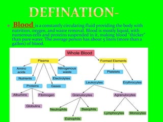
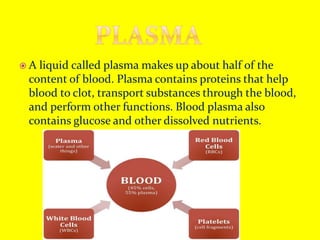
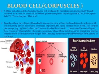
18.1.2 Formed Elements
Erythrocytes, leucocytes and platelets are collectively called formed
elements (Figure 18.1) and they constitute nearly 45 per cent of the blood.
Erythrocytes or red blood cells (RBC) are the most abundant of all
the cells in blood. A healthy adult man has, on an average, 5 millions to
5.5 millions of RBCs mm–3 of blood. RBCs are formed in the red bone
marrow in the adults. RBCs are devoid of nucleus in most of the mammals
and are biconcave in shape. They have a red coloured, iron containing
complex protein called haemoglobin, hence the colour and name of these
cells. A healthy individual has 12-16 gms of haemoglobin in every
100 ml of blood. These molecules play a significant role in transport of
respiratory gases. RBCs have an average life span of 120 days after which
they are destroyed in the spleen (graveyard of RBCs).
Leucocytes are also known as white blood cells (WBC) as they are
colourless due to the lack of haemoglobin. They are nucleated and are
relatively lesser in number which averages 6000-8000 mm–3 of blood.
Leucocytes are generally short lived. We have two main categories of WBCs
– granulocytes and agranulocytes. Neutrophils, eosinophils and basophils
are different types of granulocytes, while lymphocytes and monocytes
are the agranulocytes. Neutrophils are the most abundant cells (60-65
per cent) of the total WBCs and basophils are the least (0.5-1 per cent)
among them. Neutrophils and monocytes (6-8 per cent) are phagocytic
cells which destroy foreign organisms entering the body. Basophils secrete
histamine, serotonin, heparin, etc., and are involved in inflammatory
reactions. Eosinophils (2-3 per cent) resist infections and are also
blood and its functions/B.pharmacy 2 semesterKondal Reddy
Blood Provides the Body's Cells with Oxygen and Removes Carbon Dioxide.
Blood absorbs oxygen from air in the lungs.
It transports the oxygen to cells throughout the body, and it removes waste carbon dioxide from the cells.
Circulation involves the movement of blood in the body which carries nutrients, enzyme etc. to the respective cells and tissues.Moreover the slide is focused on the different parts involved the process of circulation, along with blood grouping and blood coagulation.
Observation of Io’s Resurfacing via Plume Deposition Using Ground-based Adapt...Sérgio Sacani
Since volcanic activity was first discovered on Io from Voyager images in 1979, changes
on Io’s surface have been monitored from both spacecraft and ground-based telescopes.
Here, we present the highest spatial resolution images of Io ever obtained from a groundbased telescope. These images, acquired by the SHARK-VIS instrument on the Large
Binocular Telescope, show evidence of a major resurfacing event on Io’s trailing hemisphere. When compared to the most recent spacecraft images, the SHARK-VIS images
show that a plume deposit from a powerful eruption at Pillan Patera has covered part
of the long-lived Pele plume deposit. Although this type of resurfacing event may be common on Io, few have been detected due to the rarity of spacecraft visits and the previously low spatial resolution available from Earth-based telescopes. The SHARK-VIS instrument ushers in a new era of high resolution imaging of Io’s surface using adaptive
optics at visible wavelengths.
(May 29th, 2024) Advancements in Intravital Microscopy- Insights for Preclini...Scintica Instrumentation
Intravital microscopy (IVM) is a powerful tool utilized to study cellular behavior over time and space in vivo. Much of our understanding of cell biology has been accomplished using various in vitro and ex vivo methods; however, these studies do not necessarily reflect the natural dynamics of biological processes. Unlike traditional cell culture or fixed tissue imaging, IVM allows for the ultra-fast high-resolution imaging of cellular processes over time and space and were studied in its natural environment. Real-time visualization of biological processes in the context of an intact organism helps maintain physiological relevance and provide insights into the progression of disease, response to treatments or developmental processes.
In this webinar we give an overview of advanced applications of the IVM system in preclinical research. IVIM technology is a provider of all-in-one intravital microscopy systems and solutions optimized for in vivo imaging of live animal models at sub-micron resolution. The system’s unique features and user-friendly software enables researchers to probe fast dynamic biological processes such as immune cell tracking, cell-cell interaction as well as vascularization and tumor metastasis with exceptional detail. This webinar will also give an overview of IVM being utilized in drug development, offering a view into the intricate interaction between drugs/nanoparticles and tissues in vivo and allows for the evaluation of therapeutic intervention in a variety of tissues and organs. This interdisciplinary collaboration continues to drive the advancements of novel therapeutic strategies.
Earliest Galaxies in the JADES Origins Field: Luminosity Function and Cosmic ...Sérgio Sacani
We characterize the earliest galaxy population in the JADES Origins Field (JOF), the deepest
imaging field observed with JWST. We make use of the ancillary Hubble optical images (5 filters
spanning 0.4−0.9µm) and novel JWST images with 14 filters spanning 0.8−5µm, including 7 mediumband filters, and reaching total exposure times of up to 46 hours per filter. We combine all our data
at > 2.3µm to construct an ultradeep image, reaching as deep as ≈ 31.4 AB mag in the stack and
30.3-31.0 AB mag (5σ, r = 0.1” circular aperture) in individual filters. We measure photometric
redshifts and use robust selection criteria to identify a sample of eight galaxy candidates at redshifts
z = 11.5 − 15. These objects show compact half-light radii of R1/2 ∼ 50 − 200pc, stellar masses of
M⋆ ∼ 107−108M⊙, and star-formation rates of SFR ∼ 0.1−1 M⊙ yr−1
. Our search finds no candidates
at 15 < z < 20, placing upper limits at these redshifts. We develop a forward modeling approach to
infer the properties of the evolving luminosity function without binning in redshift or luminosity that
marginalizes over the photometric redshift uncertainty of our candidate galaxies and incorporates the
impact of non-detections. We find a z = 12 luminosity function in good agreement with prior results,
and that the luminosity function normalization and UV luminosity density decline by a factor of ∼ 2.5
from z = 12 to z = 14. We discuss the possible implications of our results in the context of theoretical
models for evolution of the dark matter halo mass function.
A brief information about the SCOP protein database used in bioinformatics.
The Structural Classification of Proteins (SCOP) database is a comprehensive and authoritative resource for the structural and evolutionary relationships of proteins. It provides a detailed and curated classification of protein structures, grouping them into families, superfamilies, and folds based on their structural and sequence similarities.
This pdf is about the Schizophrenia.
For more details visit on YouTube; @SELF-EXPLANATORY;
https://www.youtube.com/channel/UCAiarMZDNhe1A3Rnpr_WkzA/videos
Thanks...!
2. Blood is a constantly circulating fluid providing the body with
nutrition, oxygen, and waste removal. Blood is mostly liquid, with
numerous cells and proteins suspended in it, making blood "thicker"
than pure water. The average person has about 5 liters (more than a
gallon) of blood.
3. A liquid called plasma makes up about half of the
content of blood. Plasma contains proteins that help
blood to clot, transport substances through the blood,
and perform other functions. Blood plasma also
contains glucose and other dissolved nutrients.
4. A blood cell, also called a hematocyte, is a cell produced by hematopoiesis and normally found
in blood. In mammals, these fall into three general categories: Erythrocytes (RBC’S) , Leukocytes (
WBC’S) ,Thrombocytes ( Platelets).
Together, these three kinds of blood cells add up to a total 45% of the blood tissue by volume, with
the remaining 55% of the volume composed of plasma, the liquid component of blood. This volume
percentage (e.g., 45%) of cells to total volume is called hematocrit, determined by centrifuge or
flow cytometry .Hemoglobin (the main component of red blood cells) is an iron-containing protein
that facilitates transportation of oxygen from the lungs to tissues and carbon dioxide from tissues to
the lungs.
5. Erythrocytes(Red Blood Corpuscles)
A cell that contains hemoglobin and can carry oxygen to the body. Also called
a red blood cell (RBC). The reddish color is due to the hemoglobin.
Erythrocytes are biconcave in shape, which increases the cell's surface area
and facilitates the diffusion of oxygen and carbon dioxide. This shape is
maintained by a cytoskeleton composed of several proteins. Erythrocytes are
very flexible and change shape when flowing through capillaries. Immature
erythrocytes, called reticulocytes, normally account for 1-2 percent of red cells
in the blood.
6. Leukocytes ( White Blood Corpuscles)
They are a part of the immune system and help our bodies
fight infection. They circulate in the blood so that they can be
transported to an area where an infection has developed. In
a normal adult body there are 4,000 to 10,000 (average
7,000) WBCs per micro liter of blood. When the number of
WBCs in your blood increases, this is a sign of an infection
somewhere in your body.
7. Thrombocytes (Blood Platelets)
The blood platelets are the smallest cells of the blood, averaging about
two to four micrometers in diameter. Although much more numerous
(150,000 to 400,000 per cubic millimeter) than the white cells, they
occupy a much smaller fraction of the volume of the blood because of
their relatively minute size. Like the red cells, they lack a nucleus and
are incapable of cell division (mitosis), but they have a more complex
metabolism and internal structure than have the red cells. When seen in
fresh blood they appear spheroid, but they have a tendency to extrude
hair like filaments from their membranes. They adhere to each other but
not to red cells and white cells. Tiny granules within platelets contain
substances important for the clot-promoting activity of platelets.