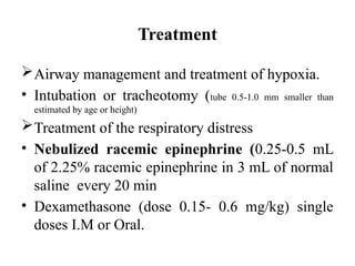
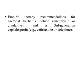
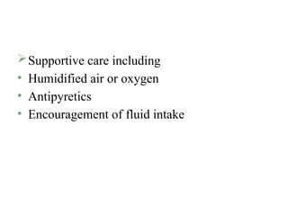
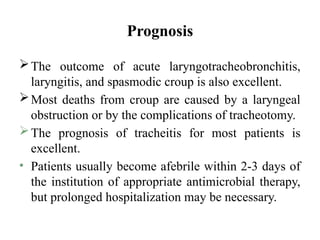
Lecture: Bacterial Tracheitis
Lecturer: Dr Abdirashid Dahir Herow
Paediatric Resident
Class 3.2
Bacterial Tracheitis
• Bacterial tracheitis is an acute bacterial
infection of the upper airway that is potentially
life-threatening.
• The mean age is between 5 and 7 yr. There is a
slight male predominance.
• Male predominance.
• It is often follows a viral respiratory
infection(Laryngotracheitis)
ETIOLOGY
• S. aureus
• S. pneumoniae
• S. pyogenes
• Moraxella catarrhalis
• nontypeable H. influenzae
• Anaerobic organisms
Clinical Manifestations
• Typically the child has a brassy cough,
apparently as part of a viral
laryngotracheobronchitis. High fever and
toxicity with respiratory distress can occur
immediately or after a few days of apparent
improvement. The patient can lie flat, does not
drool, and does not have the dysphagia
associated with epiglottitis.
• The major pathologic feature appears to be
mucosal swelling at the level of the cricoid
cartilage, complicated by copious, thick,
purulent secretions, sometimes causing
pseudomembranes
Diagnosis
• The diagnosis is clinical and based on
evidence of bacterial upper airway disease,
which includes high fever, purulent airway
secretions, and an absence of the classic
findings of epiglottitis. X-rays are not needed
but can show the classic findings purulent
material is noted below the cords during
endotracheal intubation.
• Lateral radiograph of the neck of a patient with bacterial tracheitis, showing pseudomembrane detachment in the trachea
Complications
• Cardiorespiratory arrest
• Toxic shock syndrome .
Treatment
Airway management and treatment of hypoxia.
• Intubation or tracheotomy (tube 0.5-1.0 mm smaller than
estimated by age or height)
Treatment of the respiratory distress
• Nebulized racemic epinephrine (0.25-0.5 mL
of 2.25% racemic epinephrine in 3 mL of normal
saline every 20 min
• Dexamethasone (dose 0.15- 0.6 mg/kg) single
doses I.M or Oral.
• Empiric therapy recommendations for
bacterial tracheitis include vancomycin or
clindamycin and a 3rd-generation
cephalosporin (e.g., ceftriaxone or cefepime).
Supportive care including
• Humidified air or oxygen
• Antipyretics
• Encouragement of fluid intake
Prognosis
The outcome of acute laryngotracheobronchitis,
laryngitis, and spasmodic croup is also excellent.
Most deaths from croup are caused by a laryngeal
obstruction or by the complications of tracheotomy.
The prognosis of tracheitis for most patients is
excellent.
• Patients usually become afebrile within 2-3 days of
the institution of appropriate antimicrobial therapy,
but prolonged hospitalization may be necessary.
•END

Bacterial Tracheitis Dr Herow.pptx document

  • 1.
    Lecture: Bacterial Tracheitis Lecturer:Dr Abdirashid Dahir Herow Paediatric Resident Class 3.2
  • 2.
    Bacterial Tracheitis • Bacterialtracheitis is an acute bacterial infection of the upper airway that is potentially life-threatening. • The mean age is between 5 and 7 yr. There is a slight male predominance.
  • 3.
    • Male predominance. •It is often follows a viral respiratory infection(Laryngotracheitis)
  • 4.
    ETIOLOGY • S. aureus •S. pneumoniae • S. pyogenes • Moraxella catarrhalis • nontypeable H. influenzae • Anaerobic organisms
  • 5.
    Clinical Manifestations • Typicallythe child has a brassy cough, apparently as part of a viral laryngotracheobronchitis. High fever and toxicity with respiratory distress can occur immediately or after a few days of apparent improvement. The patient can lie flat, does not drool, and does not have the dysphagia associated with epiglottitis.
  • 6.
    • The majorpathologic feature appears to be mucosal swelling at the level of the cricoid cartilage, complicated by copious, thick, purulent secretions, sometimes causing pseudomembranes
  • 7.
    Diagnosis • The diagnosisis clinical and based on evidence of bacterial upper airway disease, which includes high fever, purulent airway secretions, and an absence of the classic findings of epiglottitis. X-rays are not needed but can show the classic findings purulent material is noted below the cords during endotracheal intubation.
  • 8.
    • Lateral radiographof the neck of a patient with bacterial tracheitis, showing pseudomembrane detachment in the trachea
  • 9.
  • 10.
    Treatment Airway management andtreatment of hypoxia. • Intubation or tracheotomy (tube 0.5-1.0 mm smaller than estimated by age or height) Treatment of the respiratory distress • Nebulized racemic epinephrine (0.25-0.5 mL of 2.25% racemic epinephrine in 3 mL of normal saline every 20 min • Dexamethasone (dose 0.15- 0.6 mg/kg) single doses I.M or Oral.
  • 11.
    • Empiric therapyrecommendations for bacterial tracheitis include vancomycin or clindamycin and a 3rd-generation cephalosporin (e.g., ceftriaxone or cefepime).
  • 12.
    Supportive care including •Humidified air or oxygen • Antipyretics • Encouragement of fluid intake
  • 13.
    Prognosis The outcome ofacute laryngotracheobronchitis, laryngitis, and spasmodic croup is also excellent. Most deaths from croup are caused by a laryngeal obstruction or by the complications of tracheotomy. The prognosis of tracheitis for most patients is excellent. • Patients usually become afebrile within 2-3 days of the institution of appropriate antimicrobial therapy, but prolonged hospitalization may be necessary.
  • 14.