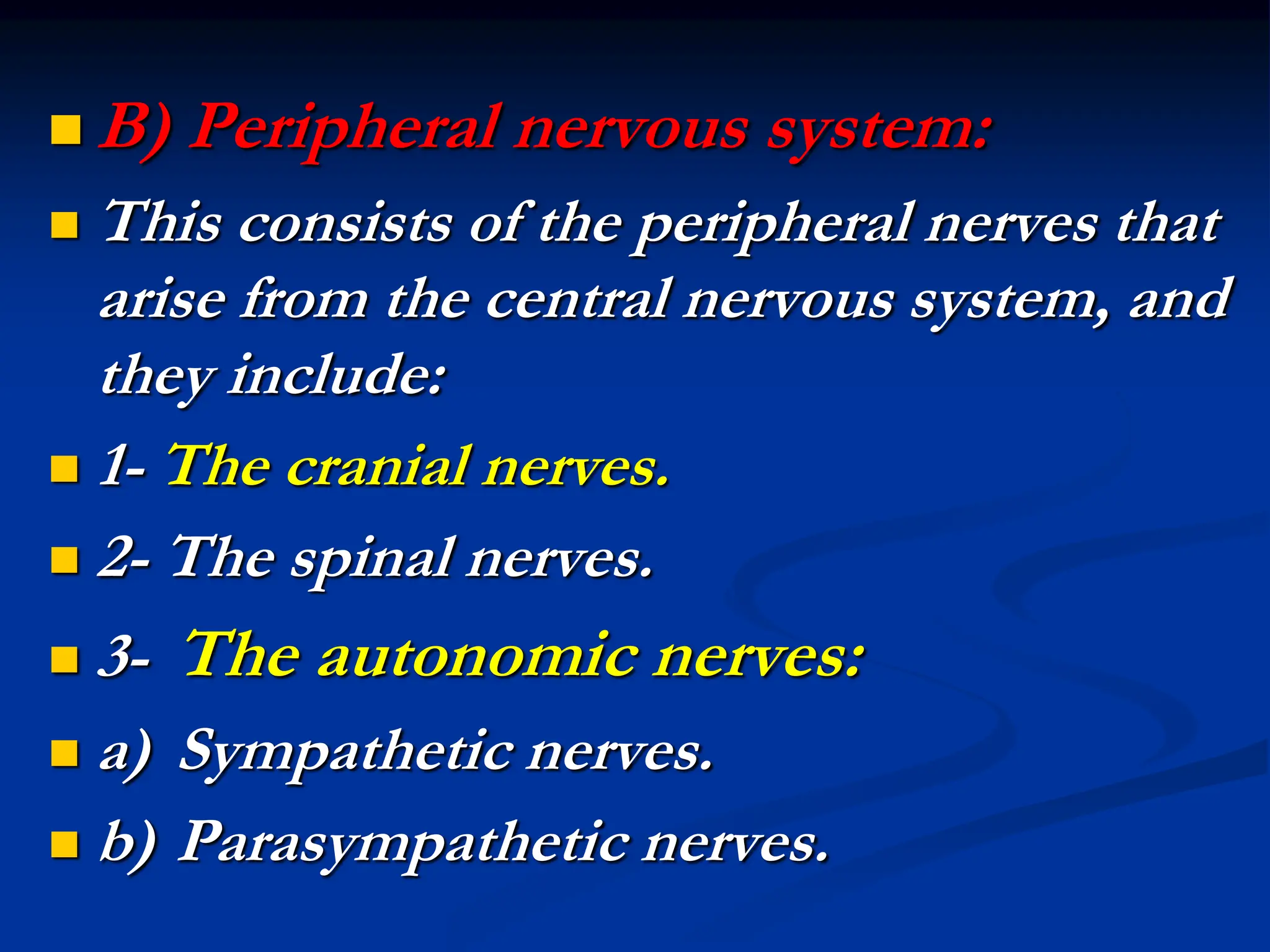
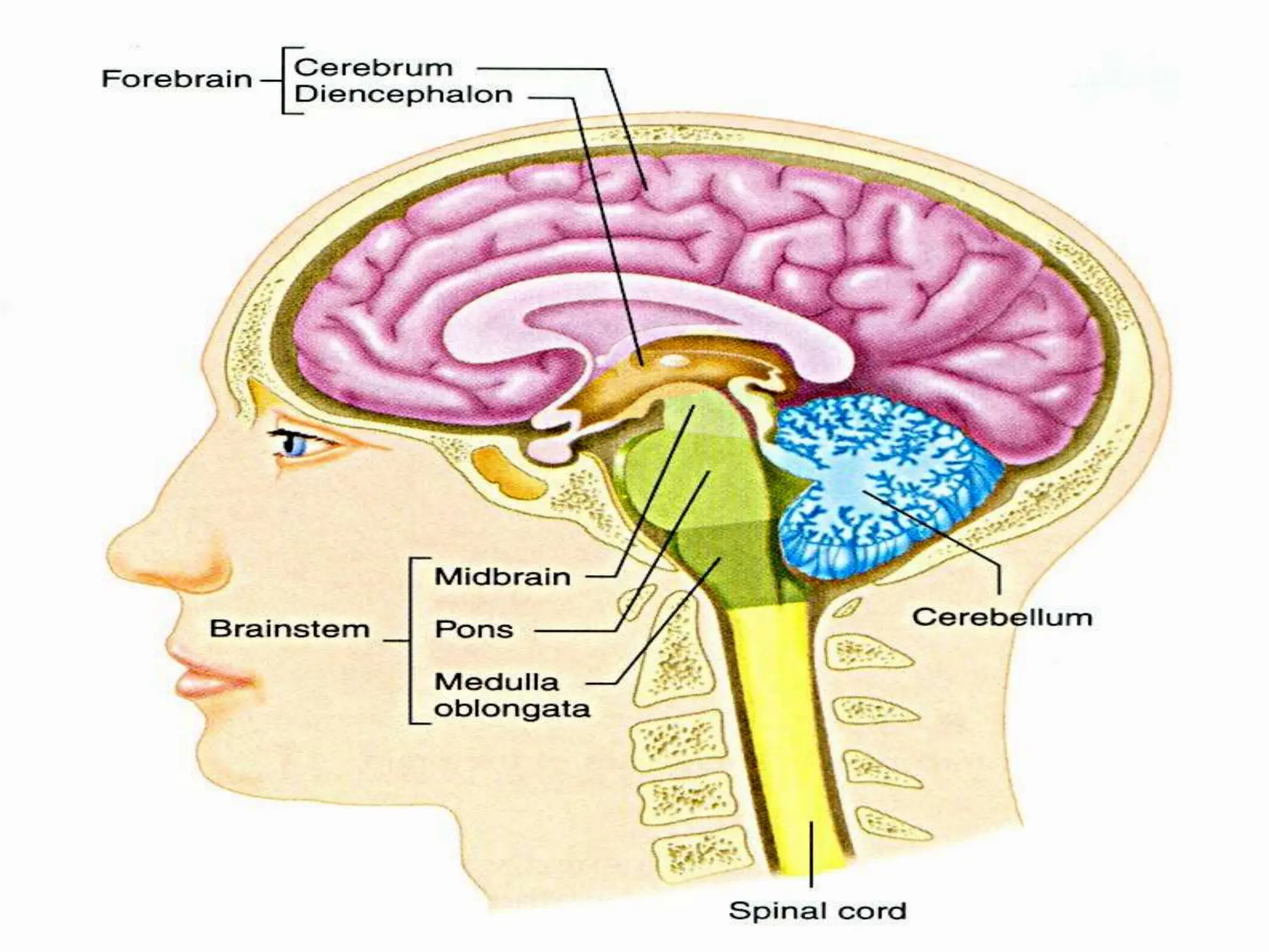
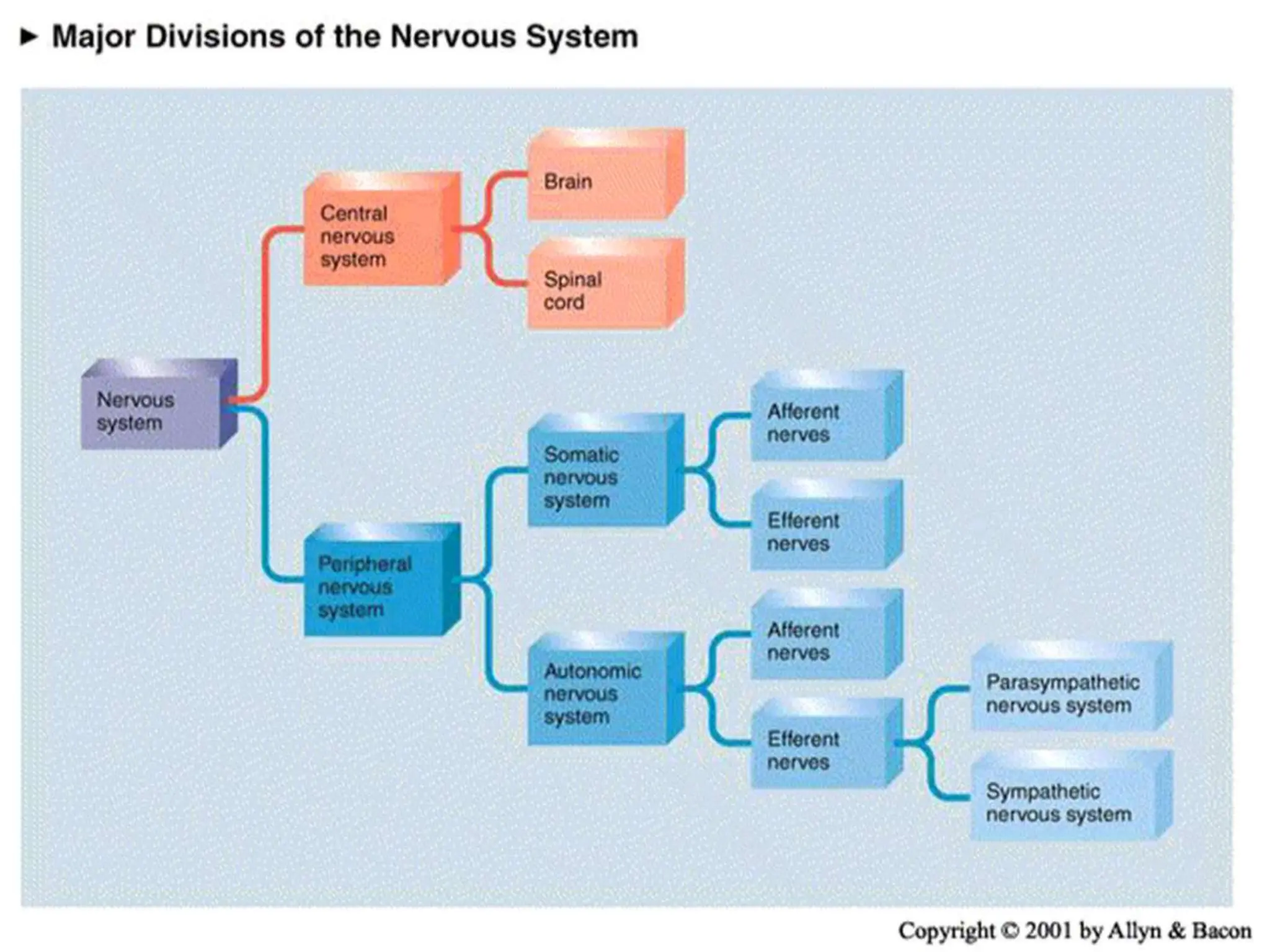
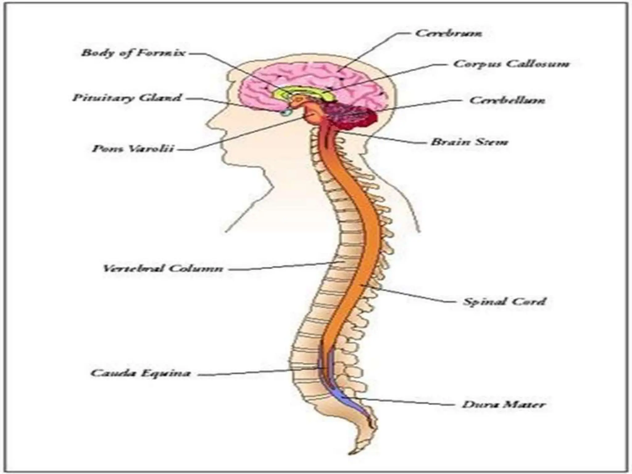
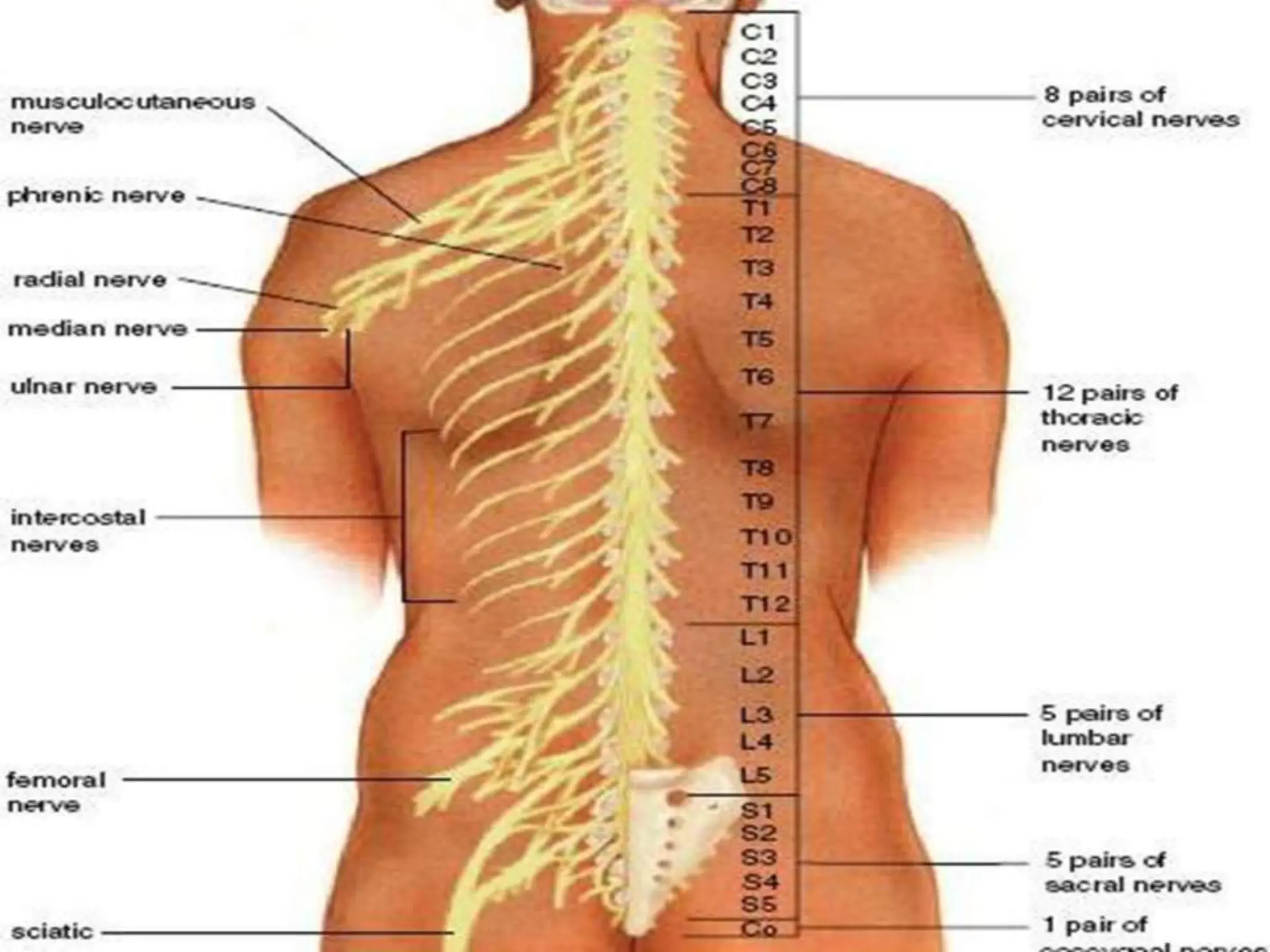
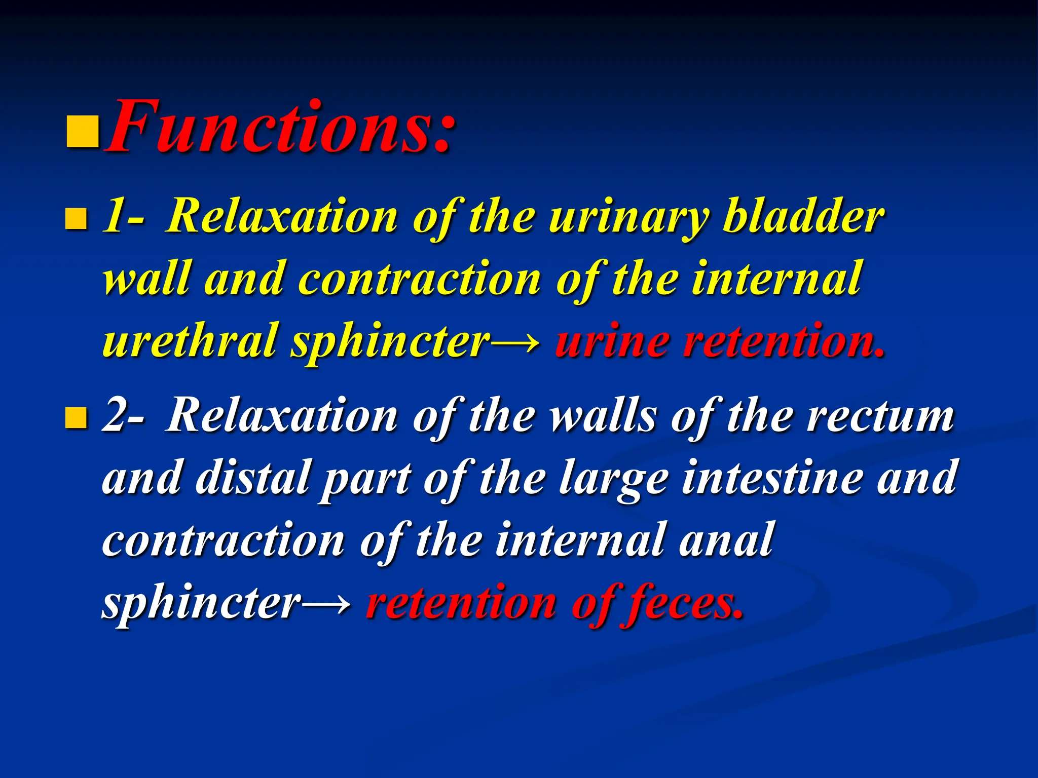
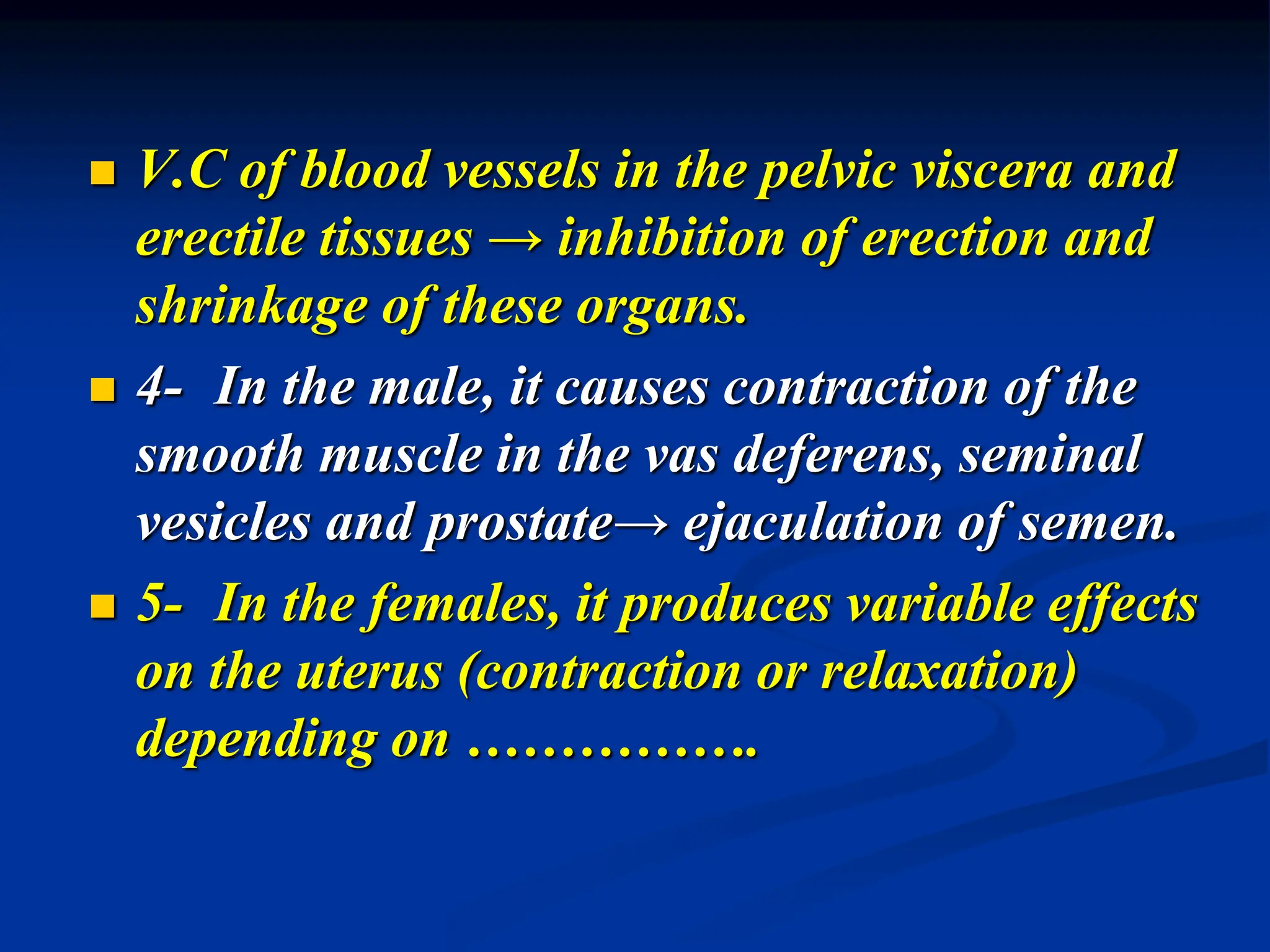
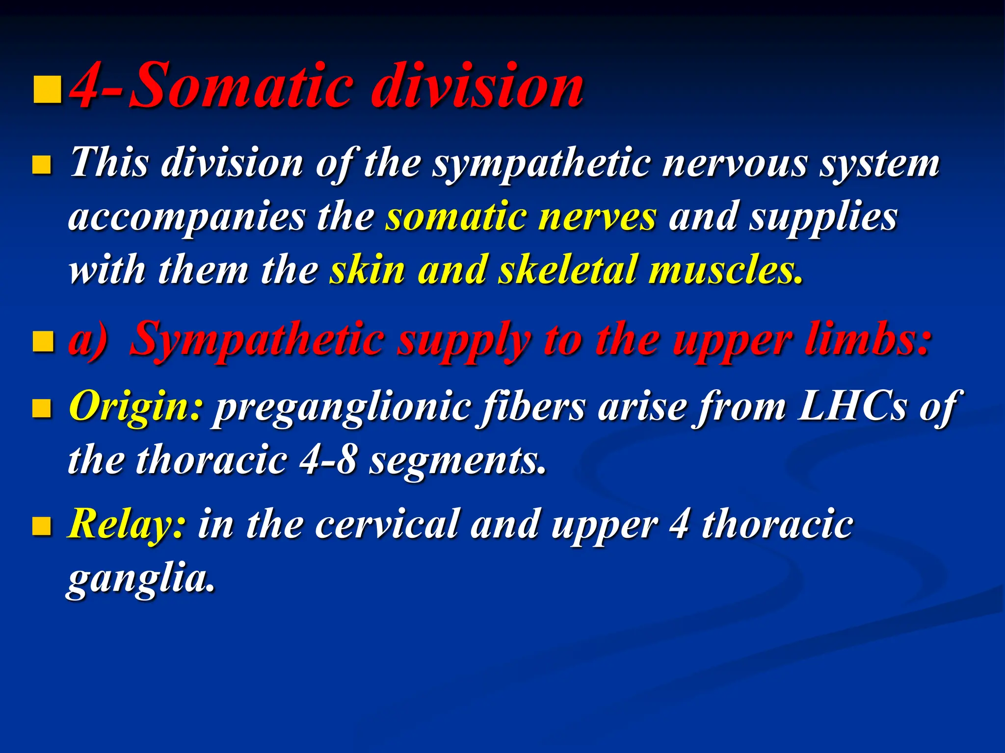
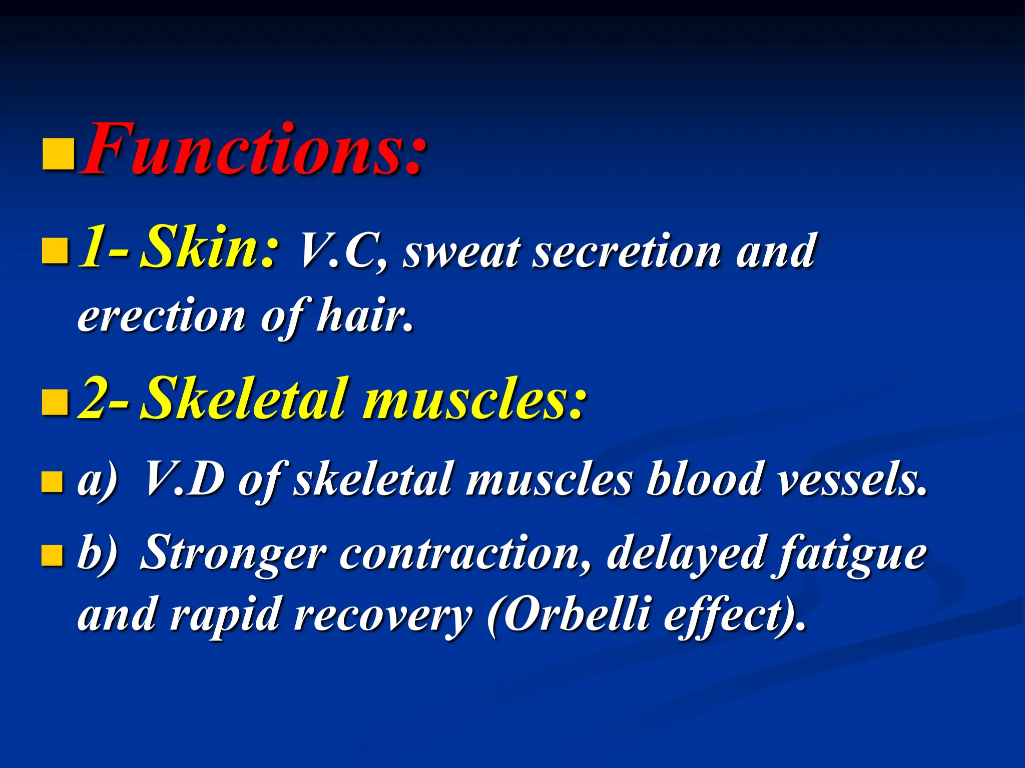
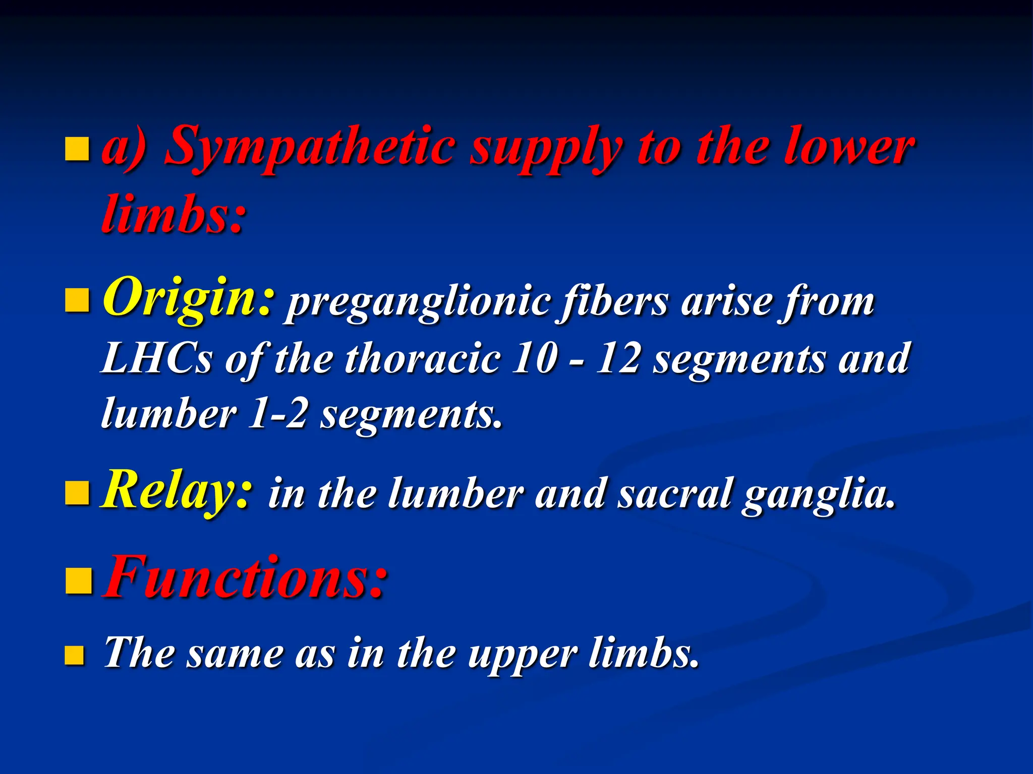
Autonomic nervous system
(A.N.S.) is the part of the nervous
system that regulates the activity of
the viscera, glands, heart and blood
vessels.
 It is also called the visceral or
involuntary nervous system.
The voluntary functions are
controlled by another part of
the nervous system called the
somatic nervous system
(central nervous system).
Anatomical divisions of the
nervous system:
 A) Central nervous system (CNS):
 1- Brain :
 a) Cerebrum.
 b) Cerebellum.
 c) Brain stem.
 2- Spinal cord (31 segments).
 B) Peripheral nervous system:
 This consists of the peripheral nerves that
arise from the central nervous system, and
they include:
 1- The cranial nerves.
 2- The spinal nerves.
 3- The autonomic nerves:
 a) Sympathetic nerves.
 b) Parasympathetic nerves.
1-The sympathetic nervous
system:
This arises from the lateral horn
cells (LHCs) of all thoracic and
upper 3 lumbar segments of the
spinal cord (thoracolumbar
outflow).
 2-The parasympathetic nervous
system:
 This arises from LHCs of the 2nd, 3rd and 4th
sacral segments of the spinal cord as well as
from the nuclei of certain cranial nerves, which
include:
 a) The 3rd cranial nerve (oculomotor).
 b) The 7th cranial nerve (facial).
 c) The 9th cranial nerve (glossopharyngeal).
 d) The 10th cranial nerve (vagus).
Reflex action:
 It is an involuntary response to a stimulus.
 It is the functional unit of the nervous
system.
 1- Somatic reflex actions: regulate the
activity of skeletal muscles.
 2- Autonomic reflex actions: regulate the
activity of the viscera.
Autonomic ganglia
 Autonomic ganglion is a collection of
nerve cells outside the C.N.S.
 It contains the postganglionic neurons.
 Preganglionic fibers may pass through
several autonomic ganglia but it relays
only in one ganglion.
Types of autonomic ganglia:
 1- Lateral (paravertebral) ganglia:
These ganglia are the main sites for relay
of preganglionic sympathetic fibers.
 2- Collateral (prevertebral) ganglia:
These are present mainly in the abdomen
in close relation to the aorta. These ganglia
are sympathetic.
3-Terminal ganglia:
These are present near or in the
walls of the effector organs.
These ganglia are mostly the
sites for relay of preganglionic
parasympathetic fibers.
Functions of autonomic ganglia:
Distributing centre: the preganglionic
fibers arise from limited regions of brain
stem and spinal cord. By the aid of
autonomic ganglia, each preganglionic
fiber diverges to average 8-9 postganglionic
neurons.
The sympathetic nervous
system
 The sympathetic nervous system can
be divided into 4 divisions:
 1- Cervical.
 2- Cardio-pulmonary.
 3- Splanchnic.
 4- Somatic.
1-Cervical division
 This division supplies different
structures in the region of the head and
neck.
 a) Origin: preganglionic fibers arise
from the lateral horn cells (L.H.Cs) of
the upper 2 thoracic segments.
 b) Relay: in the superior cervical
sympathetic ganglion.
c)Functions:
1-Eye:
 a) Contraction of the dilator pupillae muscle
leading to pupil dilatation (mydriasis).
 b) Contraction of muscles in the eyelids
(called superior and inferior tarsal muscles)
→ lid retraction→ widening of the palpebral
fissure.
 c) Contraction of Muller's muscle
behind the eyeballs → exophthalmos.
 d) Vasoconstriction (VC) of the blood
vessels of the eye.
 e) Relaxation of the ciliary muscle
during far vision.
2-Salivary glands:
 a) Trophic secretion (small in volume,
viscous and rich in organic material).
 b) Motor to the myoepithelial cells
surrounding the acini → squeeze saliva.
 c) Vasoconstriction of the salivary blood
vessels.
3- Skin:
 a) Vasoconstriction of the cutaneous blood
vessels.
 b) Sweat secretion.
 c) Hair erection as a result of contraction of
certain muscles in the skin called pilomotor
muscles.
 4- Brain:
 Slight vasoconstriction to the cerebral blood
vessels.
Horner's syndrome
 An injury to the cervical sympathetic
nerve fibers produces a syndrome
(group of symptoms) on the same side
called Horner's syndrome.
 The symptoms of Horner's syndrome:
 1- Ptosis: drooping of the upper eyelid
due to paralysis of the superior tarsal
muscle.
 2- Miosis (pupilloconstriction) due to
paralysis of the dilator pupillae muscle.
 3-Anhydrosis (absence of sweat secretion).
 4-Vasodilatation (VD) due to loss of
sympathetic V.C effect, so it becomes warmer
and more red than the healthy side.
 5- Enophthalmos (sinking of the eyeball
backward in the orbit) due to paralysis of
Muller's muscle.
2-Cardiopulmonary division
 This division supplies the heart and
lungs.
a) Origin: preganglionic fibers
arise from L.H.Cs of the upper 4
thoracic segments.
b) Relay: in the cervical and upper
4 thoracic lateral ganglia.
c) Functions:
1-heart:
 a) Stimulates all properties of the cardiac
muscle.
 b) Vasodilatation of the coronary vessels
(indirect effect).
 c) Increases the O2 consumption of the
heart.
2-Lungs:
a) Inhibits mucus secretion of
bronchial glands.
b) Produces bronchodilatation.
c) V.C of the pulmonary vessels.
3- Splanchnic (visceral) division
 This division supplies different abdominal
and pelvic viscera.
 a) Sympathetic supply to the abdomen
(greater splanchnic nerve):
 Origin: preganglionic fibers arise from the
L.H.Cs of the lower 6 thoracic segments.
 Relay: in the collateral ganglia in the
abdomen.
Functions:
 1- V.C of the majority of the arterioles in the
stomach, small intestine, proximal part of the
large intestine, kidney, liver and pancreas.
 2- Relaxation of the walls of the stomach,
small intestine and proximal part of the large
intestine and contraction their corresponding
sphincters.
 3- Contraction of the smooth muscle in the
capsule of spleen→ squeeze the stored blood
into circulation.
 4- Stimulation of glycogenolsis in the liver→
increased blood glucose level.
 5- Stimulation of adrenal medulla to secrete its
hormones (catecholamine).
 6- Inhibits insulin hormone secretion.
b)Sympathetic supply to the
pelvis (lesser splanchnic nerve):
Origin: preganglionic fibers arise
from the L.H.Cs of the upper 3 lumber
segments.
Relay: in the collateral ganglia in the
abdomen.
Functions:
 1- Relaxation of the urinary bladder
wall and contraction of the internal
urethral sphincter→ urine retention.
 2- Relaxation of the walls of the rectum
and distal part of the large intestine and
contraction of the internal anal
sphincter→ retention of feces.
 V.C of blood vessels in the pelvic viscera and
erectile tissues → inhibition of erection and
shrinkage of these organs.
 4- In the male, it causes contraction of the
smooth muscle in the vas deferens, seminal
vesicles and prostate→ ejaculation of semen.
 5- In the females, it produces variable effects
on the uterus (contraction or relaxation)
depending on …………….
4-Somatic division
 This division of the sympathetic nervous system
accompanies the somatic nerves and supplies
with them the skin and skeletal muscles.
 a) Sympathetic supply to the upper limbs:
 Origin: preganglionic fibers arise from LHCs of
the thoracic 4-8 segments.
 Relay: in the cervical and upper 4 thoracic
ganglia.
Functions:
1- Skin: V.C, sweat secretion and
erection of hair.
2- Skeletal muscles:
 a) V.D of skeletal muscles blood vessels.
 b) Stronger contraction, delayed fatigue
and rapid recovery (Orbelli effect).
 a) Sympathetic supply to the lower
limbs:
 Origin: preganglionic fibers arise from
LHCs of the thoracic 10 - 12 segments and
lumber 1-2 segments.
 Relay: in the lumber and sacral ganglia.
Functions:
 The same as in the upper limbs.
autonomddddddddddddddddddddddddic 1 .ppt

autonomddddddddddddddddddddddddic 1 .ppt

  • 1.
    Autonomic nervous system (A.N.S.)is the part of the nervous system that regulates the activity of the viscera, glands, heart and blood vessels.  It is also called the visceral or involuntary nervous system.
  • 2.
    The voluntary functionsare controlled by another part of the nervous system called the somatic nervous system (central nervous system).
  • 3.
    Anatomical divisions ofthe nervous system:  A) Central nervous system (CNS):  1- Brain :  a) Cerebrum.  b) Cerebellum.  c) Brain stem.  2- Spinal cord (31 segments).
  • 4.
     B) Peripheralnervous system:  This consists of the peripheral nerves that arise from the central nervous system, and they include:  1- The cranial nerves.  2- The spinal nerves.  3- The autonomic nerves:  a) Sympathetic nerves.  b) Parasympathetic nerves.
  • 9.
    1-The sympathetic nervous system: Thisarises from the lateral horn cells (LHCs) of all thoracic and upper 3 lumbar segments of the spinal cord (thoracolumbar outflow).
  • 10.
     2-The parasympatheticnervous system:  This arises from LHCs of the 2nd, 3rd and 4th sacral segments of the spinal cord as well as from the nuclei of certain cranial nerves, which include:  a) The 3rd cranial nerve (oculomotor).  b) The 7th cranial nerve (facial).  c) The 9th cranial nerve (glossopharyngeal).  d) The 10th cranial nerve (vagus).
  • 11.
    Reflex action:  Itis an involuntary response to a stimulus.  It is the functional unit of the nervous system.  1- Somatic reflex actions: regulate the activity of skeletal muscles.  2- Autonomic reflex actions: regulate the activity of the viscera.
  • 13.
    Autonomic ganglia  Autonomicganglion is a collection of nerve cells outside the C.N.S.  It contains the postganglionic neurons.  Preganglionic fibers may pass through several autonomic ganglia but it relays only in one ganglion.
  • 14.
    Types of autonomicganglia:  1- Lateral (paravertebral) ganglia: These ganglia are the main sites for relay of preganglionic sympathetic fibers.  2- Collateral (prevertebral) ganglia: These are present mainly in the abdomen in close relation to the aorta. These ganglia are sympathetic.
  • 15.
    3-Terminal ganglia: These arepresent near or in the walls of the effector organs. These ganglia are mostly the sites for relay of preganglionic parasympathetic fibers.
  • 16.
    Functions of autonomicganglia: Distributing centre: the preganglionic fibers arise from limited regions of brain stem and spinal cord. By the aid of autonomic ganglia, each preganglionic fiber diverges to average 8-9 postganglionic neurons.
  • 17.
    The sympathetic nervous system The sympathetic nervous system can be divided into 4 divisions:  1- Cervical.  2- Cardio-pulmonary.  3- Splanchnic.  4- Somatic.
  • 19.
    1-Cervical division  Thisdivision supplies different structures in the region of the head and neck.  a) Origin: preganglionic fibers arise from the lateral horn cells (L.H.Cs) of the upper 2 thoracic segments.  b) Relay: in the superior cervical sympathetic ganglion.
  • 20.
    c)Functions: 1-Eye:  a) Contractionof the dilator pupillae muscle leading to pupil dilatation (mydriasis).  b) Contraction of muscles in the eyelids (called superior and inferior tarsal muscles) → lid retraction→ widening of the palpebral fissure.
  • 21.
     c) Contractionof Muller's muscle behind the eyeballs → exophthalmos.  d) Vasoconstriction (VC) of the blood vessels of the eye.  e) Relaxation of the ciliary muscle during far vision.
  • 22.
    2-Salivary glands:  a)Trophic secretion (small in volume, viscous and rich in organic material).  b) Motor to the myoepithelial cells surrounding the acini → squeeze saliva.  c) Vasoconstriction of the salivary blood vessels.
  • 23.
    3- Skin:  a)Vasoconstriction of the cutaneous blood vessels.  b) Sweat secretion.  c) Hair erection as a result of contraction of certain muscles in the skin called pilomotor muscles.  4- Brain:  Slight vasoconstriction to the cerebral blood vessels.
  • 24.
    Horner's syndrome  Aninjury to the cervical sympathetic nerve fibers produces a syndrome (group of symptoms) on the same side called Horner's syndrome.  The symptoms of Horner's syndrome:  1- Ptosis: drooping of the upper eyelid due to paralysis of the superior tarsal muscle.
  • 25.
     2- Miosis(pupilloconstriction) due to paralysis of the dilator pupillae muscle.  3-Anhydrosis (absence of sweat secretion).  4-Vasodilatation (VD) due to loss of sympathetic V.C effect, so it becomes warmer and more red than the healthy side.  5- Enophthalmos (sinking of the eyeball backward in the orbit) due to paralysis of Muller's muscle.
  • 26.
    2-Cardiopulmonary division  Thisdivision supplies the heart and lungs. a) Origin: preganglionic fibers arise from L.H.Cs of the upper 4 thoracic segments. b) Relay: in the cervical and upper 4 thoracic lateral ganglia.
  • 27.
    c) Functions: 1-heart:  a)Stimulates all properties of the cardiac muscle.  b) Vasodilatation of the coronary vessels (indirect effect).  c) Increases the O2 consumption of the heart.
  • 28.
    2-Lungs: a) Inhibits mucussecretion of bronchial glands. b) Produces bronchodilatation. c) V.C of the pulmonary vessels.
  • 29.
    3- Splanchnic (visceral)division  This division supplies different abdominal and pelvic viscera.  a) Sympathetic supply to the abdomen (greater splanchnic nerve):  Origin: preganglionic fibers arise from the L.H.Cs of the lower 6 thoracic segments.  Relay: in the collateral ganglia in the abdomen.
  • 30.
    Functions:  1- V.Cof the majority of the arterioles in the stomach, small intestine, proximal part of the large intestine, kidney, liver and pancreas.  2- Relaxation of the walls of the stomach, small intestine and proximal part of the large intestine and contraction their corresponding sphincters.
  • 31.
     3- Contractionof the smooth muscle in the capsule of spleen→ squeeze the stored blood into circulation.  4- Stimulation of glycogenolsis in the liver→ increased blood glucose level.  5- Stimulation of adrenal medulla to secrete its hormones (catecholamine).  6- Inhibits insulin hormone secretion.
  • 32.
    b)Sympathetic supply tothe pelvis (lesser splanchnic nerve): Origin: preganglionic fibers arise from the L.H.Cs of the upper 3 lumber segments. Relay: in the collateral ganglia in the abdomen.
  • 33.
    Functions:  1- Relaxationof the urinary bladder wall and contraction of the internal urethral sphincter→ urine retention.  2- Relaxation of the walls of the rectum and distal part of the large intestine and contraction of the internal anal sphincter→ retention of feces.
  • 34.
     V.C ofblood vessels in the pelvic viscera and erectile tissues → inhibition of erection and shrinkage of these organs.  4- In the male, it causes contraction of the smooth muscle in the vas deferens, seminal vesicles and prostate→ ejaculation of semen.  5- In the females, it produces variable effects on the uterus (contraction or relaxation) depending on …………….
  • 35.
    4-Somatic division  Thisdivision of the sympathetic nervous system accompanies the somatic nerves and supplies with them the skin and skeletal muscles.  a) Sympathetic supply to the upper limbs:  Origin: preganglionic fibers arise from LHCs of the thoracic 4-8 segments.  Relay: in the cervical and upper 4 thoracic ganglia.
  • 36.
    Functions: 1- Skin: V.C,sweat secretion and erection of hair. 2- Skeletal muscles:  a) V.D of skeletal muscles blood vessels.  b) Stronger contraction, delayed fatigue and rapid recovery (Orbelli effect).
  • 37.
     a) Sympatheticsupply to the lower limbs:  Origin: preganglionic fibers arise from LHCs of the thoracic 10 - 12 segments and lumber 1-2 segments.  Relay: in the lumber and sacral ganglia. Functions:  The same as in the upper limbs.