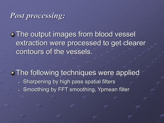
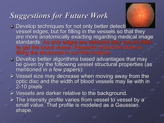
SlideShare a Scribd company logo
1 of 29
Automatic Detection of Blood Vessels
in Digital Retinal Image using
CVIP Tools
Krishna Praveena Mandava
Sri Swetha Kantamaneni
Robert LeAnder
Overview
The Devastation
 Diabetic retinopathy – 4.1 million US Adults
National Health Interview Survey and US Census
Population
 Glaucoma – 2 million individuals in the US.
Ophthalmologic images
 Important structures – Blood Vessels
 Help detect and treat Eye Diseases affecting
blood vessels
Overview
Damaged blood vessels indicate retinal disease.
 Blood clots indicate diabetic retinopathy.
 Narrow blood vessels indicate Central Retinal Artery
Occlusion.
Observation of blood vessels in retinal images
 Shows presence of disease
 Helps prevent vision loss by early detection
The Need for the Study
Automated Blood Vessel Extraction algorithms
can save time, patients’ vision and medical
costs.
Effects of Diseases on Blood Vessels
Image of Diseased Retina Due to Diabetes
Disease produces
hemorrhages,
exudates and micro
aneurysms (dark red
spots).
Central Retinal Artery Occlusion (CRAO)
Results in
narrowing blood
vessels.
Effects of Diseases on Blood Vessels
Branch Retinal Artery Occlusion (BRAO)
Where artery
branch points
are occluded or
blocked
Effects of Diseases on Blood Vessels
6 Approaches to Blood Vessel Extraction
1. Pattern recognition techniques
2. Model based approaches
3. Tracking based approaches
4. Artificial intelligence based approaches
5. Neural network based approaches
6. Miscellaneous tube-like object detection
approaches.
1. Pattern recognition techniques
Deals with automatic detection or classification of objects or features.
A.Multi scale approaches
Based on image resolution. Major vessels are extracted from low
resolution images and minor vessels from high resolution images.
B.Skeleton based approaches
Vessel centerlines are extracted and then connected to create a
vessel tree.
C. Ridge-Based Approaches
This is specialized skeleton based approaches. Ridges are peaks.
6 Approaches to Blood Vessel Extraction
D. Region growing approaches…
• Assume that pixels are close to each other and have
similar intensity values and are likely to belong to same
objects.
• Start region growth from a seed point, then segment
the image based on some predefined criterion.
• Have the Disadvantage that the seed point should be
selected manually.
E. Differential-Geometry-based approaches…
• Utilizes techniques developed from the complex
mathematical field of Differential Geometry
• Are based on blood-vessel structural properties
1. Pattern recognition techniques
F. Matched-Filter Approaches
• Are signal processing approaches where new images with un-
extracted vessels are convolved with known profiles of vessels.
• Matched filters are followed by image processing operations like
thresholding to get the final vessel contours.
G. Morphology Schemes…
• Apply structuring elements to images to effect dilation and
erosion are two main operations.
• Include Top Hat and Watershed algorithms.
6 Approaches to Blood Vessel Extraction
2. Model-Based Approaches…
• Include Snakes algorithms, which are the primary types of algorithms used for
vessel extraction.
• A “Snake” is an active (deformable) contour with a set of Control Points
connecting the segments of the contour to each other.
• It is a user interactive algorithm.
3. Tracking-Based Approaches…
 Are similar to pattern recognition approaches except they apply local, instead
of global operator
analyzing the pixels orthogonal to the tracking direction.
4. Artificial intelligence-based approaches…
 Use prior knowledge of model vessel structures to determine vessel structures
in the “unextracted” (unsegmented) image.
 Some applications may use a general blood vessel model for extraction .
5. Neural Network-Based approaches…
 Use neural networks as a classification method. The system is trained using a
set of images having blood vessel contours. The target image is
segmented using the trained system
6. Miscellaneous Tube-Like Object Detection
Approaches…
• Deals with the extraction of tubular structures from images.
• Are not designed for vessel extraction.
RETINAL BLOOD VESSEL EXTRACTION
(SEGMENTATION)
Available Image Databases
 DRIVE and STARE databases are available for the public.
http://www.ces.clemson.edu/~ahoover/stare/
http://www.parl.clemson.edu/stare/nerve/
 We worked on 50 fundus images from the STARE database.
 How the Images Were Taken
An Optical camera is used to see through the pupil of the eye to the inner
surface
of the eyeball. The resulting retinal image shows the optic nerve, fovea, and
the blood vessels.
Methods
Steps used blood vessel extraction…
 Preprocessing
 Extraction (segmentation)
 Post processing
Software:
We used Computer Vision and Image Processing Tools to apply various
algorithms to extract (segment) blood vessels.
Our Project
Preprocessing:
Preprocessing will eliminate errors caused
during taking the image and to reduce
brightness effects on the image .
The original images are resized from
150*130 to 256*256 to use in CVIP tools.
Images in green bands show vessel
structures most reliably. So, the green
band was extracted.
Extraction of blood vessels:
Tools that we applied:
 Median filters
 Laplacian filters
 Image enhancement methods like Adaptive Contrast
Enhancement, Histogram equalization.
 Edge detection like Canny edge detection.
Post processing:
The output images from blood vessel
extraction were processed to get clearer
contours of the vessels.
The following techniques were applied
 Sharpening by high pass spatial filters
 Smoothing by FFT smoothing, Ypmean filter
Original Image and Expected Output:
Our final images for different algorithms:
Exp 1 Exp 2 Exp 3
Exp 4 Exp 5
Summary:
NEED AND USE: Extraction of blood vessels
Research is ongoing and there is still a great
need to develop for an easier, more accurate
and useful algorithms.
We were able to detect major blood vessels
Better algorithms can be developed using CVIP
tools for the extraction of minor blood vessels.
Suggestions for Future Work
Develop techniques for not only better detection of
vessel edges, but for filling in the vessels so that they
are more anatomically exacting regarding medical image
standards. As only edges are detected they can be filled
to get the blood vessel. Research should be done in
filling the structures in our final outputs.
Develop better algorithms based advantages that may
be given by the following vessel structural properties (as
mentioned in a few papers):
 Vessel size may decrease when moving away from the
optic disc and the width of blood vessels may lie with in
2-10 pixels
 Vessels are darker relative to the background.
 The intensity profile varies from vessel to vessel by a
small value. That profile is modeled as a Gaussian
shape.
More Suggestions for Future Work
Extraction of Minute blood vessels.
Extracted outputs can be verified by an ophthalmologist
Extraction outputs may also be calculated of sensitivity
and specificity of blood vessels will give you better final
results.
Detection of the optic disc is also needed as the border
of the disc appears as a blood vessel. To prevent this the
optic disc should be detected and removed before blood
vessels are extracted.
Blood vessels should be separated from hemorrhages,
and micro aneurysms.
Conclusion:
CVIPtools is a very handy method for
applying extraction techniques. There is a
dire need for easier methods of blood
vessel extraction. CVIPtools may provide
accurate automatic detection algorithms
for clinical applications in retinopathy.
Reference:
1. Computer Imaging Digital Image Analysis and Processing
- Dr. Scott E Umbaugh
2. Digital Image Processing - Rafael C .Gonzalez, Richard
E .Woods
3. A Review of Vessel Extraction Techniques and Algorithms
– Cemil Kirbas and Francis Quek, Wright State University, Dayton,
Ohio
4. Automated Diagnosis and Image understanding with Object
Extraction, Object Classification and Inferencing in Retinal Images
–Micheal Goldbaum, Saied Moezzi, Adam Taylor, Shankar
Chatterjee, Edward Hunter and Ramesh Jain ,University of
California ,USA.
Reference:
5. Characterization of the optic disc in retinal imagery using a probalistic
approach
– Kenneth W.Tobin, Edward Chaum, Priya Govindaswami, Thomas
P.Karnowski, Omer Sezer, University of Tennessee, Knoxville, Tennessee.
6. Blood Vessel Segmentation in Retinal Images
– P.Echevarria, T.Miller, J.O Meara
7. An improved matched filter for blood vessel detection of digital retinal images
– Mohammed Al-Rawi, Munib Qutaishat, Mohammed Arrar, University of
Jordon,
Jordan.
8. Towards vessel characterization in the vicinity of the optic disc in digital
retinal images – H.F.Jelinek,C.Lucas, D.J.Cornforth, W.Huang and
M.J.Cree.
9. Retinal vessel segmentation using the 2-D Morlet Wavelet and Supervised
classification
– Joao V.B.Soares, Jorge J.G. Leandro ,Robert M. Cesar-Jr., Herbert F.
Jelinek and Micheal J.Cree, Senior Member IEEE
10. Locating blood vessels in retinal images by piece-wise threshold probing of
a matched filter response
– Adam Hoover, Valentina Kouznetsova, Micheal Goldbaum
Reference:
11. Automated identification of diabetic retinal exudates in digital color images
– A Osareh, M Mirmehdi, B Thomas, R Markham.
12.Survey of Retinal Image Segmentation and Registration
– Mai S. Mabrouk, Nahed H. Solouma and Yasser M.Kadah.
13.Automated detection of diabetic retinopathy on digital fundus images
– C. Sinthanayothin, J.F. Boyce, T.H. Williamson, H.L. Cook, E. Mensah, S.
Lal and D. Usher.
14.Segmentation of retinal blood vessels by combining the detection of
centerlines and morphological reconstruction
–Ana Maria Mendonca, Aurelio Campilho members IEEE.
15. The Eye Diseases Prevalence Research Group. The prevalence of diabetic
retinopathy among adults in the united states. Archives of Ophthalmology,
122(4):552–563, 2004.
16. The Eye Diseases Prevalence Research Group. Prevalence of open-angle
glaucoma among adults in the united states. Archives of Ophthalmology,
122(4):532–538, 2004.
17. Retinal Vessel Extraction Using Multiscale Matched Filters, Confidence and
Edge Measures
Michal Sofka, and Charles V. Stewart
THANK
YOU

More Related Content

Similar to 12539 Image Processing BV Extraction.ppt

Detection of Macular Edema by using Various Techniques of Feature Extraction ...
Detection of Macular Edema by using Various Techniques of Feature Extraction ...Detection of Macular Edema by using Various Techniques of Feature Extraction ...
Detection of Macular Edema by using Various Techniques of Feature Extraction ...IRJET Journal
 
An Automated Systems for the Detection of Macular Ischemia based-Diabetic Ret...
An Automated Systems for the Detection of Macular Ischemia based-Diabetic Ret...An Automated Systems for the Detection of Macular Ischemia based-Diabetic Ret...
An Automated Systems for the Detection of Macular Ischemia based-Diabetic Ret...iosrjce
 
Automatic Blood Vessels Segmentation of Retinal Images
Automatic Blood Vessels Segmentation of Retinal ImagesAutomatic Blood Vessels Segmentation of Retinal Images
Automatic Blood Vessels Segmentation of Retinal ImagesHarish Rajula
 
IRJET- Retinal Blood Vessel Tree Segmentation using Fast Marching Method
IRJET- Retinal Blood Vessel Tree Segmentation using Fast Marching MethodIRJET- Retinal Blood Vessel Tree Segmentation using Fast Marching Method
IRJET- Retinal Blood Vessel Tree Segmentation using Fast Marching MethodIRJET Journal
 
RETINA DISEASE IDENTIFICATION USING IMAGE PROCESSING
RETINA DISEASE IDENTIFICATION USING IMAGE PROCESSINGRETINA DISEASE IDENTIFICATION USING IMAGE PROCESSING
RETINA DISEASE IDENTIFICATION USING IMAGE PROCESSINGIRJET Journal
 
Performance analysis of retinal image blood vessel segmentation
Performance analysis of retinal image blood vessel segmentationPerformance analysis of retinal image blood vessel segmentation
Performance analysis of retinal image blood vessel segmentationacijjournal
 
AUTOMATED SEGMENTATION OF FLUORESCENT AND FUNDS IMAGES BASED ON RETINAL BLOOD...
AUTOMATED SEGMENTATION OF FLUORESCENT AND FUNDS IMAGES BASED ON RETINAL BLOOD...AUTOMATED SEGMENTATION OF FLUORESCENT AND FUNDS IMAGES BASED ON RETINAL BLOOD...
AUTOMATED SEGMENTATION OF FLUORESCENT AND FUNDS IMAGES BASED ON RETINAL BLOOD...acijjournal
 
Computational Biomedicine Lab: Current Members, pumpsandpipesmdhc
Computational Biomedicine Lab: Current Members, pumpsandpipesmdhcComputational Biomedicine Lab: Current Members, pumpsandpipesmdhc
Computational Biomedicine Lab: Current Members, pumpsandpipesmdhctmhsweb
 
Diabetic-Retinopathy
Diabetic-RetinopathyDiabetic-Retinopathy
Diabetic-Retinopathyanish h
 
C LASSIFICATION O F D IABETES R ETINA I MAGES U SING B LOOD V ESSEL A REAS
C LASSIFICATION  O F D IABETES  R ETINA I MAGES  U SING B LOOD V ESSEL A REASC LASSIFICATION  O F D IABETES  R ETINA I MAGES  U SING B LOOD V ESSEL A REAS
C LASSIFICATION O F D IABETES R ETINA I MAGES U SING B LOOD V ESSEL A REASIJCI JOURNAL
 
Human recognition system based on retina vascular network characteristics
Human recognition system based on retina vascular network characteristicsHuman recognition system based on retina vascular network characteristics
Human recognition system based on retina vascular network characteristicscbnaikodi
 
Digitech Ophthalmologist (Techlead Image Processing)
Digitech Ophthalmologist (Techlead Image Processing)Digitech Ophthalmologist (Techlead Image Processing)
Digitech Ophthalmologist (Techlead Image Processing)techlead-india
 
Efficient Diabetic Retinopathy Space Subjective Fuzzy Clustering In Digital F...
Efficient Diabetic Retinopathy Space Subjective Fuzzy Clustering In Digital F...Efficient Diabetic Retinopathy Space Subjective Fuzzy Clustering In Digital F...
Efficient Diabetic Retinopathy Space Subjective Fuzzy Clustering In Digital F...IOSR Journals
 
Retinal Vessels Segmentation Using Supervised Classifiers for Identification ...
Retinal Vessels Segmentation Using Supervised Classifiers for Identification ...Retinal Vessels Segmentation Using Supervised Classifiers for Identification ...
Retinal Vessels Segmentation Using Supervised Classifiers for Identification ...IOSR Journals
 
Retinal Vessel Segmentation in U-Net Using Deep Learning
Retinal Vessel Segmentation in U-Net Using Deep LearningRetinal Vessel Segmentation in U-Net Using Deep Learning
Retinal Vessel Segmentation in U-Net Using Deep LearningIRJET Journal
 
Diabetic Retinopathy detection using Machine learning
Diabetic Retinopathy detection using Machine learningDiabetic Retinopathy detection using Machine learning
Diabetic Retinopathy detection using Machine learningIRJET Journal
 

Similar to 12539 Image Processing BV Extraction.ppt (20)

Detection of Macular Edema by using Various Techniques of Feature Extraction ...
Detection of Macular Edema by using Various Techniques of Feature Extraction ...Detection of Macular Edema by using Various Techniques of Feature Extraction ...
Detection of Macular Edema by using Various Techniques of Feature Extraction ...
 
An Automated Systems for the Detection of Macular Ischemia based-Diabetic Ret...
An Automated Systems for the Detection of Macular Ischemia based-Diabetic Ret...An Automated Systems for the Detection of Macular Ischemia based-Diabetic Ret...
An Automated Systems for the Detection of Macular Ischemia based-Diabetic Ret...
 
Automatic Blood Vessels Segmentation of Retinal Images
Automatic Blood Vessels Segmentation of Retinal ImagesAutomatic Blood Vessels Segmentation of Retinal Images
Automatic Blood Vessels Segmentation of Retinal Images
 
IRJET- Retinal Blood Vessel Tree Segmentation using Fast Marching Method
IRJET- Retinal Blood Vessel Tree Segmentation using Fast Marching MethodIRJET- Retinal Blood Vessel Tree Segmentation using Fast Marching Method
IRJET- Retinal Blood Vessel Tree Segmentation using Fast Marching Method
 
RETINA DISEASE IDENTIFICATION USING IMAGE PROCESSING
RETINA DISEASE IDENTIFICATION USING IMAGE PROCESSINGRETINA DISEASE IDENTIFICATION USING IMAGE PROCESSING
RETINA DISEASE IDENTIFICATION USING IMAGE PROCESSING
 
Performance analysis of retinal image blood vessel segmentation
Performance analysis of retinal image blood vessel segmentationPerformance analysis of retinal image blood vessel segmentation
Performance analysis of retinal image blood vessel segmentation
 
AUTOMATED SEGMENTATION OF FLUORESCENT AND FUNDS IMAGES BASED ON RETINAL BLOOD...
AUTOMATED SEGMENTATION OF FLUORESCENT AND FUNDS IMAGES BASED ON RETINAL BLOOD...AUTOMATED SEGMENTATION OF FLUORESCENT AND FUNDS IMAGES BASED ON RETINAL BLOOD...
AUTOMATED SEGMENTATION OF FLUORESCENT AND FUNDS IMAGES BASED ON RETINAL BLOOD...
 
AUTOMATED SEGMENTATION OF FLUORESCENT AND FUNDS IMAGES BASED ON RETINAL BLOOD...
AUTOMATED SEGMENTATION OF FLUORESCENT AND FUNDS IMAGES BASED ON RETINAL BLOOD...AUTOMATED SEGMENTATION OF FLUORESCENT AND FUNDS IMAGES BASED ON RETINAL BLOOD...
AUTOMATED SEGMENTATION OF FLUORESCENT AND FUNDS IMAGES BASED ON RETINAL BLOOD...
 
Computational Biomedicine Lab: Current Members, pumpsandpipesmdhc
Computational Biomedicine Lab: Current Members, pumpsandpipesmdhcComputational Biomedicine Lab: Current Members, pumpsandpipesmdhc
Computational Biomedicine Lab: Current Members, pumpsandpipesmdhc
 
Diabetic-Retinopathy
Diabetic-RetinopathyDiabetic-Retinopathy
Diabetic-Retinopathy
 
139406323-F
139406323-F139406323-F
139406323-F
 
C LASSIFICATION O F D IABETES R ETINA I MAGES U SING B LOOD V ESSEL A REAS
C LASSIFICATION  O F D IABETES  R ETINA I MAGES  U SING B LOOD V ESSEL A REASC LASSIFICATION  O F D IABETES  R ETINA I MAGES  U SING B LOOD V ESSEL A REAS
C LASSIFICATION O F D IABETES R ETINA I MAGES U SING B LOOD V ESSEL A REAS
 
Human recognition system based on retina vascular network characteristics
Human recognition system based on retina vascular network characteristicsHuman recognition system based on retina vascular network characteristics
Human recognition system based on retina vascular network characteristics
 
Digitech Ophthalmologist (Techlead Image Processing)
Digitech Ophthalmologist (Techlead Image Processing)Digitech Ophthalmologist (Techlead Image Processing)
Digitech Ophthalmologist (Techlead Image Processing)
 
Efficient Diabetic Retinopathy Space Subjective Fuzzy Clustering In Digital F...
Efficient Diabetic Retinopathy Space Subjective Fuzzy Clustering In Digital F...Efficient Diabetic Retinopathy Space Subjective Fuzzy Clustering In Digital F...
Efficient Diabetic Retinopathy Space Subjective Fuzzy Clustering In Digital F...
 
Retinal Vessels Segmentation Using Supervised Classifiers for Identification ...
Retinal Vessels Segmentation Using Supervised Classifiers for Identification ...Retinal Vessels Segmentation Using Supervised Classifiers for Identification ...
Retinal Vessels Segmentation Using Supervised Classifiers for Identification ...
 
SYSTEM BASED ON THE NEURAL NETWORK FOR THE DIAGNOSIS OF DIABETIC RETINOPATHY
SYSTEM BASED ON THE NEURAL NETWORK FOR THE DIAGNOSIS OF DIABETIC RETINOPATHYSYSTEM BASED ON THE NEURAL NETWORK FOR THE DIAGNOSIS OF DIABETIC RETINOPATHY
SYSTEM BASED ON THE NEURAL NETWORK FOR THE DIAGNOSIS OF DIABETIC RETINOPATHY
 
Ijebea14 222
Ijebea14 222Ijebea14 222
Ijebea14 222
 
Retinal Vessel Segmentation in U-Net Using Deep Learning
Retinal Vessel Segmentation in U-Net Using Deep LearningRetinal Vessel Segmentation in U-Net Using Deep Learning
Retinal Vessel Segmentation in U-Net Using Deep Learning
 
Diabetic Retinopathy detection using Machine learning
Diabetic Retinopathy detection using Machine learningDiabetic Retinopathy detection using Machine learning
Diabetic Retinopathy detection using Machine learning
 

More from RamithaDevi

ML edddddddddddddddddddddddddxduated detection.pptx
ML edddddddddddddddddddddddddxduated detection.pptxML edddddddddddddddddddddddddxduated detection.pptx
ML edddddddddddddddddddddddddxduated detection.pptxRamithaDevi
 
cccccccccccccccccccccccccccmlppt-171107181444.pptx
cccccccccccccccccccccccccccmlppt-171107181444.pptxcccccccccccccccccccccccccccmlppt-171107181444.pptx
cccccccccccccccccccccccccccmlppt-171107181444.pptxRamithaDevi
 
sdfdfdsdddddddddddddddddddfffffffffffffffffdpl ppt.pdf
sdfdfdsdddddddddddddddddddfffffffffffffffffdpl ppt.pdfsdfdfdsdddddddddddddddddddfffffffffffffffffdpl ppt.pdf
sdfdfdsdddddddddddddddddddfffffffffffffffffdpl ppt.pdfRamithaDevi
 
Tdffffffffffffffffffffffffffffffffffffffehranipoor.pdf
Tdffffffffffffffffffffffffffffffffffffffehranipoor.pdfTdffffffffffffffffffffffffffffffffffffffehranipoor.pdf
Tdffffffffffffffffffffffffffffffffffffffehranipoor.pdfRamithaDevi
 
iot-full-notes-iot-for-smart-systems.pdf
iot-full-notes-iot-for-smart-systems.pdfiot-full-notes-iot-for-smart-systems.pdf
iot-full-notes-iot-for-smart-systems.pdfRamithaDevi
 
DeepDRImageGuidedDiabeticRetinopathyDetectionUsingAttentionBasedDeepLearningS...
DeepDRImageGuidedDiabeticRetinopathyDetectionUsingAttentionBasedDeepLearningS...DeepDRImageGuidedDiabeticRetinopathyDetectionUsingAttentionBasedDeepLearningS...
DeepDRImageGuidedDiabeticRetinopathyDetectionUsingAttentionBasedDeepLearningS...RamithaDevi
 

More from RamithaDevi (6)

ML edddddddddddddddddddddddddxduated detection.pptx
ML edddddddddddddddddddddddddxduated detection.pptxML edddddddddddddddddddddddddxduated detection.pptx
ML edddddddddddddddddddddddddxduated detection.pptx
 
cccccccccccccccccccccccccccmlppt-171107181444.pptx
cccccccccccccccccccccccccccmlppt-171107181444.pptxcccccccccccccccccccccccccccmlppt-171107181444.pptx
cccccccccccccccccccccccccccmlppt-171107181444.pptx
 
sdfdfdsdddddddddddddddddddfffffffffffffffffdpl ppt.pdf
sdfdfdsdddddddddddddddddddfffffffffffffffffdpl ppt.pdfsdfdfdsdddddddddddddddddddfffffffffffffffffdpl ppt.pdf
sdfdfdsdddddddddddddddddddfffffffffffffffffdpl ppt.pdf
 
Tdffffffffffffffffffffffffffffffffffffffehranipoor.pdf
Tdffffffffffffffffffffffffffffffffffffffehranipoor.pdfTdffffffffffffffffffffffffffffffffffffffehranipoor.pdf
Tdffffffffffffffffffffffffffffffffffffffehranipoor.pdf
 
iot-full-notes-iot-for-smart-systems.pdf
iot-full-notes-iot-for-smart-systems.pdfiot-full-notes-iot-for-smart-systems.pdf
iot-full-notes-iot-for-smart-systems.pdf
 
DeepDRImageGuidedDiabeticRetinopathyDetectionUsingAttentionBasedDeepLearningS...
DeepDRImageGuidedDiabeticRetinopathyDetectionUsingAttentionBasedDeepLearningS...DeepDRImageGuidedDiabeticRetinopathyDetectionUsingAttentionBasedDeepLearningS...
DeepDRImageGuidedDiabeticRetinopathyDetectionUsingAttentionBasedDeepLearningS...
 

Recently uploaded

306MTAMount UCLA University Bachelor's Diploma in Social Media
306MTAMount UCLA University Bachelor's Diploma in Social Media306MTAMount UCLA University Bachelor's Diploma in Social Media
306MTAMount UCLA University Bachelor's Diploma in Social MediaD SSS
 
call girls in Harsh Vihar (DELHI) 🔝 >༒9953330565🔝 genuine Escort Service 🔝✔️✔️
call girls in Harsh Vihar (DELHI) 🔝 >༒9953330565🔝 genuine Escort Service 🔝✔️✔️call girls in Harsh Vihar (DELHI) 🔝 >༒9953330565🔝 genuine Escort Service 🔝✔️✔️
call girls in Harsh Vihar (DELHI) 🔝 >༒9953330565🔝 genuine Escort Service 🔝✔️✔️9953056974 Low Rate Call Girls In Saket, Delhi NCR
 
2024新版美国旧金山州立大学毕业证成绩单pdf电子版制作修改#毕业文凭制作#回国入职#diploma#degree
2024新版美国旧金山州立大学毕业证成绩单pdf电子版制作修改#毕业文凭制作#回国入职#diploma#degree2024新版美国旧金山州立大学毕业证成绩单pdf电子版制作修改#毕业文凭制作#回国入职#diploma#degree
2024新版美国旧金山州立大学毕业证成绩单pdf电子版制作修改#毕业文凭制作#回国入职#diploma#degreeyuu sss
 
Call In girls Bhikaji Cama Place 🔝 ⇛8377877756 FULL Enjoy Delhi NCR
Call In girls Bhikaji Cama Place 🔝 ⇛8377877756 FULL Enjoy Delhi NCRCall In girls Bhikaji Cama Place 🔝 ⇛8377877756 FULL Enjoy Delhi NCR
Call In girls Bhikaji Cama Place 🔝 ⇛8377877756 FULL Enjoy Delhi NCRdollysharma2066
 
(办理学位证)埃迪斯科文大学毕业证成绩单原版一比一
(办理学位证)埃迪斯科文大学毕业证成绩单原版一比一(办理学位证)埃迪斯科文大学毕业证成绩单原版一比一
(办理学位证)埃迪斯科文大学毕业证成绩单原版一比一Fi sss
 
Passbook project document_april_21__.pdf
Passbook project document_april_21__.pdfPassbook project document_april_21__.pdf
Passbook project document_april_21__.pdfvaibhavkanaujia
 
Architecture case study India Habitat Centre, Delhi.pdf
Architecture case study India Habitat Centre, Delhi.pdfArchitecture case study India Habitat Centre, Delhi.pdf
Architecture case study India Habitat Centre, Delhi.pdfSumit Lathwal
 
Top 10 Modern Web Design Trends for 2025
Top 10 Modern Web Design Trends for 2025Top 10 Modern Web Design Trends for 2025
Top 10 Modern Web Design Trends for 2025Rndexperts
 
Revit Understanding Reference Planes and Reference lines in Revit for Family ...
Revit Understanding Reference Planes and Reference lines in Revit for Family ...Revit Understanding Reference Planes and Reference lines in Revit for Family ...
Revit Understanding Reference Planes and Reference lines in Revit for Family ...Narsimha murthy
 
办理学位证(SFU证书)西蒙菲莎大学毕业证成绩单原版一比一
办理学位证(SFU证书)西蒙菲莎大学毕业证成绩单原版一比一办理学位证(SFU证书)西蒙菲莎大学毕业证成绩单原版一比一
办理学位证(SFU证书)西蒙菲莎大学毕业证成绩单原版一比一F dds
 
Kindergarten Assessment Questions Via LessonUp
Kindergarten Assessment Questions Via LessonUpKindergarten Assessment Questions Via LessonUp
Kindergarten Assessment Questions Via LessonUpmainac1
 
3D Printing And Designing Final Report.pdf
3D Printing And Designing Final Report.pdf3D Printing And Designing Final Report.pdf
3D Printing And Designing Final Report.pdfSwaraliBorhade
 
shot list for my tv series two steps back
shot list for my tv series two steps backshot list for my tv series two steps back
shot list for my tv series two steps back17lcow074
 
PORTFOLIO DE ARQUITECTURA CRISTOBAL HERAUD 2024
PORTFOLIO DE ARQUITECTURA CRISTOBAL HERAUD 2024PORTFOLIO DE ARQUITECTURA CRISTOBAL HERAUD 2024
PORTFOLIO DE ARQUITECTURA CRISTOBAL HERAUD 2024CristobalHeraud
 
Call Girls In Safdarjung Enclave 24/7✡️9711147426✡️ Escorts Service
Call Girls In Safdarjung Enclave 24/7✡️9711147426✡️ Escorts ServiceCall Girls In Safdarjung Enclave 24/7✡️9711147426✡️ Escorts Service
Call Girls In Safdarjung Enclave 24/7✡️9711147426✡️ Escorts Servicejennyeacort
 
VIP Call Girls Service Bhagyanagar Hyderabad Call +91-8250192130
VIP Call Girls Service Bhagyanagar Hyderabad Call +91-8250192130VIP Call Girls Service Bhagyanagar Hyderabad Call +91-8250192130
VIP Call Girls Service Bhagyanagar Hyderabad Call +91-8250192130Suhani Kapoor
 
VIP Call Girls Service Mehdipatnam Hyderabad Call +91-8250192130
VIP Call Girls Service Mehdipatnam Hyderabad Call +91-8250192130VIP Call Girls Service Mehdipatnam Hyderabad Call +91-8250192130
VIP Call Girls Service Mehdipatnam Hyderabad Call +91-8250192130Suhani Kapoor
 
Call Us ✡️97111⇛47426⇛Call In girls Vasant Vihar༒(Delhi)
Call Us ✡️97111⇛47426⇛Call In girls Vasant Vihar༒(Delhi)Call Us ✡️97111⇛47426⇛Call In girls Vasant Vihar༒(Delhi)
Call Us ✡️97111⇛47426⇛Call In girls Vasant Vihar༒(Delhi)jennyeacort
 
PORTAFOLIO 2024_ ANASTASIYA KUDINOVA
PORTAFOLIO   2024_  ANASTASIYA  KUDINOVAPORTAFOLIO   2024_  ANASTASIYA  KUDINOVA
PORTAFOLIO 2024_ ANASTASIYA KUDINOVAAnastasiya Kudinova
 
办理学位证(NUS证书)新加坡国立大学毕业证成绩单原版一比一
办理学位证(NUS证书)新加坡国立大学毕业证成绩单原版一比一办理学位证(NUS证书)新加坡国立大学毕业证成绩单原版一比一
办理学位证(NUS证书)新加坡国立大学毕业证成绩单原版一比一Fi L
 

Recently uploaded (20)

306MTAMount UCLA University Bachelor's Diploma in Social Media
306MTAMount UCLA University Bachelor's Diploma in Social Media306MTAMount UCLA University Bachelor's Diploma in Social Media
306MTAMount UCLA University Bachelor's Diploma in Social Media
 
call girls in Harsh Vihar (DELHI) 🔝 >༒9953330565🔝 genuine Escort Service 🔝✔️✔️
call girls in Harsh Vihar (DELHI) 🔝 >༒9953330565🔝 genuine Escort Service 🔝✔️✔️call girls in Harsh Vihar (DELHI) 🔝 >༒9953330565🔝 genuine Escort Service 🔝✔️✔️
call girls in Harsh Vihar (DELHI) 🔝 >༒9953330565🔝 genuine Escort Service 🔝✔️✔️
 
2024新版美国旧金山州立大学毕业证成绩单pdf电子版制作修改#毕业文凭制作#回国入职#diploma#degree
2024新版美国旧金山州立大学毕业证成绩单pdf电子版制作修改#毕业文凭制作#回国入职#diploma#degree2024新版美国旧金山州立大学毕业证成绩单pdf电子版制作修改#毕业文凭制作#回国入职#diploma#degree
2024新版美国旧金山州立大学毕业证成绩单pdf电子版制作修改#毕业文凭制作#回国入职#diploma#degree
 
Call In girls Bhikaji Cama Place 🔝 ⇛8377877756 FULL Enjoy Delhi NCR
Call In girls Bhikaji Cama Place 🔝 ⇛8377877756 FULL Enjoy Delhi NCRCall In girls Bhikaji Cama Place 🔝 ⇛8377877756 FULL Enjoy Delhi NCR
Call In girls Bhikaji Cama Place 🔝 ⇛8377877756 FULL Enjoy Delhi NCR
 
(办理学位证)埃迪斯科文大学毕业证成绩单原版一比一
(办理学位证)埃迪斯科文大学毕业证成绩单原版一比一(办理学位证)埃迪斯科文大学毕业证成绩单原版一比一
(办理学位证)埃迪斯科文大学毕业证成绩单原版一比一
 
Passbook project document_april_21__.pdf
Passbook project document_april_21__.pdfPassbook project document_april_21__.pdf
Passbook project document_april_21__.pdf
 
Architecture case study India Habitat Centre, Delhi.pdf
Architecture case study India Habitat Centre, Delhi.pdfArchitecture case study India Habitat Centre, Delhi.pdf
Architecture case study India Habitat Centre, Delhi.pdf
 
Top 10 Modern Web Design Trends for 2025
Top 10 Modern Web Design Trends for 2025Top 10 Modern Web Design Trends for 2025
Top 10 Modern Web Design Trends for 2025
 
Revit Understanding Reference Planes and Reference lines in Revit for Family ...
Revit Understanding Reference Planes and Reference lines in Revit for Family ...Revit Understanding Reference Planes and Reference lines in Revit for Family ...
Revit Understanding Reference Planes and Reference lines in Revit for Family ...
 
办理学位证(SFU证书)西蒙菲莎大学毕业证成绩单原版一比一
办理学位证(SFU证书)西蒙菲莎大学毕业证成绩单原版一比一办理学位证(SFU证书)西蒙菲莎大学毕业证成绩单原版一比一
办理学位证(SFU证书)西蒙菲莎大学毕业证成绩单原版一比一
 
Kindergarten Assessment Questions Via LessonUp
Kindergarten Assessment Questions Via LessonUpKindergarten Assessment Questions Via LessonUp
Kindergarten Assessment Questions Via LessonUp
 
3D Printing And Designing Final Report.pdf
3D Printing And Designing Final Report.pdf3D Printing And Designing Final Report.pdf
3D Printing And Designing Final Report.pdf
 
shot list for my tv series two steps back
shot list for my tv series two steps backshot list for my tv series two steps back
shot list for my tv series two steps back
 
PORTFOLIO DE ARQUITECTURA CRISTOBAL HERAUD 2024
PORTFOLIO DE ARQUITECTURA CRISTOBAL HERAUD 2024PORTFOLIO DE ARQUITECTURA CRISTOBAL HERAUD 2024
PORTFOLIO DE ARQUITECTURA CRISTOBAL HERAUD 2024
 
Call Girls In Safdarjung Enclave 24/7✡️9711147426✡️ Escorts Service
Call Girls In Safdarjung Enclave 24/7✡️9711147426✡️ Escorts ServiceCall Girls In Safdarjung Enclave 24/7✡️9711147426✡️ Escorts Service
Call Girls In Safdarjung Enclave 24/7✡️9711147426✡️ Escorts Service
 
VIP Call Girls Service Bhagyanagar Hyderabad Call +91-8250192130
VIP Call Girls Service Bhagyanagar Hyderabad Call +91-8250192130VIP Call Girls Service Bhagyanagar Hyderabad Call +91-8250192130
VIP Call Girls Service Bhagyanagar Hyderabad Call +91-8250192130
 
VIP Call Girls Service Mehdipatnam Hyderabad Call +91-8250192130
VIP Call Girls Service Mehdipatnam Hyderabad Call +91-8250192130VIP Call Girls Service Mehdipatnam Hyderabad Call +91-8250192130
VIP Call Girls Service Mehdipatnam Hyderabad Call +91-8250192130
 
Call Us ✡️97111⇛47426⇛Call In girls Vasant Vihar༒(Delhi)
Call Us ✡️97111⇛47426⇛Call In girls Vasant Vihar༒(Delhi)Call Us ✡️97111⇛47426⇛Call In girls Vasant Vihar༒(Delhi)
Call Us ✡️97111⇛47426⇛Call In girls Vasant Vihar༒(Delhi)
 
PORTAFOLIO 2024_ ANASTASIYA KUDINOVA
PORTAFOLIO   2024_  ANASTASIYA  KUDINOVAPORTAFOLIO   2024_  ANASTASIYA  KUDINOVA
PORTAFOLIO 2024_ ANASTASIYA KUDINOVA
 
办理学位证(NUS证书)新加坡国立大学毕业证成绩单原版一比一
办理学位证(NUS证书)新加坡国立大学毕业证成绩单原版一比一办理学位证(NUS证书)新加坡国立大学毕业证成绩单原版一比一
办理学位证(NUS证书)新加坡国立大学毕业证成绩单原版一比一
 

12539 Image Processing BV Extraction.ppt

  • 1. Automatic Detection of Blood Vessels in Digital Retinal Image using CVIP Tools Krishna Praveena Mandava Sri Swetha Kantamaneni Robert LeAnder
  • 2. Overview The Devastation  Diabetic retinopathy – 4.1 million US Adults National Health Interview Survey and US Census Population  Glaucoma – 2 million individuals in the US. Ophthalmologic images  Important structures – Blood Vessels  Help detect and treat Eye Diseases affecting blood vessels
  • 3. Overview Damaged blood vessels indicate retinal disease.  Blood clots indicate diabetic retinopathy.  Narrow blood vessels indicate Central Retinal Artery Occlusion. Observation of blood vessels in retinal images  Shows presence of disease  Helps prevent vision loss by early detection
  • 4. The Need for the Study Automated Blood Vessel Extraction algorithms can save time, patients’ vision and medical costs.
  • 5. Effects of Diseases on Blood Vessels Image of Diseased Retina Due to Diabetes Disease produces hemorrhages, exudates and micro aneurysms (dark red spots).
  • 6. Central Retinal Artery Occlusion (CRAO) Results in narrowing blood vessels. Effects of Diseases on Blood Vessels
  • 7. Branch Retinal Artery Occlusion (BRAO) Where artery branch points are occluded or blocked Effects of Diseases on Blood Vessels
  • 8. 6 Approaches to Blood Vessel Extraction 1. Pattern recognition techniques 2. Model based approaches 3. Tracking based approaches 4. Artificial intelligence based approaches 5. Neural network based approaches 6. Miscellaneous tube-like object detection approaches.
  • 9. 1. Pattern recognition techniques Deals with automatic detection or classification of objects or features. A.Multi scale approaches Based on image resolution. Major vessels are extracted from low resolution images and minor vessels from high resolution images. B.Skeleton based approaches Vessel centerlines are extracted and then connected to create a vessel tree. C. Ridge-Based Approaches This is specialized skeleton based approaches. Ridges are peaks. 6 Approaches to Blood Vessel Extraction
  • 10. D. Region growing approaches… • Assume that pixels are close to each other and have similar intensity values and are likely to belong to same objects. • Start region growth from a seed point, then segment the image based on some predefined criterion. • Have the Disadvantage that the seed point should be selected manually. E. Differential-Geometry-based approaches… • Utilizes techniques developed from the complex mathematical field of Differential Geometry • Are based on blood-vessel structural properties 1. Pattern recognition techniques
  • 11. F. Matched-Filter Approaches • Are signal processing approaches where new images with un- extracted vessels are convolved with known profiles of vessels. • Matched filters are followed by image processing operations like thresholding to get the final vessel contours. G. Morphology Schemes… • Apply structuring elements to images to effect dilation and erosion are two main operations. • Include Top Hat and Watershed algorithms. 6 Approaches to Blood Vessel Extraction
  • 12. 2. Model-Based Approaches… • Include Snakes algorithms, which are the primary types of algorithms used for vessel extraction. • A “Snake” is an active (deformable) contour with a set of Control Points connecting the segments of the contour to each other. • It is a user interactive algorithm. 3. Tracking-Based Approaches…  Are similar to pattern recognition approaches except they apply local, instead of global operator analyzing the pixels orthogonal to the tracking direction. 4. Artificial intelligence-based approaches…  Use prior knowledge of model vessel structures to determine vessel structures in the “unextracted” (unsegmented) image.  Some applications may use a general blood vessel model for extraction .
  • 13. 5. Neural Network-Based approaches…  Use neural networks as a classification method. The system is trained using a set of images having blood vessel contours. The target image is segmented using the trained system 6. Miscellaneous Tube-Like Object Detection Approaches… • Deals with the extraction of tubular structures from images. • Are not designed for vessel extraction.
  • 14. RETINAL BLOOD VESSEL EXTRACTION (SEGMENTATION) Available Image Databases  DRIVE and STARE databases are available for the public. http://www.ces.clemson.edu/~ahoover/stare/ http://www.parl.clemson.edu/stare/nerve/  We worked on 50 fundus images from the STARE database.  How the Images Were Taken An Optical camera is used to see through the pupil of the eye to the inner surface of the eyeball. The resulting retinal image shows the optic nerve, fovea, and the blood vessels.
  • 15. Available Image Databases  DRIVE and STARE databases are available for the public. http://www.ces.clemson.edu/~ahoover/stare/ http://www.parl.clemson.edu/stare/nerve/  We worked on 50 fundus images from the STARE database.  How the Images Were Taken An Optical camera is used to see through the pupil of the eye to the inner surface of the eyeball. The resulting retinal image shows the optic nerve, fovea, and the blood vessels.
  • 16. Methods Steps used blood vessel extraction…  Preprocessing  Extraction (segmentation)  Post processing Software: We used Computer Vision and Image Processing Tools to apply various algorithms to extract (segment) blood vessels. Our Project
  • 17. Preprocessing: Preprocessing will eliminate errors caused during taking the image and to reduce brightness effects on the image . The original images are resized from 150*130 to 256*256 to use in CVIP tools. Images in green bands show vessel structures most reliably. So, the green band was extracted.
  • 18. Extraction of blood vessels: Tools that we applied:  Median filters  Laplacian filters  Image enhancement methods like Adaptive Contrast Enhancement, Histogram equalization.  Edge detection like Canny edge detection.
  • 19. Post processing: The output images from blood vessel extraction were processed to get clearer contours of the vessels. The following techniques were applied  Sharpening by high pass spatial filters  Smoothing by FFT smoothing, Ypmean filter
  • 20. Original Image and Expected Output:
  • 21. Our final images for different algorithms: Exp 1 Exp 2 Exp 3 Exp 4 Exp 5
  • 22. Summary: NEED AND USE: Extraction of blood vessels Research is ongoing and there is still a great need to develop for an easier, more accurate and useful algorithms. We were able to detect major blood vessels Better algorithms can be developed using CVIP tools for the extraction of minor blood vessels.
  • 23. Suggestions for Future Work Develop techniques for not only better detection of vessel edges, but for filling in the vessels so that they are more anatomically exacting regarding medical image standards. As only edges are detected they can be filled to get the blood vessel. Research should be done in filling the structures in our final outputs. Develop better algorithms based advantages that may be given by the following vessel structural properties (as mentioned in a few papers):  Vessel size may decrease when moving away from the optic disc and the width of blood vessels may lie with in 2-10 pixels  Vessels are darker relative to the background.  The intensity profile varies from vessel to vessel by a small value. That profile is modeled as a Gaussian shape.
  • 24. More Suggestions for Future Work Extraction of Minute blood vessels. Extracted outputs can be verified by an ophthalmologist Extraction outputs may also be calculated of sensitivity and specificity of blood vessels will give you better final results. Detection of the optic disc is also needed as the border of the disc appears as a blood vessel. To prevent this the optic disc should be detected and removed before blood vessels are extracted. Blood vessels should be separated from hemorrhages, and micro aneurysms.
  • 25. Conclusion: CVIPtools is a very handy method for applying extraction techniques. There is a dire need for easier methods of blood vessel extraction. CVIPtools may provide accurate automatic detection algorithms for clinical applications in retinopathy.
  • 26. Reference: 1. Computer Imaging Digital Image Analysis and Processing - Dr. Scott E Umbaugh 2. Digital Image Processing - Rafael C .Gonzalez, Richard E .Woods 3. A Review of Vessel Extraction Techniques and Algorithms – Cemil Kirbas and Francis Quek, Wright State University, Dayton, Ohio 4. Automated Diagnosis and Image understanding with Object Extraction, Object Classification and Inferencing in Retinal Images –Micheal Goldbaum, Saied Moezzi, Adam Taylor, Shankar Chatterjee, Edward Hunter and Ramesh Jain ,University of California ,USA.
  • 27. Reference: 5. Characterization of the optic disc in retinal imagery using a probalistic approach – Kenneth W.Tobin, Edward Chaum, Priya Govindaswami, Thomas P.Karnowski, Omer Sezer, University of Tennessee, Knoxville, Tennessee. 6. Blood Vessel Segmentation in Retinal Images – P.Echevarria, T.Miller, J.O Meara 7. An improved matched filter for blood vessel detection of digital retinal images – Mohammed Al-Rawi, Munib Qutaishat, Mohammed Arrar, University of Jordon, Jordan. 8. Towards vessel characterization in the vicinity of the optic disc in digital retinal images – H.F.Jelinek,C.Lucas, D.J.Cornforth, W.Huang and M.J.Cree. 9. Retinal vessel segmentation using the 2-D Morlet Wavelet and Supervised classification – Joao V.B.Soares, Jorge J.G. Leandro ,Robert M. Cesar-Jr., Herbert F. Jelinek and Micheal J.Cree, Senior Member IEEE 10. Locating blood vessels in retinal images by piece-wise threshold probing of a matched filter response – Adam Hoover, Valentina Kouznetsova, Micheal Goldbaum
  • 28. Reference: 11. Automated identification of diabetic retinal exudates in digital color images – A Osareh, M Mirmehdi, B Thomas, R Markham. 12.Survey of Retinal Image Segmentation and Registration – Mai S. Mabrouk, Nahed H. Solouma and Yasser M.Kadah. 13.Automated detection of diabetic retinopathy on digital fundus images – C. Sinthanayothin, J.F. Boyce, T.H. Williamson, H.L. Cook, E. Mensah, S. Lal and D. Usher. 14.Segmentation of retinal blood vessels by combining the detection of centerlines and morphological reconstruction –Ana Maria Mendonca, Aurelio Campilho members IEEE. 15. The Eye Diseases Prevalence Research Group. The prevalence of diabetic retinopathy among adults in the united states. Archives of Ophthalmology, 122(4):552–563, 2004. 16. The Eye Diseases Prevalence Research Group. Prevalence of open-angle glaucoma among adults in the united states. Archives of Ophthalmology, 122(4):532–538, 2004. 17. Retinal Vessel Extraction Using Multiscale Matched Filters, Confidence and Edge Measures Michal Sofka, and Charles V. Stewart