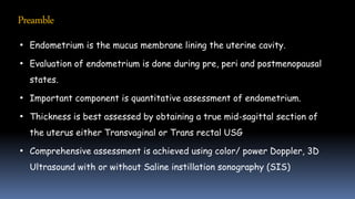
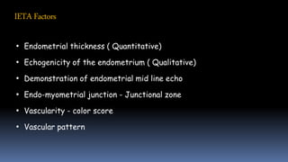
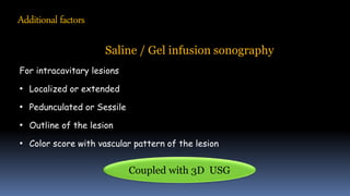
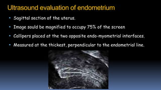
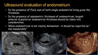
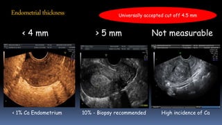
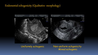
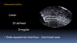
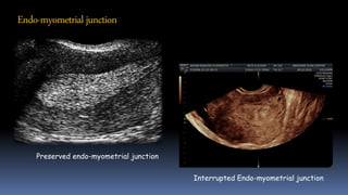
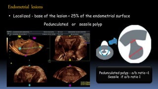
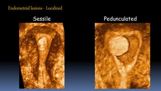
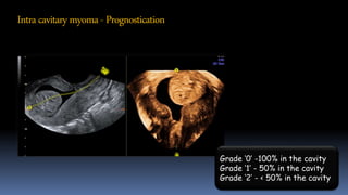
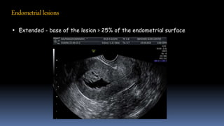
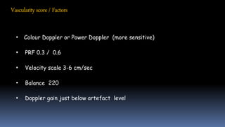
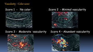
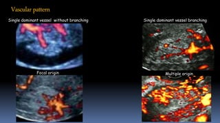
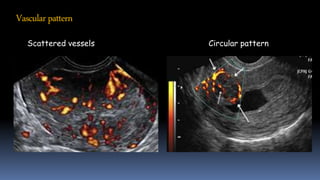
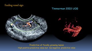
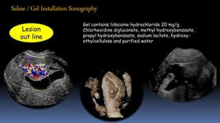
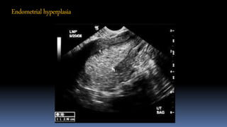
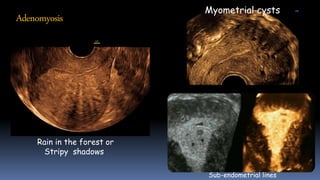
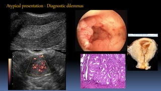
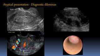
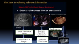
Evaluation of Abnormal Uterine Bleeding (AUB)
Dr. T L N Praveen
Director, Consultant Radiologist
Abhisheks Institute of Imageology
Hyderabad
IETA Model
Abnormal uterine bleeding - AUB
AUB - is considered when there is significant deviation from a women’s
established menstrual pattern
AUB can occur in the pre, peri, and postmenopausal states
AUB is the commonest (20%) clinical presentation in the Gynec OP
FIGO - Classification of AUB
PALM - COEIN
P – Polyp ( AUB -P)
A - Adenomyosis (AUB- A)
L – Leiomyoma (AUB – L)
M – Malignancy & hyperplasia (AUB- M)
C – Coagulopathy (AUB – C)
O – Ovulatory dysfunction (AUB- O)
E – Endometrial pathologies ( AUB -E)
I – Iatrogenic (AUB – I)
N – Not yet classified ( AUB – N)
PALM Group of causes –Discrete,
Structural, which can be measured
and evaluated by imaging techniques
COEIN Group related to entities
that are not defined by imaging
techniques (non-structural)
Preamble
• Endometrium is the mucus membrane lining the uterine cavity.
• Evaluation of endometrium is done during pre, peri and postmenopausal
states.
• Important component is quantitative assessment of endometrium.
• Thickness is best assessed by obtaining a true mid-sagittal section of
the uterus either Transvaginal or Trans rectal USG
• Comprehensive assessment is achieved using color/ power Doppler, 3D
Ultrasound with or without Saline instillation sonography (SIS)
Technical requirements
• High resolution intra cavitary USG
• Color / power Doppler USG
• 3D Ultrasound
• Saline / Gel infusion sonography
IETAFactors
• Endometrial thickness ( Quantitative)
• Echogenicity of the endometrium ( Qualitative)
• Demonstration of endometrial mid line echo
• Endo-myometrial junction - Junctional zone
• Vascularity - color score
• Vascular pattern
Additional factors
Saline / Gel infusion sonography
For intracavitary lesions
• Localized or extended
• Pedunculated or Sessile
• Outline of the lesion
• Color score with vascular pattern of the lesion
Coupled with 3D USG
Ultrasound evaluation of endometrium
• Sagittal section of the uterus.
• Image sould be magnified to occupy 75% of the screen
• Callipers placed at the two opposite endo-myometrial interfaces.
• Measured at the thickest, perpendicular to the endometrial line.
Ultrasound evaluation of endometrium
• In the presence of fluid, sum of both single endometrial lining gives the
thickness.
• In the presence of asymmetric thickness of endometrium, largest
anterior & posterior endometrial thickness should be taken into
consideration.
• When endometrium is not clearly delineated – it should be reported as “
non-measurable”
Endometrialthickness
< 4 mm > 5 mm Not measurable
< 1% Ca Endometrium 10% - Biopsy recommended High incidence of Ca
Universally accepted cut off 4.5 mm
Endometrialechogenicity
• Endometrial echogenicity in relation to the echogenicity of the
myometrium
Isoechoic
Hypoechoic
Hyperechoic
Uniformly echogenic Non-uniform echgenicity
Mixed echogenic
Endometrialechogenicity(Qualitative -morphology)
Endometrialmid-line
Linear
Ill defined
Irregular
• Endo-myometrial interface - Junctional zone
Interrupted Endo-myometrial junction
Preserved endo-myometrial junction
Endo-myometrial junction
Endometrial lesions
• Localized - base of the lesion < 25% of the endometrial surface
Pedunculated or sessile polyp
Pedunculated polyp - a/b ratio <1
Sessile if a/b ratio 1
Endometriallesions - Localized
Sessile Pedunculated
Intracavitarymyoma- Prognostication
Grade ‘0’ -100% in the cavity
Grade ‘1’ - 50% in the cavity
Grade ‘2’ - < 50% in the cavity
Endometriallesions
• Extended - base of the lesion > 25% of the endometrial surface
Vascularityscore/Factors
• Colour Doppler or Power Doppler (more sensitive)
• PRF 0.3 / 0.6
• Velocity scale 3-6 cm/sec
• Balance 220
• Doppler gain just below artefact level
Vascularity- Colorscore
Score 1 - No color Score 2 - Minimal vascularity
Score 3 - Moderate vascularity Score 4 – Abundant vascularity
Vascularpattern
Single dominant vessel without branching Single dominant vessel branching
Focal origin Multiple origin
Vascularpattern
Scattered vessels Circular pattern
Predictive of focally growing lesion
High positive predictive value but low negative predictive value
Timmerman 2003 UOG
Feeding vessel sign
Gel contains lidocaine hydrochloride 20 mg/g,
Chlorhexidine digluconate, methyl hydroxybenzoate,
propyl hydroxybenzoate, sodium lactate, hydroxy-
ethylcellulose and purified water
Lesion
out line
Saline/Gel Installation Sonography
Endometritis
• Due to deficiency in the molecular mechanism of endometrial repair
• USG - Thick, irregular endometrium
• Obliterated endo-myometrial junction
Endometrialhyperplasia
Adenomyosis
Rain in the forest or
Stripy shadows
Myometrial cysts
Sub-endometrial lines
• Endometrial thickness - > 5mm
• Hyper or mixed echoic endometrium.
• Irregular interrupted endo-myometrial interface
• Multiple, densely packed, large, irregular branching
vessels.
• High color scoring.
• SIS showing irregular surface
Endometrial thickness is the
most specific parameter
Featuresof Endometrialcancer
Atypicalpresentation- Diagnosticdilemmas
Atypical presentation - Diagnosticdilemmas
• Endometrial thickness >5mm or unmeasurable
Global
Simple Endometrial
biopsy
Failed/ Insufficient
Sample
Diagnostic
/operative
Hysteroscopy
Feeding vessel
Failed
Inconclusive
Exam
Diagnostic
Hysteroscopy
Flowchart in evaluatingendometrialabnormality
(Epstein 2002 ACTA Obstet Gynecol Scandinavica)
Ultrasound in Obstetrics & Gynecology
Volume 43, Issue 5, pages 557-568, 2 MAY 2014
Risk of endometrial cancer ( REC) score –
9 variables – value between 0 to 9
REC score 4 or more – Malignancy.
REC score 3 (not including GIS) Malignancy
Risk ofendometrialcancer Dueholmetal
• Endometrial thickness < 4mm – no further investigation.
• “ Unmeasurable “ endometrium should be evaluated.
• Vascularity of the endometrium and colour score helps in
grading the lesions.
• SIS /GIS outlines the lesions and its surface
irregularities.
• Endometrial and myometrial junction helps in identifying
focal lesions.
• Pre-operative assessment of myometrial and cervical
invasion is effectively done by ultrasound.
Summary
• Trans vaginal ultrasound is the imaging modality of choice.
• IETA guideline are useful in early detection of endometrial
abnormalities and “ Score the risk of Endometrial cancer”.
• Flow charts are useful in managing these patients.
• Newer techniques such as elastography & Contrast enhanced
ultrasound are complimentary to the conventional techniques
Takehomepoints
Thank you

Aub ieta -lucknow

  • 1.
    Evaluation of AbnormalUterine Bleeding (AUB) Dr. T L N Praveen Director, Consultant Radiologist Abhisheks Institute of Imageology Hyderabad IETA Model
  • 2.
    Abnormal uterine bleeding- AUB AUB - is considered when there is significant deviation from a women’s established menstrual pattern AUB can occur in the pre, peri, and postmenopausal states AUB is the commonest (20%) clinical presentation in the Gynec OP
  • 3.
    FIGO - Classificationof AUB PALM - COEIN P – Polyp ( AUB -P) A - Adenomyosis (AUB- A) L – Leiomyoma (AUB – L) M – Malignancy & hyperplasia (AUB- M) C – Coagulopathy (AUB – C) O – Ovulatory dysfunction (AUB- O) E – Endometrial pathologies ( AUB -E) I – Iatrogenic (AUB – I) N – Not yet classified ( AUB – N)
  • 4.
    PALM Group ofcauses –Discrete, Structural, which can be measured and evaluated by imaging techniques COEIN Group related to entities that are not defined by imaging techniques (non-structural)
  • 5.
    Preamble • Endometrium isthe mucus membrane lining the uterine cavity. • Evaluation of endometrium is done during pre, peri and postmenopausal states. • Important component is quantitative assessment of endometrium. • Thickness is best assessed by obtaining a true mid-sagittal section of the uterus either Transvaginal or Trans rectal USG • Comprehensive assessment is achieved using color/ power Doppler, 3D Ultrasound with or without Saline instillation sonography (SIS)
  • 6.
    Technical requirements • Highresolution intra cavitary USG • Color / power Doppler USG • 3D Ultrasound • Saline / Gel infusion sonography
  • 7.
    IETAFactors • Endometrial thickness( Quantitative) • Echogenicity of the endometrium ( Qualitative) • Demonstration of endometrial mid line echo • Endo-myometrial junction - Junctional zone • Vascularity - color score • Vascular pattern
  • 8.
    Additional factors Saline /Gel infusion sonography For intracavitary lesions • Localized or extended • Pedunculated or Sessile • Outline of the lesion • Color score with vascular pattern of the lesion Coupled with 3D USG
  • 9.
    Ultrasound evaluation ofendometrium • Sagittal section of the uterus. • Image sould be magnified to occupy 75% of the screen • Callipers placed at the two opposite endo-myometrial interfaces. • Measured at the thickest, perpendicular to the endometrial line.
  • 10.
    Ultrasound evaluation ofendometrium • In the presence of fluid, sum of both single endometrial lining gives the thickness. • In the presence of asymmetric thickness of endometrium, largest anterior & posterior endometrial thickness should be taken into consideration. • When endometrium is not clearly delineated – it should be reported as “ non-measurable”
  • 11.
    Endometrialthickness < 4 mm> 5 mm Not measurable < 1% Ca Endometrium 10% - Biopsy recommended High incidence of Ca Universally accepted cut off 4.5 mm
  • 12.
    Endometrialechogenicity • Endometrial echogenicityin relation to the echogenicity of the myometrium Isoechoic Hypoechoic Hyperechoic
  • 13.
    Uniformly echogenic Non-uniformechgenicity Mixed echogenic Endometrialechogenicity(Qualitative -morphology)
  • 14.
  • 15.
    Interrupted Endo-myometrial junction Preservedendo-myometrial junction Endo-myometrial junction
  • 16.
    Endometrial lesions • Localized- base of the lesion < 25% of the endometrial surface Pedunculated or sessile polyp Pedunculated polyp - a/b ratio <1 Sessile if a/b ratio 1
  • 17.
  • 18.
    Intracavitarymyoma- Prognostication Grade ‘0’-100% in the cavity Grade ‘1’ - 50% in the cavity Grade ‘2’ - < 50% in the cavity
  • 19.
    Endometriallesions • Extended -base of the lesion > 25% of the endometrial surface
  • 20.
    Vascularityscore/Factors • Colour Doppleror Power Doppler (more sensitive) • PRF 0.3 / 0.6 • Velocity scale 3-6 cm/sec • Balance 220 • Doppler gain just below artefact level
  • 21.
    Vascularity- Colorscore Score 1- No color Score 2 - Minimal vascularity Score 3 - Moderate vascularity Score 4 – Abundant vascularity
  • 22.
    Vascularpattern Single dominant vesselwithout branching Single dominant vessel branching Focal origin Multiple origin
  • 23.
  • 24.
    Predictive of focallygrowing lesion High positive predictive value but low negative predictive value Timmerman 2003 UOG Feeding vessel sign
  • 25.
    Gel contains lidocainehydrochloride 20 mg/g, Chlorhexidine digluconate, methyl hydroxybenzoate, propyl hydroxybenzoate, sodium lactate, hydroxy- ethylcellulose and purified water Lesion out line Saline/Gel Installation Sonography
  • 26.
    Endometritis • Due todeficiency in the molecular mechanism of endometrial repair • USG - Thick, irregular endometrium • Obliterated endo-myometrial junction
  • 27.
  • 28.
    Adenomyosis Rain in theforest or Stripy shadows Myometrial cysts Sub-endometrial lines
  • 29.
    • Endometrial thickness- > 5mm • Hyper or mixed echoic endometrium. • Irregular interrupted endo-myometrial interface • Multiple, densely packed, large, irregular branching vessels. • High color scoring. • SIS showing irregular surface Endometrial thickness is the most specific parameter Featuresof Endometrialcancer
  • 30.
  • 31.
    Atypical presentation -Diagnosticdilemmas
  • 32.
    • Endometrial thickness>5mm or unmeasurable Global Simple Endometrial biopsy Failed/ Insufficient Sample Diagnostic /operative Hysteroscopy Feeding vessel Failed Inconclusive Exam Diagnostic Hysteroscopy Flowchart in evaluatingendometrialabnormality (Epstein 2002 ACTA Obstet Gynecol Scandinavica)
  • 33.
    Ultrasound in Obstetrics& Gynecology Volume 43, Issue 5, pages 557-568, 2 MAY 2014 Risk of endometrial cancer ( REC) score – 9 variables – value between 0 to 9 REC score 4 or more – Malignancy. REC score 3 (not including GIS) Malignancy Risk ofendometrialcancer Dueholmetal
  • 34.
    • Endometrial thickness< 4mm – no further investigation. • “ Unmeasurable “ endometrium should be evaluated. • Vascularity of the endometrium and colour score helps in grading the lesions. • SIS /GIS outlines the lesions and its surface irregularities. • Endometrial and myometrial junction helps in identifying focal lesions. • Pre-operative assessment of myometrial and cervical invasion is effectively done by ultrasound. Summary
  • 35.
    • Trans vaginalultrasound is the imaging modality of choice. • IETA guideline are useful in early detection of endometrial abnormalities and “ Score the risk of Endometrial cancer”. • Flow charts are useful in managing these patients. • Newer techniques such as elastography & Contrast enhanced ultrasound are complimentary to the conventional techniques Takehomepoints
  • 36.