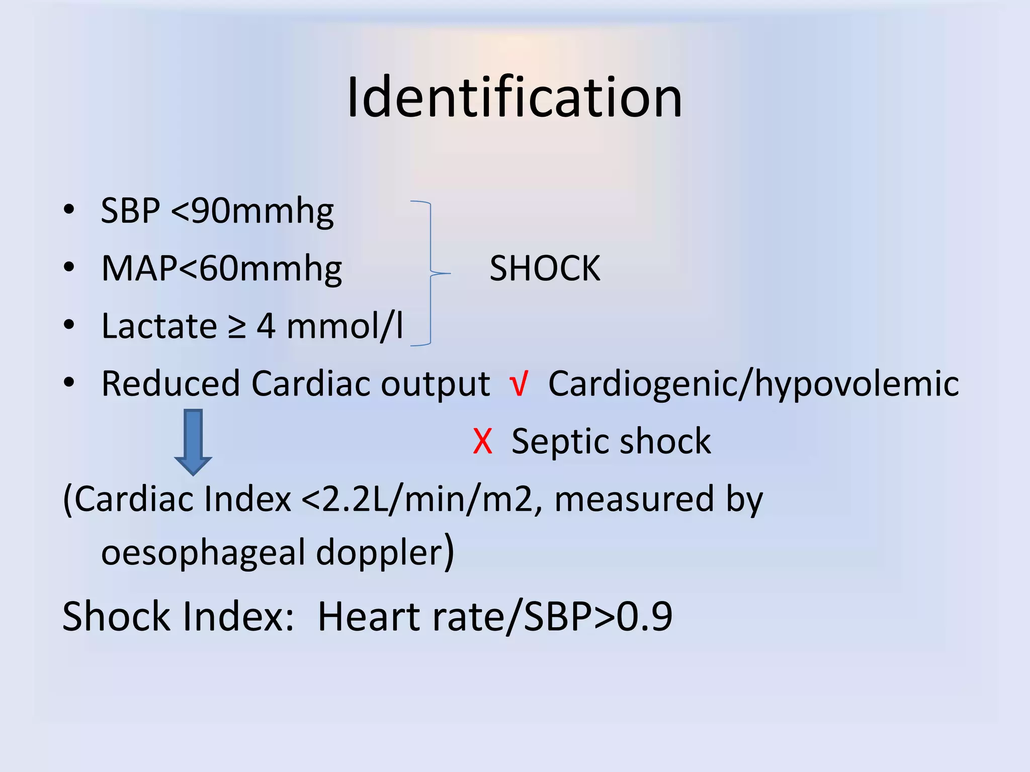
1) Shock is defined as inadequate tissue perfusion and is usually accompanied by hypotension, with a mean arterial pressure less than 60.
2) The first step in approaching shock is to identify the cause, with goals of reversing tissue hypoperfusion.
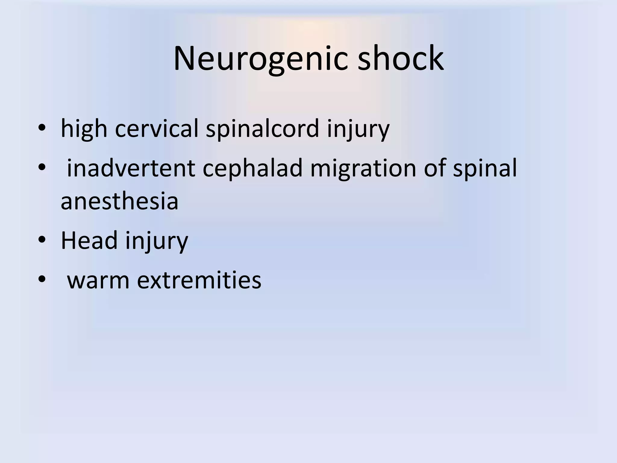
3) The main types of shock are hypovolemic, cardiogenic, septic, neurogenic, and anaphylactic. Each type has distinguishing clinical features and targeted treatment approaches.


![SHOCK
• Inadequate tissue perfusion
• Clinical shock is usually accompanied by
hypotension ( meanarterial pressure [MAP]
<60)
• First step- Cause
• Goal: reverse tissue hypoperfusion
•](https://image.slidesharecdn.com/approachtoshock-190427165338/75/Approach-to-shock-4-2048.jpg)