Download to read offline
![Research Article
Antiobesity and Hypolipidemic Activity of Moringa oleifera
Leaves against High Fat Diet-Induced Obesity in Rats
Souravh Bais,1
Guru Sewak Singh,2
and Ramica Sharma2
1
Department of Pharmacology, Rayat Institute of Pharmacy, Railmajra, SBS Nagar District, Punjab 144506, India
2
Rayat Institute of Pharmacy, Railmajra, SBS Nagar District, Punjab 144506, India
Correspondence should be addressed to Souravh Bais; souravh2008.123@rediffmail.com
Received 19 May 2014; Revised 21 June 2014; Accepted 21 June 2014; Published 10 July 2014
Academic Editor: Octavio Franco
Copyright © 2014 Souravh Bais et al. This is an open access article distributed under the Creative Commons Attribution License,
which permits unrestricted use, distribution, and reproduction in any medium, provided the original work is properly cited.
In the present study, the methanolic extract of Moringa oleifera leaves (MEMOL) was evaluated for antiobesity activity in rats.
The antiobesity potential of MEMOL was studied against high fat diet-induced obesity (HFD) in rats. In this study, chronic
administration of HFD in rats produced hypercholesterolemia (116.2 ± 0.27 mg/dL), which led to an increase in the body weight
(225 gr), total cholesterol, triglycerides (263.0 ± 4.69 mg/dL), and attenuation in the levels of HDL (34.51 ± 2.20 mg/dL) as well as
changes in body temperature of animals. Treatment of obese rats with MEMOL for 49 days resulted in a significant (𝑃 < 0.001)
change in body weight, total cholesterol, triglycerides, and LDL level along with a significant (𝑃 < 0.001) increase in body
temperature as compared to the HFD-induced obesity. MEMOL treated rats also showed a significant decrease in the level of
liver biomarkers, organ weight, and blood glucose level. Further, rats treated with MEMOL (200 mg and 400 mg/kg) show reduced
atherogenic index (1.7 ± 0.6 and 0.87 ± 0.76). The results indicate that the rats treated with Moringa oleifera (MO) have significantly
attenuated the body weight without any change in the feed intake and also elicited significant thermogenic effect and to act as
hypolipidemic and thermogenic property in obesity related disorders.
1. Introduction
Nowadays Obesity has emerged as a major health problem
and risk factor for various disorders worldwide [1]. Over-
weight and obesity are defined as abnormal or excessive fat
accumulation triggered by disproportion in energy intake
and expenditure [2–4]. In addition to this attenuation in adi-
pogenesis and over expression of pancreatic lipase enzyme
which plays a pivotal role in progression of obesity [5]. The
literature review revealed that alteration in dietary habit and
less physical exercises, too, increase the frequency of obesity
and related disorders [6, 7]. Further, obesity has been found to
be associated with various disorders such as osteoarthritis [8],
ischemic heart diseases (IHD) [8], atherosclerosis, diabetes,
and hypertension [9–11]. A streak of evidence indicates that
serotonin, histamine, dopamine, and their associated recep-
tor activities are closely associated with obesity regulation [5].
Most importantly, strong evidences are available that elicited
the role of leptin, ghrelin, and neuropeptides in obesity
[12–14]. Currently, no pharmacological treatment provides
sustained weight loss with minimal adverse effects [15, 16].
Thus, attempts have been made to reduce body weight with
such pharmacological intervention that possesses minimal
side effects. Plants have been used as traditional natural
medicines for healing many diseases. In particular, various
oriental medicinal plants are reported to have biological
activity [17]. Literature review has revealed that various
herbal plants such as Fucus vesiculosus, Citrus aurantium [18],
Yacon syrup [19], Curcumin [20], Nigella Sativa, Camellia
Synensis, Green Tea, and Black Chinese Tea [21] are used in the
management of obesity. M. O. (M. oleifera) Lam that belongs
to Moringaceae family is commonly known as Drumstick
tree that possesses various nutritional and medicinal values
attributed to its roots, bark, leaves, flowers, fruits, and seeds
[22–24]. Data revealed that most of the parts of the plant pos-
sess antimicrobial activity [25, 26], antidiabetic [27–29], hep-
atoprotective [30], and for cardiac stimulation [31]. Recently,
hypocholesterolemic activity of crude extract of M. oleifera
Hindawi Publishing Corporation
Advances in Biology
Volume 2014,Article ID 162914, 9 pages
http://dx.doi.org/10.1155/2014/162914](https://image.slidesharecdn.com/antiobesity-activity-of-moringa-oleifera-150210053712-conversion-gate01/85/Antiobesity-activity-of-moringa-oleifera-1-320.jpg)
![2 Advances in Biology
crude extract was explored [32], but its thermogenic and
antiobesity activity has not been investigated; hence, the study
delineated with antiobesity property of methanolic extract M.
oleifera leaves in experimentally induced obesity.
2. Materials and Methods
Age matched young wistar albino rats of either sex, weighing
120–150 gr, were housed in room temperature of 25 ± 1∘
C
and 12 hrs light and dark cycles (9:00 AM). Animals of con-
trol group were feed on a standard chow diet (Ashirwad
Industries, Ropar, India) and water ad libitum, whereas
animals used for evaluation of obesity are feed on a HFD and
water ad libitum. The experimental protocols were approved
by the Institutional Animal Ethics Committee (IAEC) and
conducted according to the guidelines of the Committee for
the Purpose of Control and Supervision of Experiments on
Animals (CPCSEA), New Delhi, India {CPCSEA Approval
No.: RIP/IAEC/2012-2013/13}. The animals were distributed
in five groups of 10 animals each and were fed a HFD. Rats
receiving the MEMOL extract were dosed for 7 weeks in
parallel as detailed in Table 2.
2.1. Drugs and Chemicals. Sibutramine was purchased from
Lupin (Bhopal), Pyridine from S.D. Fine Chemicals Ltd.,
Mumbai. Solvents like methanol, chloroform, petroleum
ether, acetone, di-ethyl ether were of analytical grade (AR).
Serum Cholesterol kit, Serum Triglyceride kit, Serum HDL-
Cholesterol kit, Serum LDL-Cholesterol kit, Serum VLDL-
Cholesterol kit, Blood-Glucose kits were provided from
Spruce Enterprises Ambala.
2.2. Collection and Preparation of Plant Extract. Leaves of
Moringa oleifera (M. O.) were collected after proper iden-
tification and authentication by the chief scientist of
NISCAIR, New Delhi, with voucher number (NISCAIR/
RHMD/Consult/2013/2279/54) and kept in the herbarium.
The fresh leaves were air-dried for a period of two weeks,
crushed in a mortar, and later pulverized into fine powder
using electric blender. The powder was sieved through a mesh
(2 mm) and used for preparation of methanolic extract. The
extract was prepared by adding 100 gm of the plant powder
in 200 mL of petroleum ether to remove fatty materials in
a conical flask. The mark obtained from petroleum ether
extraction was again extracted with 200 mL of methanol. The
solvent was completely removed under reduced pressure till
the dried extract was obtained. A dark greenish semisolid
residue was obtained with a yield of 7%. The extract was
stored in desiccators and a weighed (1 gm) amount was
suspended in distilled water using carboxyl methyl cellulose
(CMC) (2%) as suspending agent prior to administration.
2.3. Dose Selection. In the present study, two doses of the
Moringa oleifera leaf extract were selected as 200 mg and
400 mg/kg, p.o. These doses were selected on the basis of
previous reports of the acute toxicity study performed using
the single dose of orally administered 2 g/kg of methanolic
extracts of M. oleifera (leaf) which shows no signs of toxicity
in rats [33].
2.4. High-Fat Diet Formula. HFD that consists of 58% fat,
25% protein and 17% carbohydrate, lard (13%), cholesterol
(1%), vitamin, and minerals (0.6%) as a percentage of total
kcal ad libitum, respectively, was administered every day
[34]. Food intake was calculated every day and body weight
was measured once in every two days. The composition
of normal pellet diet (NPD) and HFD diets is shown in
Table 1.
2.5. Preliminary Phytochemical Screening. The preliminary
phytochemical screening of MEMO was carried out accord-
ing to the methods described by Khandelwal [35] and Kokate
[36]. Phytochemical analysis of the extract was performed for
the identification of phytochemicals such as carbohydrates,
alkaloids, tannins, saponins, flavonoids, triterpenoids, and
steroids.
2.6. Methodology. High-fat diet (HFD) induced obesity in
rats is considered to be a reliable tool for the evaluation
of antiobesity activity. The study comprises 5 groups with
10 animals in each group. Group 1 represented the normal
control in which the animals were feed on a normal diet
(NPD) and had free access to water. Group 2 represented
a negative control in which the rats were feed on high-fat
diet (HFD) for a period of 49 days. Group 3 represented
standard control in which rats were treated with simvas-
tatin (3 mg/kg, p.o). Group 4 represented test treatment in
which rats were treated with the 1st dose of methanolic
extract of M. oleifera (200 mg/kg) along with high-fat diet.
Group 5 represented test treatment in which rats were
treated with the 2nd dose of methanolic extract of M.
oleifera (400 mg/kg) along with high-fat diet for 49 days
(Table 2). Various parameters like cholesterol (TC), high
density lipoproteins (HDL-C), triglycerides (TG), low density
lipoproteins (LDL-C), very low density lipoproteins (VLDL-
C), atherogenic index, percentage protection, serum glutamic
oxaloacetic transaminase (SGOT), serum glutamic pyrubic
transaminase (SGPT), and total bilirubin were also recorded
weekly. At the end of the protocol, animals were sacrificed
by cervical dislocation and liver and kidney were removed
to measure the change in weight and for histopathological
evaluation [34].
2.7. In Vivo Pharmacological Evaluation
2.7.1. Body Weight and Food Intake. The body weight (gm)
was recorded on day one and then weekly consecutively for
49 days using a digital weighing balance. In addition to this,
the daily food intake for each group was measured weekly for
49 days.
2.7.2. Body Temperature. The body temperature was noted
using a rectal thermometer before and after drug adminis-
tration at 0, 30, 60, 90, and 120 minutes with a contact time
of 1 minute.](https://image.slidesharecdn.com/antiobesity-activity-of-moringa-oleifera-150210053712-conversion-gate01/85/Antiobesity-activity-of-moringa-oleifera-2-320.jpg)
![Advances in Biology 3
Table 1: Composition of the control and test diets.
Control (%)∗
HFD (%)∗∗
Fat 58% 58%
Protein 12.4% 12.4%
Carbohydrate 17% 17%
Lard 12% 11%
Cholesterol 0 1%
Vitamin and mineral 0.6% 0.6%
Total (gm)
∗
Lab chow diet (NPD). ∗∗
High-fat diet (cholesterol: 1%).
Table 2: Treatment Groups.
Groups Treatment (mg/kg body weight)
1 Control
2 High-fat diet (HFD)
3 Standard group (simvastatin-3 mg/kg, p.o)
4 HFD + MEMOL (200 mg/kg, p.o)
5 HFD + MEMOL (400 mg/kg, p.o)
2.8. Biochemical Estimations. On the 49th day of the experi-
ment all the animals were sacrificed by cervical dislocation,
and blood samples were collected by carotid bleeding sep-
arately into sterilized dry centrifugation tubes and allowed
to stand for 30 minutes at 20–25∘
C. The clear serum was
separated at 2500 rpm for 10 min using a centrifuge. The levels
of serum glucose, total TC, HDL-C, TG, LDL-C, VLDL-
C, atherogenic index, percentage protection, SGOT, SGPT,
and total bilirubin were determined to analyze using auto-
analyzer (semi-autoanalyzer star 21 plus), with commercial
kits (autospan diagnostics Pvt. Ltd.). The Friedewald formula
[37] was used to calculate serum low-density lipoprotein
cholesterol (LDL-C) values and atherogenic index as follows:
LDL-C = TC-(HDL-C +TG/5) and atherogenic index = (TC-
HDL-C)/HDL-C.
2.9. Statistics. Data were analysed with one-way ANOVAs
followed by Tukey’s post hoc tests, with significance set to
𝑃 < 0.001. Food consumption was analysed using two-way
ANOVA repeated measures tests followed by Tukey’s post hoc
tests, with significance set to 𝑃 < 0.001. All analyses were
performed using Sigma Stat (10.0).
3. Results
3.1. Preliminary Phytochemical Analysis. The results of the
preliminary phytochemical screening of methanolic extract
of M. oleifera leaves (MEMOL) showed the presence of
alkaloids, tannins, flavonoids and terpenoids, and steroids.
3.2. Effects of High-Fat Diet and MEMOL on Food Intake and
Body Weight. There was no momentous difference in all of
the treatment groups at the commencement of the study.
However, animals feed with high-fat diet showed significant
increase in body weight compared to those feed with normal
pellet diet (NPD) (𝑃 < 0.001). Similarly, the average daily
250
200
150
100
1 32 4 5 6 7
a
Weeks
Bodyweight(gm)
Control group
HFD
Standard
HFD + MEMOL 200 (a, b)
HFD + MEMOL 400 (a, b)
a, b
a, b
Figure 1: Body weight of rats fed a high-fat diet and treated with
MEMOL extracts for 7 weeks; all values are expressed as Mean ±
SEM, (𝑛 = 10). (a) Significant difference compared to control;
(b) significant difference compared to HFD, 𝑃 < 0.001 (one-way
ANOVA, Tukey’s post hoc test, 𝑛 = 10 per group).
12.0
10.5
9.0
7.5
6.0
1 32 4 5 6 7
a, b
a, b
a, b
Weeks
Control group
HFD
Simvastatin HFD + MEMOL 200
HFD + MEMOL 400
Food consumption
Foodintake(gm)
Figure 2: Food consumption (kcal) in rats fed a high-fat diet and
treated with MEMOL for 7 weeks; all values are expressed as Mean
± SEM, (𝑛 = 10) (a) significant difference compared to control;
(b) significant difference compared to HFF, 𝑃 < 0.001 (one-way
ANOVA, Tukey’s post hoc test).
feed intake of all the groups was the same at the start of
the study; however, 21-day treatment with HFD resulted in
a slight increase in food intake (Figure 1).
Following 2 week treatment with MEMOL supplementa-
tion, low food intake was observed in the MEMOL 200 mg/kg
and MEMOL 400 mg/kg groups (𝑃 < 0.001) when compared
to the HFD group. Subsequently, the body weight gain (at
days 0, 7, 14, 21, 28, 35, 42, and 49) in these groups was
significantly lower than the rats feed with HFD (Figure 2).](https://image.slidesharecdn.com/antiobesity-activity-of-moringa-oleifera-150210053712-conversion-gate01/85/Antiobesity-activity-of-moringa-oleifera-3-320.jpg)
![Advances in Biology 5
Table 5: Effect of MEMOL on Body temperature.
S. No Groups
Body temperature ∘
C
0 min 30 min 60 min 90 min 120 min
I Control 39.3 ± 1.117 37.2 ± 1.219 39.7 ± 0.653 38.3 ± 2.240 38.5 ± 1.364
II High-fat diet 32.9 ± 2.295ns
27.3 ± 2.508a∗
25.0 ± 2.87a∗∗
25.1 ± 2.84a∗∗
23.3 ± 1.83a∗∗∗
III Standard (3 mg/kg) 36.1 ± 1.411ns
39.9 ± 1.161b∗∗
35.4 ± 1.161b∗
35.6 ± 1.340b∗
34.6 ± 2.228b∗
IV MEMOL (200 mg/kg) 39.5 ± 0.81ns
38.4 ± 0.934b∗∗
35.4 ± 1.391b∗∗∗
38.9 ± 0.817b∗∗c ns
36.1 ± 1.554b∗c ns
V MEMOL (400 mg/kg) 36.2 ± 1.53ns
34.0 ± 1.455b∗
39.1 ± 2.026b∗∗
41.3 ± 0.497b∗∗∗c∗
40.6 ± 0.702b∗∗∗c∗
All values are expressed as Mean ± SEM (𝑛 = 10). Statistical significance testing for the comparisons was made by ANOVA, followed by Tukey’s post hoc test.
∗
𝑃 < 0.05, ∗∗
𝑃 < 0.01, ∗∗∗
𝑃 < 0.001, ns
𝑃 > 0.001. a
versus Normal Control; b
versus HFD-induced obesity; c
versus standard control (one-way ANOVA,
Tukey’s post hoc test, 𝑛 = 10 per group).
Table 6: Effect of MEMOL supplementation on organ weight.
Organ weight (g) Normal control HFD Standard (3 mg/kg) MEMOL
200 mg/kg
MEMOL
400 mg/kg
Liver 5.737 ± 0.088 8.845 ± 0.982a∗∗∗
6.158 ± 0.892b∗∗∗
7.573 ± 0.779b∗
6.947 ± 1.754b∗∗
Right Kidney 0.765 ± 0.012 1.76 ± 0.095a∗∗∗
0.801 ± 0.053b ns
0.874 ± 0.073b ns
0.842 ± 0.017b ns
Left Kidney 0.748 ± 0.017 0.958 ± 0.075a∗∗
0.758 ± 0.019b∗∗
0.813 ± 0.012b ns
0.804 ± 0.009b∗
All values are expressed as Mean ± SEM (𝑛 = 10) ∗∗∗
𝑃 < 0.001 when compared with the normal control group, ∗
𝑃 < 0.05; ∗∗∗
𝑃 < 0.001; ∗∗
𝑃 < 0.01; ns
𝑃 >
0.05. a
versus high fat diet-induced obesity; b
versus standard control, one-way ANOVA followed by Tukey’s post hoc test.
4. Discussion
The lethal dose (LD50) value of the M. oleifera indicated that
the extract (methanol) was safe and nontoxic up to 5 g/kg
[33, 38]. Previous experiments have reported the antiulcer,
diuretic, anti-inflammatory, antifertility, CNS depressant,
and wound healing properties of leaves of M. oleifera [26, 38–
41]. However, the results of the previous laboratory animal
study indicated that crude extract of M. oleifera possesses
hypocholesterolemic activity in rats [32]. Still no evidences
are available for antiobesity potential of methanolic extract
of M. oleifera. Hence, the study has been designed to demon-
strate the effect of M. oleifera in high fat diet-induced obesity.
Obesity is a major risk factor for augmented morbidity
and mortality and is associated with various medical ailments
[42]. High fat diet-induced obesity has been considered as
the most popular model among researchers due to its high
similarity of mimicking the usual route of obesity episodes
in human [43] and so why it is considered as a reliable
tool for studying obesity as they will readily gain weight
when feed high-fat diets [44]. Human studies have revealed
that increased fat intake is associated with body weight
gain, which can lead to obesity and other related metabolic
diseases. This study thus proved that rats exposed to high-
fat diet for 2 weeks cause a significant increase of animals’
body weight, thus verifying the obese status [45]. Although
there was a significant difference in the body weights between
the high-fat and normal diet groups, no significant difference
was observed in the daily food intake of animals. This
observation provides us with the fact that an increase in
body weight is independent of the amount of food consumed
by the animals. Treatment of HFD rats with MEMOL at
200 mg/kg and 400 mg/kg p.o conversely causes a remarkable
reduction of body weights when compared to the high-fat
diet administered rats. The result also suggests that MEMOL
supplementation at 200 mg/kg and 400 mg/kg are capable
of preventing body weight gain, concomitantly helping in
maintaining the current body weight. This result was in
accordance with the results reported from the previous study
where a dose dependent decrease in the body weight was
observed [33]. Further, treatment with MEMOL remarkably
decreases the organ weight of rats feed on high-fat diet. Thus
it proved the weight reducing potential of MEMOL.
Further, dyslipidemia is another important hallmark in
the pathogenesis of obesity characterized by hypertriglyc-
eridemia with decreased level of LDL and VLDL [46, 47].
Chronic dyslipidemia has been characterized as a major risk
factor for cardiovascular risk, including atherosclerosis [48,
49]. In the present study apart from reduction in weight,
supplementation with MEMOL was observed to attenuate
significantly the levels of total cholesterol and LDL and
increased the level of HDL level in rats feed with HFD. The
increase in the level of HDL was found to be in a dose
dependent manner; that is, supplementation with MEMOL
at a dose of 400 mg/kg shows a better effect in comparison
to 200 mg/kg. Similar results were obtained by Ghasi et al.
[32], where treatment with crude extract of M. oleifera led to
an increased serum HDL level and decreased levels of total
cholesterol, LDL, and triglyceride. Thus, it can be concluded
that leaves of M. oleifera possess cardioprotective potential
[50]. Further, atherogenic index is regarded as a marker
for various cardiovascular disorders; the higher the value,
the higher the risk of developing cardiovascular disease and
vice versa [51, 52]. High-fat diet exposure resulted in the
increased atherogenic index. Treatment with 200 mg/kg and
400 mg/kg significantly attenuated the atherogenic index and](https://image.slidesharecdn.com/antiobesity-activity-of-moringa-oleifera-150210053712-conversion-gate01/85/Antiobesity-activity-of-moringa-oleifera-5-320.jpg)
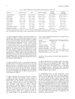
![6 Advances in Biology
(a) Normal
FDH
VCD
(b) High-fat diet (HFD)
CD
CV
(c) HFD + simvastatin (3 mg/kg)
CD
FDH
(d) HFD + MEMO (200 mg/kg)
FDH
CD
(e) HFD + MEMO (400 mg/kg)
Figure 3: Histopathology of liver showing ballooning degeneration and inflammation. (CD: cellular degeneration, VCD: vascular congestion
fatty deposition, FDH: foamy degeneration of hepatocytes, CV: central vein, and N: necrosis).
thus provides cardioprotection. The decreased atherogenic
index by MEMOL thus supports the cardioprotectant nature
of M. oleifera.
In order to supplement the results, the histopatho-
logical studies were also performed. The literature review
revealed that high fat diet-induced obesity and abnormal lipid
metabolism all collectively are associated with inflammation,
congestion, and nonalcoholic fatty liver disease (NAFLD)
leading to hepatic failure causing a boost in SGOT, SGPT,
and total bilirubin level in the serum [52–54]. Our results
showed that consumption of high-fat diet may play a crucial
role in the pathogenesis of fatty liver or hepatic steatosis
associated with obesity depicted via ballooning degeneration.
Elevated levels of liver enzymes are a monitor of hepatocel-
lular damage and correlate with increased liver weight [55].
The results obtained in the present study established that](https://image.slidesharecdn.com/antiobesity-activity-of-moringa-oleifera-150210053712-conversion-gate01/85/Antiobesity-activity-of-moringa-oleifera-6-320.jpg)
![Advances in Biology 7
high-fat diet causes hepatocellular damage, as clearly seen by
the marked elevation of serum enzymes (SGOT, SGPT, and
total bilirubin) activities and histopathological studies of liver
exaggerated with hepatic steatosis. However, treatment with
MEMOL causes a momentary reduction in the enzyme levels,
signifying the role of MEMOL in preventing liver damage
caused by high-fat diet.
Insulin resistance is associated with a number of metabol-
ic disorders such as obesity, hyperlipidemia, and hyperten-
sion. HFD intakes were shown to contribute to syndromes
such as hyperlipidemia, glucose intolerance, hypertension,
and atherosclerosis [56]. Numerous evidences indicated
that in experimental animals, high-fat diets resulted in
disturbance in glucose metabolism and impaired glucose
tolerance [57, 58], and the present study also demonstrate
the reduction in blood glucose level those treated with
MEMOL (200 and 400 mg/kg). It has been reported that
thermogenesis plays a crucial role in weight management
[59, 60]. Changes in body temperature are associated with
significant changes in metabolic rate [61]. In support for
this, the theory has been shown in different animal models
which were obese, and leptin-deficient ob/ob mouse and the
polyphonic obese mouse exhibited hyperphagia, a decreased
metabolic rate, and a decreased core body temperature [62,
63]. This contention is supported in our results where rats
feed on HFD show decreased body temperature in compari-
son with normal rats. Treatment with MEMOL (200 mg/kg
and 400 mg/kg) reflected a sharp increase in rectal body
temperature. The increase in rectal body temperature may be
attributed to the overall stimulant and thermogenic property
of phytoconstituents of the extract.
Preliminary phytochemical studies of the extract of M.
oleifera showed the presence of alkaloids, tannins, flavonoids
and terpenoids, and steroids. Moringa leaves act as a good
source of natural antioxidant due to the presence of vari-
ous types of antioxidant compounds such as ascorbic acid,
flavonoids, phenolics, and carotenoids [23, 64]. The high
concentrations of ascorbic acid; oestrogenic substances and
𝛽-sitosterol; iron; calcium; phosphorus; copper; vitamins A,
B and C; 𝛼-tocopherol; riboflavin; nicotinic acid; folic acid;
pyridoxine; 𝛽-carotene; protein; and in particular essential
amino acids such as methionine, cysteine, tryptophan, and
lysine present in Moringa leaves and pods make it a virtually
ideal dietary supplement [64]. The hypolipedemic potential
is associated with the presence of 𝛽-sitosterol [32] in crude
extract of M. oleifera. Therefore, further study needs to be
carried out for identification of specific constituents present
in M. oleifera for its observed effects.
The present study thus concludes that the extract of
leaves of M. oleifera possess hypolipedemic and antiobesity
potential that protects the body against adverse effects of
high fat diet-induced obesity. Further, we demonstrated that
the daily supplementation of M. oleifera leaves extract may
reverse the formation of hepatic steatosis and nonalcoholic
fatty liver disorder. The results in the present study established
that high-fat diet causes elevation in body weight and reduces
lipid metabolism as clearly seen by the marked elevation
of liver enzymes and lipid level. However, supplementation
with MEMOL reverses all the parameters thus suggesting its
weight reducing potential.
5. Conclusion
Thus, from the present study it can be concluded that the
methanolic extract of M. oleifera is beneficial to the weight
management, which supports its traditional claim. Further,
studies are carried out in order to determine the active
principle of this plant, followed by the identification of
the mechanistic approach of MEMOL that helps in weight
management.
Conflict of Interests
The authors declare that there is no conflict of interests
regarding the publication of this paper.
Acknowledgment
The authors would like to thank Dr. A. C. Rana the Director
of Rayat Institute of Pharmacy (Punjab) for providing the
necessary research facilities.
References
[1] C. Roh and U. Jung, “Screening of crude plant extracts with anti-
obesity activity,” International Journal of Molecular Sciences, vol.
13, no. 2, pp. 1710–1719, 2012.
[2] B. M. Spiegelman and J. S. Flier, “Obesity and the regulation of
energy balance,” Cell, vol. 104, no. 4, pp. 531–543, 2001.
[3] P. G. Kopelman, “Obesity as a medical problem,” Nature, vol.
404, no. 6778, pp. 635–643, 2000.
[4] S. Panico and A. Iannuzzi, “Dietary fat composition and the
metabolic syndrome,” European Journal of Lipid Science and
Technology, vol. 106, no. 1, pp. 61–67, 2004.
[5] C. V. Chandrasekaran, M. A. Vijayalakshmi, K. Prakash, V.
S. Bansal, J. Meenakshi, and A. Amit, “Review Article: herbal
approach for obesity management,” American Journal of Plant
Sciences, vol. 3, no. 7, pp. 1003–1014, 2012.
[6] B. P. Latha, R. M. Reddy, S. M. Ismail, and T. Vijaya, “Medicinal
plants and their derivatives as potential source in treatment of
obesity,” Asian Journal of Experimental Biological Sciences, vol.
1, no. 4, pp. 719–727, 2010.
[7] A. Mangal and M. C. Sharma, “Evaluation of certain medicinal
plants for antiobesity properties,” Indian Journal of Traditional
Knowledge, vol. 8, no. 4, pp. 602–605, 2009.
[8] A. Azimi, M. G. Charlot, C. Torp-Pedersen et al., “Moderate
overweight is beneficial and severe obesity detrimental for
patients with documented atherosclerotic heart disease,” Heart,
vol. 99, no. 9, pp. 655–660, 2013.
[9] D. Nath, M.-T. Heemels, and L. Anson, “Obesity and diabetes,”
Nature, vol. 444, no. 7121, p. 839, 2006.
[10] J. A. N. Dorresteijn, F. L. J. Visseren, and W. Spiering, “Mecha-
nisms linking obesity to hypertension,” Obesity Reviews, vol. 13,
no. 1, pp. 17–26, 2012.
[11] M. Ouimet, “Autophagy in obesity and atherosclerosis: inter-
relationships between cholesterol homeostasis, lipoprotein
metabolism and autophagy in macrophages and other systems,”](https://image.slidesharecdn.com/antiobesity-activity-of-moringa-oleifera-150210053712-conversion-gate01/85/Antiobesity-activity-of-moringa-oleifera-7-320.jpg)
![8 Advances in Biology
Biochimica et Biophysica Acta: Molecular and Cell Biology of
Lipids, vol. 1831, no. 6, pp. 1124–1133, 2013.
[12] N. F. Berbari, R. C. Pasek, E. B. Malarkey et al., “Leptin resis-
tance is a secondary consequence of the obesity in ciliopathy
mutant mice,” Proceedings of the National Academy of Sciences
of the United States of America, vol. 110, no. 19, pp. 7796–7801,
2013.
[13] M. Khazaei and Z. Tahergorabi, “Systemic ghrelin administra-
tion alters serum biomarkers of angiogenesis in diet-induced
obese mice,” International Journal of Peptides, vol. 2013, Article
ID 249565, 5 pages, 2013.
[14] K. Singer, D. L. Morris, K. E. Oatmen et al., “Neuropeptide Y is
produced by adipose tissue macrophages and regulates obesity-
induced inflammation,” PLoS ONE, vol. 8, no. 3, Article ID
e57929, 2013.
[15] B. Gombis, “Pharmacological treatment of obesity,” Revista de
Medicina—Universidad de Navarra, vol. 48, no. 2, pp. 63–65,
2004.
[16] U. Pagotto, D. Vanuzzo, V. Vicennati, and R. Pasquali, “Pharma-
cological therapy of obesity,” Giornale Italiano di Cardiologia,
vol. 9, no. 4, pp. 83–93, 2008.
[17] L. Shizhen and L. Xiwen, Compendium of Materia Medica, vol.
6, Foreign Languages Press, 2003.
[18] C. O. Moro and G. Basile, “Obesity and medicinal plants,”
Fitoterapia, vol. 71, supplement 1, pp. S73–S82, 2007.
[19] S. Genta, W. Cabrera, N. Habib et al., “Yacon syrup: beneficial
effects on obesity and insulin resistance in humans,” Clinical
Nutrition, vol. 28, no. 2, pp. 182–187, 2009.
[20] J. Ahn, H. Lee, S. Kim, and T. Ha, “Curcumin-induced suppres-
sion of adipogenic differentiation is accompanied by activation
of Wnt/𝛽-catenin signaling,” American Journal of Physiology:
Cell Physiology, vol. 298, no. 6, pp. C1510–C1516, 2010.
[21] S. Hasani-Ranjbar, Z. Jouyandeh, and M. Abdollahi, “A sys-
tematic review of anti-obesity medicinal plants—an update,”
Journal of Diabetes and Metabolic Disorders, vol. 12, no. 1, article
28, 2013.
[22] C. Ramachandran, K. V. Peter, and P. K. Gopalakrishnan,
“Drumstick (Moringa oleifera): a multipurpose Indian veg-
etable,” Economic Botany, vol. 34, no. 3, pp. 276–283, 1980.
[23] F. Anwar, S. Latif, M. Ashraf, and A. H. Gilani, “Moringa
oleifera: a food plant with multiple medicinal uses,” Phytother-
apy Research, vol. 21, no. 1, pp. 17–25, 2007.
[24] S. P. Kumar, D. Mishra, G. Ghosh, and C. S. Panda, “Medicinal
uses and pharmacological properties of Moringa oleifera,”
International Journal of Phytomedicine, vol. 2, no. 3, pp. 210–216,
2010.
[25] G. C. Bhavasar, L. V. Guru, and A. K. Chadha, “Antibacterial
activity of some indigenous medicinal plants,” Medical-Surgical
Nursing, vol. 5, pp. 11–14, 1965.
[26] A. Caceres, A. Saravia, S. Rizzo, L. Zabala, E. De Leon, and
F. Nave, “Pharmacologic properties of Moringa oleifera. 2:
screening for antispasmodic, antiinflammatory and diuretic
activity,” Journal of Ethnopharmacology, vol. 36, no. 3, pp. 233–
237, 1992.
[27] K. K. Bhishagratna, An English Translation of the Sushruta
Samhita: Based on Original Sanskrit Text, vol. 30, part 3 of
Chowkhamba Sanskrit Studies, Chowkhamba Sanskrit Series
Office, Varanasi, India, 1991.
[28] P. V. Sharma, Charaka Samhita, vol. 1, Choukhamba Orientalia,
Varanasi, India, 1981.
[29] R. Babu and M. Chaudhuri, “Home water treatment by direct
filtration with natural coagulant,” Journal of Water and Health,
vol. 3, no. 1, pp. 27–30, 2005.
[30] K. Ruckmani, S. Kavimani, R. Anandan, and B. Jaykar, “Effect of
Moringa oleifera lam on paracetamol-induced hepatotoxicity,”
Indian Journal of Pharmaceutical Sciences, vol. 60, no. 1, pp. 33–
35, 1998.
[31] R. D. Chaudhary and R. D. Chopra, Herbal Drug Industry:
A Practical Approach to Industrial Pharmacognosy, Eastern
Publishers, New Delhi, India, 1996.
[32] S. Ghasi, E. Nwobodo, and J. O. Ofili, “Hypocholesterolemic
effects of crude extract of leaf of Moringa oleifera Lam in high-
fat diet fed wistar rats,” Journal of Ethnopharmacology, vol. 69,
no. 1, pp. 21–25, 2000.
[33] A. A. Adedapo, O. M. Mogbojuri, and B. O. Emikpe, “Safety
evaluations of the aqueous extract of the leaves of Moringa
oleifera in rats,” Journal of Medicinal Plants Research, vol. 3, no.
8, pp. 586–591, 2009.
[34] K. Srinivasan, B. Viswanad, L. Asrat, C. L. Kaul, and P. Ramarao,
“Combination of high-fat diet-fed and low-dose streptozotocin-
treated rat: A model for type 2 diabetes and pharmacological
screening,” Pharmacological Research, vol. 52, no. 4, pp. 313–320,
2005.
[35] K. R. Khandelwal, “Techniques and experiments,” in Practical
Pharmacognosy, pp. 149–156, Nirali Prakashan, 11th edition,
2004.
[36] C. K. Kokate, Practical Pharmacognosy, Vallabh Prakashan, New
Delhi, India, 2005.
[37] W. T. Friedewald, R. I. Levy, and D. S. Fredrickson, “Estimation
of the concentration of low-density lipoprotein cholesterol in
plasma, without use of the preparative ultracentrifuge,” Clinical
Chemistry, vol. 18, no. 6, pp. 499–502, 1972.
[38] K. R. Kirtikar and B. D. Basu, Indian Medicinal Plants, Bishen
Singh Mahendra Pal Singh, 1935.
[39] S. L. Udupa, A. L. Udupa, and D. R. Kulkarni, “Studies on the
anti-inflammatory and wound healing properties of Moringa
oleifera and Aegle marmelos,” Fitoterapia, vol. 65, no. 2, pp. 119–
123, 1994.
[40] S. K. Pal, P. K. Mukherjee, and B. P. Saha, “Studies on the
antiulcer activity of Moringa oleifera leaf extract on gastric ulcer
models in rats,” Phytotherapy Research, vol. 9, no. 6, pp. 463–465,
1995.
[41] S. K. Pal, P. K. Mukherjee, K. Saha, M. Pal, and B. P. Saha, “Stud-
ies on some psychopharmacological actions of Moringa oleifera
Lam. (Moringaceae) leaf extract,” Phytotherapy Research, vol. 10,
no. 5, pp. 402–405, 1996.
[42] Y. Wang and T. Lobstein, “Worldwide trends in childhood over-
weight and obesity,” International Journal of Pediatric Obesity,
vol. 1, no. 1, pp. 11–25, 2006.
[43] R. Buettner, J. Sch¨olmerich, and L. C. Bollheimer, “High-fat
diets: modeling the metabolic disorders of human obesity in
rodents,” Obesity, vol. 15, no. 4, pp. 798–808, 2007.
[44] A. M. Gajda, “High fat diets for diet-induced obesity models.
Open diet purified formula for rats,” Obesity, 9 pages, 2009.
[45] A. M. Neyrinck, L. B. Bindels, F. De Backer, B. D. Pachikian,
P. D. Cani, and N. M. Delzenne, “Dietary supplementation
with chitosan derived from mushrooms changes adipocytokine
profile in diet-induced obese mice, a phenomenon linked to its
lipid-lowering action,” International Immunopharmacology, vol.
9, no. 6, pp. 767–773, 2009.](https://image.slidesharecdn.com/antiobesity-activity-of-moringa-oleifera-150210053712-conversion-gate01/85/Antiobesity-activity-of-moringa-oleifera-8-320.jpg)
![Advances in Biology 9
[46] A. P. Haley, M. M. Gonzales, T. Tarumi, and H. Tanaka,
“Dyslipidemia links obesity to early cerebral neurochemical
alterations,” Obesity, vol. 21, no. 10, pp. 2007–2013, 2013.
[47] B. Klop, J. W. F. Elte, and M. C. Cabezas, “Dyslipidemia in
obesity: mechanisms and potential targets,” Nutrients, vol. 5, no.
4, pp. 1218–1240, 2013.
[48] I. J. Martins and T. G. Redgrave, “Obesity and post-prandial
lipid metabolism. Feast or famine?” Journal of Nutritional
Biochemistry, vol. 15, no. 3, pp. 130–141, 2004.
[49] M. Mbikay, “Therapeutic potential of Moringa oleifera leaves in
chronic hyperglycemia and dyslipidemia: a review,” Frontiers in
Pharmacology, vol. 3, no. 24, pp. 1–12, 2012.
[50] M. Nandave, S. K. Ojha, S. Joshi, S. Kumari, and D. S. Arya,
“Moringa oleifera leaf extract prevents isoproterenol-induced
myocardial damage in rats: Evidence for an antioxidant,
antiperoxidative, and cardioprotective intervention,” Journal of
Medicinal Food, vol. 12, no. 1, pp. 47–55, 2009.
[51] Y. Takasaki, “Serum lipid levels and factors affecting atherogenic
index in Japanese children,” Journal of Physiological Anthropol-
ogy and Applied Human Science, vol. 24, no. 4, pp. 511–515, 2005.
[52] Z. Altunkaynak, “Effects of high fat diet induced obesity on
female rat livers (a histochemical study),” European Journal of
General Medicine, vol. 2, no. 3, pp. 100–109, 2005.
[53] E. Conkova, A. Laciakova, B. Pastorova, H. Seidel, and G. Kovac,
“The effect of zearalenone on some enzymatic parameters in
rabbits,” Toxicology Letters, vol. 121, pp. 145–149, 2001.
[54] S. Kameshwaran, C. Jothimanivannan, R. Senthilkumar, and A.
R. Kothai, “Anti-obesity and hypolipidemic activity of methanol
extract of tecoma stans flowers on atherogenic diet induced
obesity in rats,” Pharmacologia, vol. 4, no. 2, pp. 77–81, 2013.
[55] J. K. Reddy and M. S. Rao, “Lipid metabolism and liver
inflammation. II. Fatty liver disease and fatty acid oxidation,”
The American Journal of Physiology—Gastrointestinal and Liver
Physiology, vol. 290, no. 5, pp. G852–G858, 2006.
[56] M. Sumiyoshi, M. Sakanaka, and Y. Kimura, “Chronic intake
of high-fat and high-sucrose diets differentially affects glucose
intolerance in mice,” Journal of Nutrition, vol. 136, no. 3, pp. 582–
587, 2006.
[57] B. Vessby, “Dietary fat and insulin action in humans,” British
Journal of Nutrition, vol. 83, supplement 1, pp. S91–S96, 2000.
[58] A. H. Lichtenstein and U. S. Schwab, “Relationship of dietary
fat to glucose metabolism,” Atherosclerosis, vol. 150, no. 2, pp.
227–243, 2000.
[59] K. R. Westerterp, “Diet induced thermogenesis,” Nutrition and
Metabolism, vol. 1, article 5, 2004.
[60] J. R. Arch and P. Trayhur, “Detection of thermogenesis in
rodents in response to anti-obesity drugs and genetic modifi-
cation,” Frontiers in Physiology, vol. 4, article 64, 2013.
[61] L. Landsberg, J. B. Young, W. R. Leonard, R. A. Linsenmeier,
and F. W. Turek, “Is obesity associated with lower body tempera-
tures? Core temperature: a forgotten variable in energy balance,”
Metabolism: Clinical and Experimental, vol. 58, no. 6, pp. 871–
876, 2009.
[62] H. S. J¨urgens, A. Sch¨urmann, R. Kluge et al., “Hyperphagia,
lower body temperature, and reduced running wheel activity
precede development of morbid obesity in New Zealand obese
mice,” Physiological Genomics, vol. 25, no. 2, pp. 234–241, 2006.
[63] M. J. Heikens, A. M. Gorbach, H. S. Eden et al., “Core body
temperature in obesity,” American Journal of Clinical Nutrition,
vol. 93, no. 5, pp. 963–967, 2011.
[64] H. P. S. Makkar and K. Becker, “Nutrional value and antinu-
tritional components of whole and ethanol extracted Moringa
oleifera leaves,” Animal Feed Science and Technology, vol. 63, no.
1–4, pp. 211–228, 1996.](https://image.slidesharecdn.com/antiobesity-activity-of-moringa-oleifera-150210053712-conversion-gate01/85/Antiobesity-activity-of-moringa-oleifera-9-320.jpg)


This research article studied the anti-obesity effects of Moringa oleifera leaf extract (MEMOL) in rats fed a high-fat diet. Rats fed the high-fat diet for 49 days became obese, with increased body weight, total cholesterol, triglycerides, and lowered HDL levels. Treatment with MEMOL at doses of 200 mg/kg and 400 mg/kg for 49 days significantly reduced body weight gain, total cholesterol, triglycerides, and LDL levels in obese rats compared to those fed just the high-fat diet. MEMOL treatment also increased body temperature and lowered liver enzymes, organ weights, and blood glucose levels in obese rats. The results suggest that MEMOL has anti-ob


