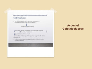
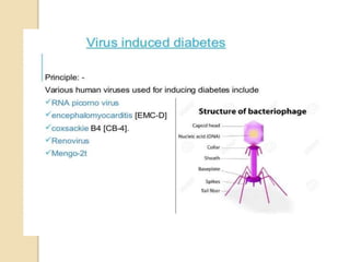
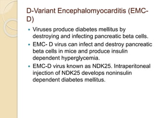
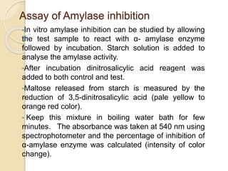
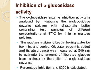
in-vivo, in-vitro models to study anti-
diabetic activity of new substances
Abu Sufiyan Chhipa
M. Pharm (Pharmacology)
DIABETES MELLITUS
Chronic metabolic disease, occurs when
pancreas is not producing enough insulin or the
produced insulin cannot be used by the body.
Two Types:
a) Type I: Inability of pancreas to produce
enough insulin.
b) Type II : Inability of body to respond to
produced insulin.
in-vivo studies
Studies in which the effects of various
biological entities are tested on whole
living organisms or cells, usually
animals (including humans).
Examples: Development of
antibiotics,
development of new
drugs.
in- vitro studies
Known as “test tube” experiments.
These are the studies performed with
microorganisms, cells or biological
molecules outside their normal
biological context. These are
performed in labwares.
Example: in-vitro fertilization
techniques
in-vivo models
CHEMICAL INDUCED
DIABETES
Alloxan induced diabetes
‱ Alloxan is a derivative of pyrimidine.
‱Alloxan produces selective necrosis of
beta cells.
‱ Alloxan is used for induction of diabetes
in experimental animals such as mice,
rats, rabbits and dogs.
‱The routes and dose of alloxan required
may vary depending upon the animal
species.
 A First short lived hypoglycemic phase lasting for 30
min from the first minutes of alloxan administration.
The hypoglycemic stage may be due to the
stimulation of insulin release and high levels of
plasma insulin levels.
 The mechanism at back of the hyperinsulinemia is
due to the short term increase of ATP availability
and glucokinase inhibition.
 The second phase is the increase in the blood
glucose levels one hour after administration of
alloxan, the plasma insulin concentration decreases.
 The pronounced hyperglycemia lasts for 2-4 hours
is due to decrease plasma insulin concentrations.
This may be due to inhibition of insulin secretion
and beta cell toxicity.
 Alloxan treatment brings out a sudden rise in insulin
secretion in the presence and absence of glucose.
The insulin release occurs until the complete
suppression of the islet response to glucose.
 Alloxan reacts with two sulfhydryl in the glucokinase
resulting in disulfide bond and inactivation of the
enzyme.
Streptozotocin induced
diabetes
 Streptozotocin (STZ) is a naturally occurring
chemical that particularly produces toxin to the
beta cells of the pancreas.
 Two hours after injection, the hyperglycemia
occurs due to the decrease in blood insulin
levels.
 STZ impairs glucose oxidation and decreases
insulin synthesis and release.
 STZ changes the DNA in pancreatic B cells.
The B cell death is due to alkylation of DNA by
STZ.
Insulin antibodies induced
diabetes
 The insulin antibodies have the affinity and
capacity to bind insulin.
 Insulin deficiency mechanism may cause
greater postprandial hyperglycemia because
antibody-bound insulin is unavailable to
tissues.
Goldthioglucose obese diabetic
mouse model
 Gold thioglucose (GTG) is a diabetogenic
compound, which manifest obesity induced Type -2
diabetes.
 The intrapertonial administration GTG in
experimental animal gradually develops obesity,
hyperinsulinemia, hyperglycemia, insulin resistance
for a period of 16- 20 weeks.
 It increases body lipid, hepatic lipogenesis and
triglyceride secretion, increased adipose tissue
lipogenesis and decreases glucose metabolism.
 The GTG is transported in particular to the cells
and causes necrotic lesions, which is responsible
for the development of hyperphagia and obesity.
Action of
Goldthioglucose
Virus Induced Diabetes
D-Variant Encephalomyocarditis (EMC-
D)
 Viruses produce diabetes mellitus by
destroying and infecting pancreatic beta cells.
 EMC- D virus can infect and destroy pancreatic
beta cells in mice and produce insulin
dependent hyperglycemia.
 EMC-D virus known as NDK25. Intraperitoneal
injection of NDK25 develops noninsulin
dependent diabetes mellitus.
Coxsackie viruses
 Coxsackie viruses cause diabetes in mice;
it can infect and destroy pancreatic acinar
cells.
 Acinar cells are responsible for synthesis,
storage and release of various pancreatic
digestive enzymes.
 Coxsackie B4 virus is strongly associated
with the development of insulin-dependent
diabetes mellitus in humans.
Hormone Induced Diabetes
Growth hormone induced
diabetes
 Repeated administration of growth hormone
in higher experimental animals induces
diabetes with ketonuria and ketonemia
(indicating that an alternate source of
energy is used especially fatty acids) .
 Prolonged administration of growth
hormone produced permanent diabetes.
Corticosteroid induced
diabetes
 Corticosteroid induces diabetes, which
is called steroid diabetes.
 The prednisolone and
dexamethasone, cause steroid
diabetes.
 Glucocorticoids stimulate
gluconeogenesis, in the liver, resulting
in increase in hepatic glucose and
induce insulin resistance and
hyperglycemia.
Spontaneous Diabetic Obese
Rodent Models
Ob/ob mouse
‱The ob/ob mouse strain, have leptin deficiency
because of the mutation in leptin gene leading to
severe insulin resistance. The ob/ob mice exhibit
rapid gain in body weight, insulin resistance and
hyperinsulinemia occurs.
‱The ob/ob or obese mouse is a mutant mouse
that eats excessively due to mutations in
the gene responsible for the production of leptin
(responsible to inhibit hunger) and becomes
profoundly obese. It is an animal model of type II
diabetes.
db/db mouse
 The db/db mouse is a genetically
mutated mouse in which leptin receptors do not
function properly. The db/db mouse is
extremely obese and has many of the
metabolic defects (hyperphagia,
hyperglycemia, hyperinsulinemia, and infertility)
found in ob/ob mouse.
Ob/ob or db/db
mouse with Leptin
hormone deficiency
Zucker Diabetic Fatty (ZDF) rat
‱Obese Zucker rats have high levels of lipids
and cholesterol in their bloodstream, are resistant to
insulin without being hyperglycemic, and gain weight
from an increase in both the size and number of fat
cells. Obesity in Zucker rats is primarily linked to
their hyperphagic nature, and excessive hunger
‱The male ZDF rat becomes fully diabetic at 12
weeks. The serum insulin levels of male ZDF rat
reach the peak at about 7 to 10 weeks, but cannot
respond to glucose stimulus and the insulin level
drops.
Zucker rat
New Zealand Obese (NZO) mouse
‱The New Zealand strain of obese mice, gains weight
at 10 weeks of life as a result of hyperphagia,
hyperglycemia and hyperinsulinemia. NZO mouse
manifests insulin resistance at an early age.
‱With the growth of NZO mouse, hyperglycemia and
glucose tolerance increase and the level of blood
glucose reaches 300-400 mg/dL at the age of 20 to
24 weeks. It is useful model for studying obesity and
diabetes.
New Zealand Obese
Mice
Otsuka Long-Evans Tokushima Fatty
(OLETF) rat
‱The OLETF rat develops hyperglycemia at
around 18 to 25 weeks age. OLETF rats exhibits
obesity,
hyperglycemia,hyperinsulinemia,hypertriglycerid
emia, hypercholesterolemia, and onset of
diabetes similar to human Type 2DM.
‱Many recessive genes on several
chromosomes including the X chromosome are
involved in the induction of diabetes in OLETF
rats.
OLETF RATS
Spontaneous Diabetic Non Obese
Rodent Models
Goto Kakizaki (GK) rat
‱The Goto-Kakizaki (GK) rat is a non-obese Wistar
substrain which develops Type 2 diabetes mellitus
early in life.
‱The GK rat is a model of T2DM with hyperglycemia,
hyperinsulinemia and insulin resistance.
‱In GK rats, a stable fasting hyperglycemia is
observed at the end of the first 2 weeks. After 8
weeks, hyperglycemia degenerates and insulin
secretion of the islets stimulated by glucose.
‱GK rats develop complications of diabetes like
peripheral neuropathy, and retinopathy.
GOTO KAKIZAKI
RAT
Cohen diabetic rat
‱Cohen diabetic rat is a genetic model derived
from diet-induced Type 2 DM model by placing
the rat on a synthetic 72% sucrose-copper-
poor diet for 2 months, manifest the human
Type 2 DM.
‱The manifestations include non-obesity,
hyperinsulinemia, and insulin resistance.
‱The Cohen diabetic rat expresses genetic
susceptibility to a carbohydrate-rich diet, a
feature of Type 2 DM in human.
Spontaneously Diabetic Torii (SDT)
rat
‱SDT rat is a new spontaneously non-obese
diabetic strain.It has characteristics like glucose
intolerance, hyperglycemia,hyperinsulinemia,
hypertriglyceridemia .
‱Because of the severe hyperglycemia, SDT rats
develop diabetic retinopathy, diabetic neuropathy,
and diabetic nephropathy.
‱This model is suitable for studying complications of
human T2DM.
Surgical model of Diabetes mellitus
‱Surgery technique used to induce diabetes, is
complete removal of the pancreas.
‱Limitation to this technique include high level of
technical expertise and adequate surgical room
environment.
in-vitro models
Assay of Amylase inhibition
‱In vitro amylase inhibition can be studied by allowing
the test sample to react with α- amylase enzyme
followed by incubation. Starch solution is added to
analyse the amylase activity.
‱After incubation dinitrosalicylic acid reagent was
added to both control and test.
‱Maltose released from starch is measured by the
reduction of 3,5-dinitrosalicylic acid (pale yellow to
orange red color).
‱ Keep this mixture in boiling water bath for few
minutes. The absorbance was taken at 540 nm using
spectrophotometer and the percentage of inhibition of
α-amylase enzyme was calculated (intensity of color
change).
Inhibition of α-glucosidase
activity
 The α-glucosidase enzyme inhibition activity is
analysed by incubating the α-glucosidase
enzyme solution with phosphate buffer
containing test samples of different
concentrations at 37°C for 1 hr in maltose
solution.
 The reaction mixture is kept in boiling water for
few min. and cooled. Glucose reagent is added
and its absorbance was measured at 540 nm
to estimate the amount of liberated glucose
from maltose by the action of α-glucosidase
enzyme.
 Percentage inhibition and IC50 is calculated.
Preclinical screening of anti diabetic drugs

Preclinical screening of anti diabetic drugs

  • 1.
    in-vivo, in-vitro modelsto study anti- diabetic activity of new substances Abu Sufiyan Chhipa M. Pharm (Pharmacology)
  • 2.
    DIABETES MELLITUS Chronic metabolicdisease, occurs when pancreas is not producing enough insulin or the produced insulin cannot be used by the body. Two Types: a) Type I: Inability of pancreas to produce enough insulin. b) Type II : Inability of body to respond to produced insulin.
  • 3.
    in-vivo studies Studies inwhich the effects of various biological entities are tested on whole living organisms or cells, usually animals (including humans). Examples: Development of antibiotics, development of new drugs.
  • 4.
    in- vitro studies Knownas “test tube” experiments. These are the studies performed with microorganisms, cells or biological molecules outside their normal biological context. These are performed in labwares. Example: in-vitro fertilization techniques
  • 5.
  • 6.
  • 7.
    Alloxan induced diabetes ‱Alloxan is a derivative of pyrimidine. ‱Alloxan produces selective necrosis of beta cells. ‱ Alloxan is used for induction of diabetes in experimental animals such as mice, rats, rabbits and dogs. ‱The routes and dose of alloxan required may vary depending upon the animal species.
  • 8.
     A Firstshort lived hypoglycemic phase lasting for 30 min from the first minutes of alloxan administration. The hypoglycemic stage may be due to the stimulation of insulin release and high levels of plasma insulin levels.  The mechanism at back of the hyperinsulinemia is due to the short term increase of ATP availability and glucokinase inhibition.  The second phase is the increase in the blood glucose levels one hour after administration of alloxan, the plasma insulin concentration decreases.
  • 9.
     The pronouncedhyperglycemia lasts for 2-4 hours is due to decrease plasma insulin concentrations. This may be due to inhibition of insulin secretion and beta cell toxicity.  Alloxan treatment brings out a sudden rise in insulin secretion in the presence and absence of glucose. The insulin release occurs until the complete suppression of the islet response to glucose.  Alloxan reacts with two sulfhydryl in the glucokinase resulting in disulfide bond and inactivation of the enzyme.
  • 10.
    Streptozotocin induced diabetes  Streptozotocin(STZ) is a naturally occurring chemical that particularly produces toxin to the beta cells of the pancreas.  Two hours after injection, the hyperglycemia occurs due to the decrease in blood insulin levels.  STZ impairs glucose oxidation and decreases insulin synthesis and release.  STZ changes the DNA in pancreatic B cells. The B cell death is due to alkylation of DNA by STZ.
  • 11.
    Insulin antibodies induced diabetes The insulin antibodies have the affinity and capacity to bind insulin.  Insulin deficiency mechanism may cause greater postprandial hyperglycemia because antibody-bound insulin is unavailable to tissues.
  • 12.
    Goldthioglucose obese diabetic mousemodel  Gold thioglucose (GTG) is a diabetogenic compound, which manifest obesity induced Type -2 diabetes.  The intrapertonial administration GTG in experimental animal gradually develops obesity, hyperinsulinemia, hyperglycemia, insulin resistance for a period of 16- 20 weeks.  It increases body lipid, hepatic lipogenesis and triglyceride secretion, increased adipose tissue lipogenesis and decreases glucose metabolism.  The GTG is transported in particular to the cells and causes necrotic lesions, which is responsible for the development of hyperphagia and obesity.
  • 13.
  • 14.
  • 16.
    D-Variant Encephalomyocarditis (EMC- D) Viruses produce diabetes mellitus by destroying and infecting pancreatic beta cells.  EMC- D virus can infect and destroy pancreatic beta cells in mice and produce insulin dependent hyperglycemia.  EMC-D virus known as NDK25. Intraperitoneal injection of NDK25 develops noninsulin dependent diabetes mellitus.
  • 17.
    Coxsackie viruses  Coxsackieviruses cause diabetes in mice; it can infect and destroy pancreatic acinar cells.  Acinar cells are responsible for synthesis, storage and release of various pancreatic digestive enzymes.  Coxsackie B4 virus is strongly associated with the development of insulin-dependent diabetes mellitus in humans.
  • 18.
  • 19.
    Growth hormone induced diabetes Repeated administration of growth hormone in higher experimental animals induces diabetes with ketonuria and ketonemia (indicating that an alternate source of energy is used especially fatty acids) .  Prolonged administration of growth hormone produced permanent diabetes.
  • 20.
    Corticosteroid induced diabetes  Corticosteroidinduces diabetes, which is called steroid diabetes.  The prednisolone and dexamethasone, cause steroid diabetes.  Glucocorticoids stimulate gluconeogenesis, in the liver, resulting in increase in hepatic glucose and induce insulin resistance and hyperglycemia.
  • 21.
  • 22.
    Ob/ob mouse ‱The ob/obmouse strain, have leptin deficiency because of the mutation in leptin gene leading to severe insulin resistance. The ob/ob mice exhibit rapid gain in body weight, insulin resistance and hyperinsulinemia occurs. ‱The ob/ob or obese mouse is a mutant mouse that eats excessively due to mutations in the gene responsible for the production of leptin (responsible to inhibit hunger) and becomes profoundly obese. It is an animal model of type II diabetes.
  • 23.
    db/db mouse  Thedb/db mouse is a genetically mutated mouse in which leptin receptors do not function properly. The db/db mouse is extremely obese and has many of the metabolic defects (hyperphagia, hyperglycemia, hyperinsulinemia, and infertility) found in ob/ob mouse.
  • 24.
    Ob/ob or db/db mousewith Leptin hormone deficiency
  • 25.
    Zucker Diabetic Fatty(ZDF) rat ‱Obese Zucker rats have high levels of lipids and cholesterol in their bloodstream, are resistant to insulin without being hyperglycemic, and gain weight from an increase in both the size and number of fat cells. Obesity in Zucker rats is primarily linked to their hyperphagic nature, and excessive hunger ‱The male ZDF rat becomes fully diabetic at 12 weeks. The serum insulin levels of male ZDF rat reach the peak at about 7 to 10 weeks, but cannot respond to glucose stimulus and the insulin level drops.
  • 26.
  • 27.
    New Zealand Obese(NZO) mouse ‱The New Zealand strain of obese mice, gains weight at 10 weeks of life as a result of hyperphagia, hyperglycemia and hyperinsulinemia. NZO mouse manifests insulin resistance at an early age. ‱With the growth of NZO mouse, hyperglycemia and glucose tolerance increase and the level of blood glucose reaches 300-400 mg/dL at the age of 20 to 24 weeks. It is useful model for studying obesity and diabetes.
  • 28.
  • 29.
    Otsuka Long-Evans TokushimaFatty (OLETF) rat ‱The OLETF rat develops hyperglycemia at around 18 to 25 weeks age. OLETF rats exhibits obesity, hyperglycemia,hyperinsulinemia,hypertriglycerid emia, hypercholesterolemia, and onset of diabetes similar to human Type 2DM. ‱Many recessive genes on several chromosomes including the X chromosome are involved in the induction of diabetes in OLETF rats.
  • 30.
  • 31.
    Spontaneous Diabetic NonObese Rodent Models
  • 32.
    Goto Kakizaki (GK)rat ‱The Goto-Kakizaki (GK) rat is a non-obese Wistar substrain which develops Type 2 diabetes mellitus early in life. ‱The GK rat is a model of T2DM with hyperglycemia, hyperinsulinemia and insulin resistance. ‱In GK rats, a stable fasting hyperglycemia is observed at the end of the first 2 weeks. After 8 weeks, hyperglycemia degenerates and insulin secretion of the islets stimulated by glucose. ‱GK rats develop complications of diabetes like peripheral neuropathy, and retinopathy.
  • 33.
  • 34.
    Cohen diabetic rat ‱Cohendiabetic rat is a genetic model derived from diet-induced Type 2 DM model by placing the rat on a synthetic 72% sucrose-copper- poor diet for 2 months, manifest the human Type 2 DM. ‱The manifestations include non-obesity, hyperinsulinemia, and insulin resistance. ‱The Cohen diabetic rat expresses genetic susceptibility to a carbohydrate-rich diet, a feature of Type 2 DM in human.
  • 35.
    Spontaneously Diabetic Torii(SDT) rat ‱SDT rat is a new spontaneously non-obese diabetic strain.It has characteristics like glucose intolerance, hyperglycemia,hyperinsulinemia, hypertriglyceridemia . ‱Because of the severe hyperglycemia, SDT rats develop diabetic retinopathy, diabetic neuropathy, and diabetic nephropathy. ‱This model is suitable for studying complications of human T2DM.
  • 36.
    Surgical model ofDiabetes mellitus ‱Surgery technique used to induce diabetes, is complete removal of the pancreas. ‱Limitation to this technique include high level of technical expertise and adequate surgical room environment.
  • 37.
  • 38.
    Assay of Amylaseinhibition ‱In vitro amylase inhibition can be studied by allowing the test sample to react with α- amylase enzyme followed by incubation. Starch solution is added to analyse the amylase activity. ‱After incubation dinitrosalicylic acid reagent was added to both control and test. ‱Maltose released from starch is measured by the reduction of 3,5-dinitrosalicylic acid (pale yellow to orange red color). ‱ Keep this mixture in boiling water bath for few minutes. The absorbance was taken at 540 nm using spectrophotometer and the percentage of inhibition of α-amylase enzyme was calculated (intensity of color change).
  • 39.
    Inhibition of α-glucosidase activity The α-glucosidase enzyme inhibition activity is analysed by incubating the α-glucosidase enzyme solution with phosphate buffer containing test samples of different concentrations at 37°C for 1 hr in maltose solution.  The reaction mixture is kept in boiling water for few min. and cooled. Glucose reagent is added and its absorbance was measured at 540 nm to estimate the amount of liberated glucose from maltose by the action of α-glucosidase enzyme.  Percentage inhibition and IC50 is calculated.