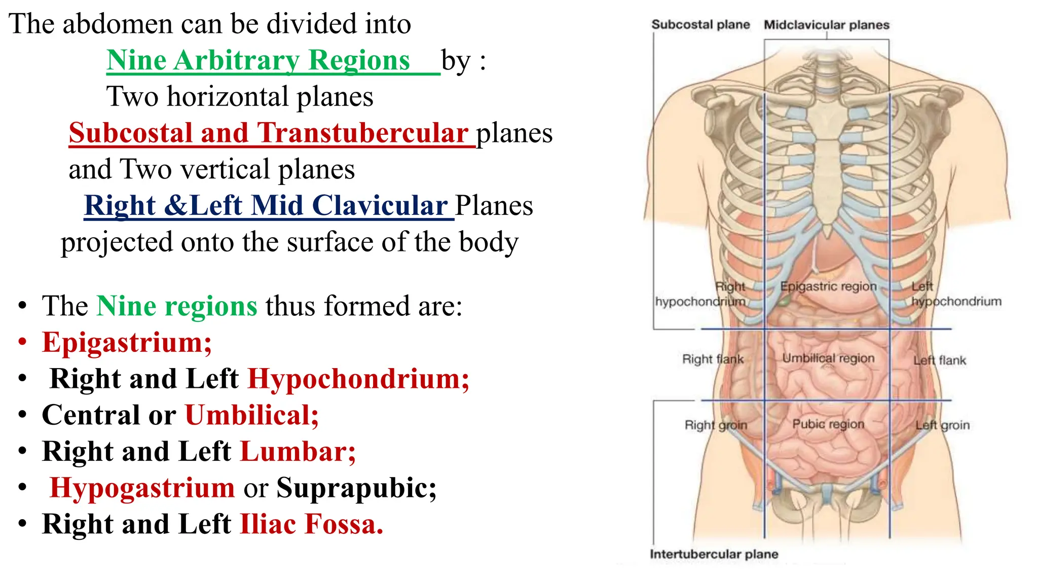
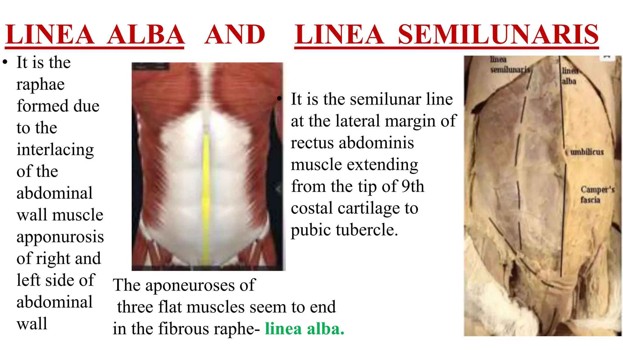
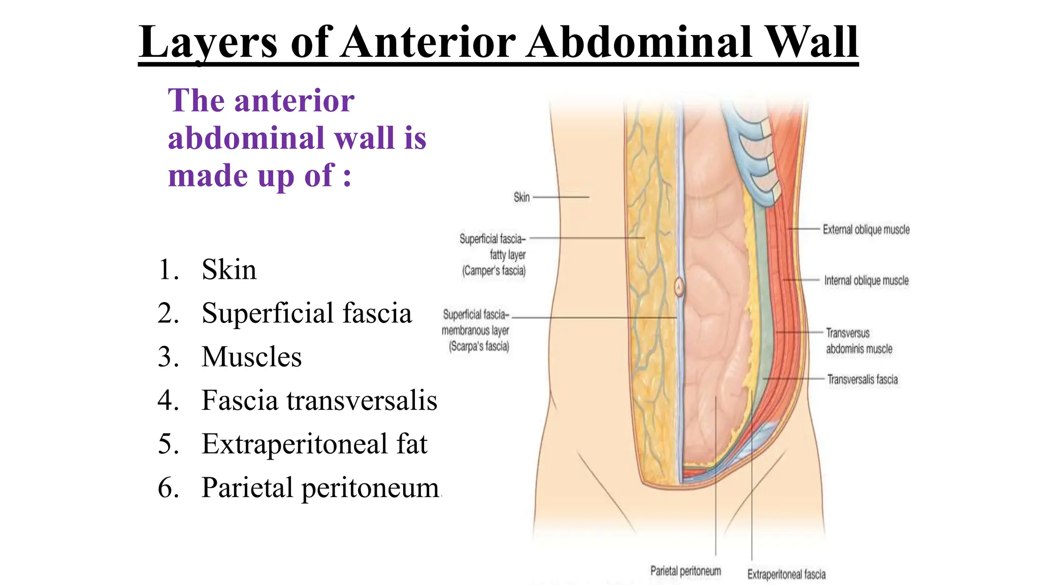
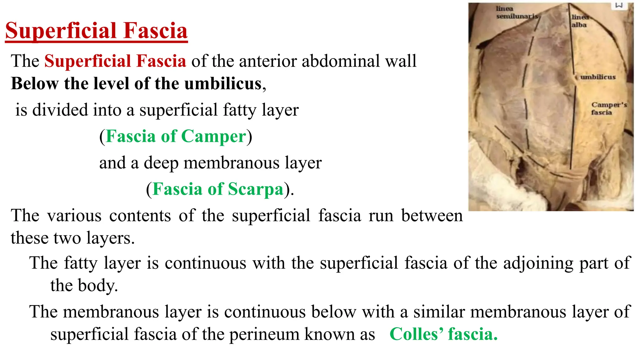
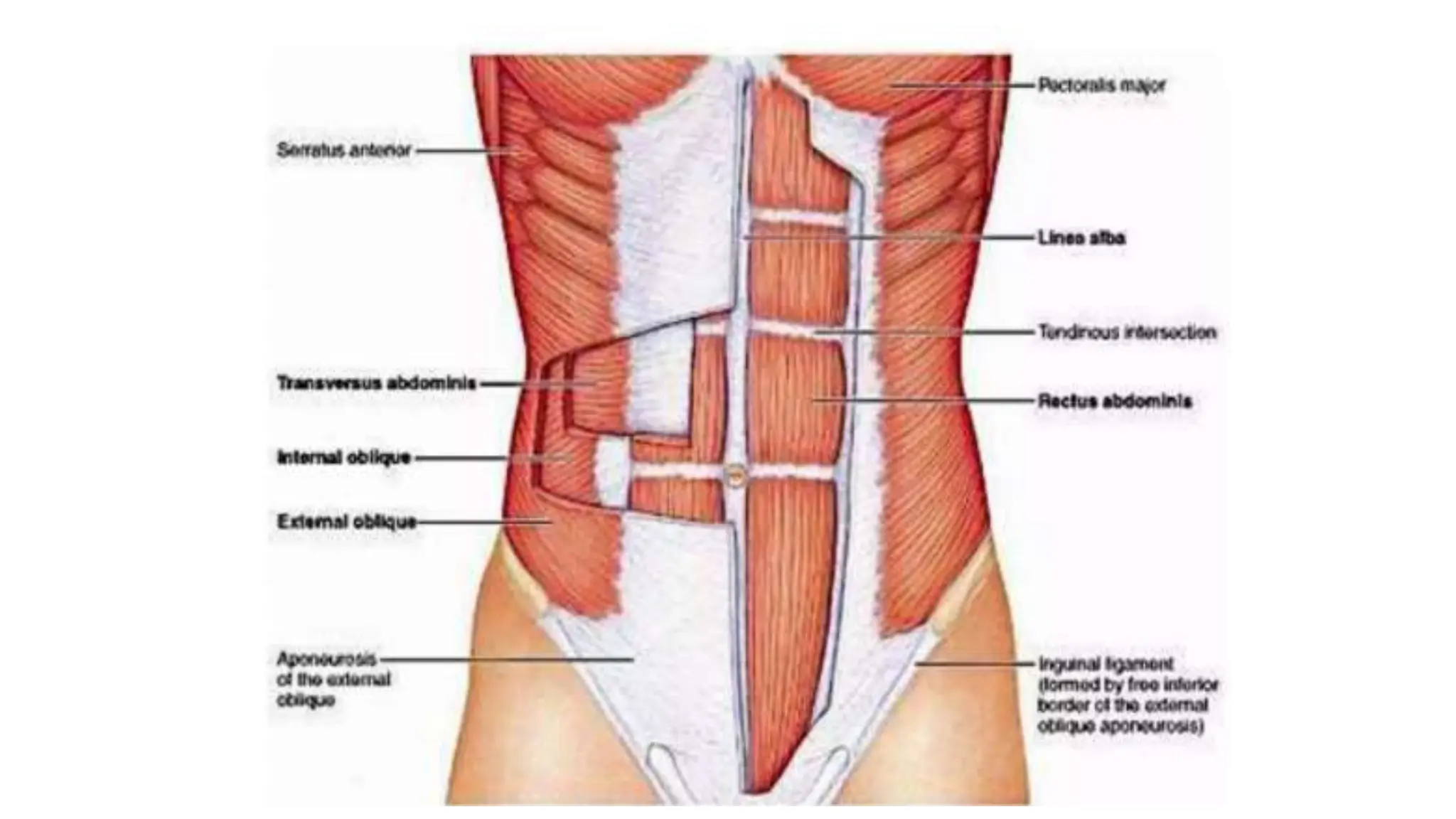
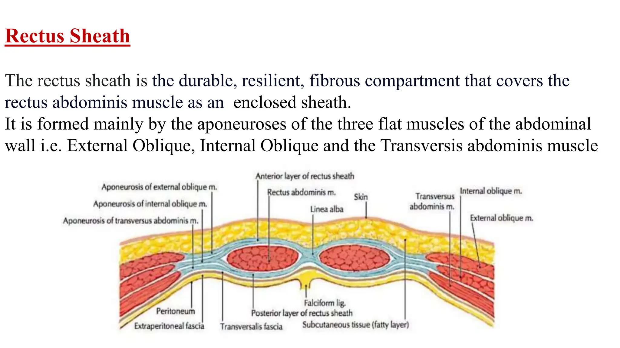
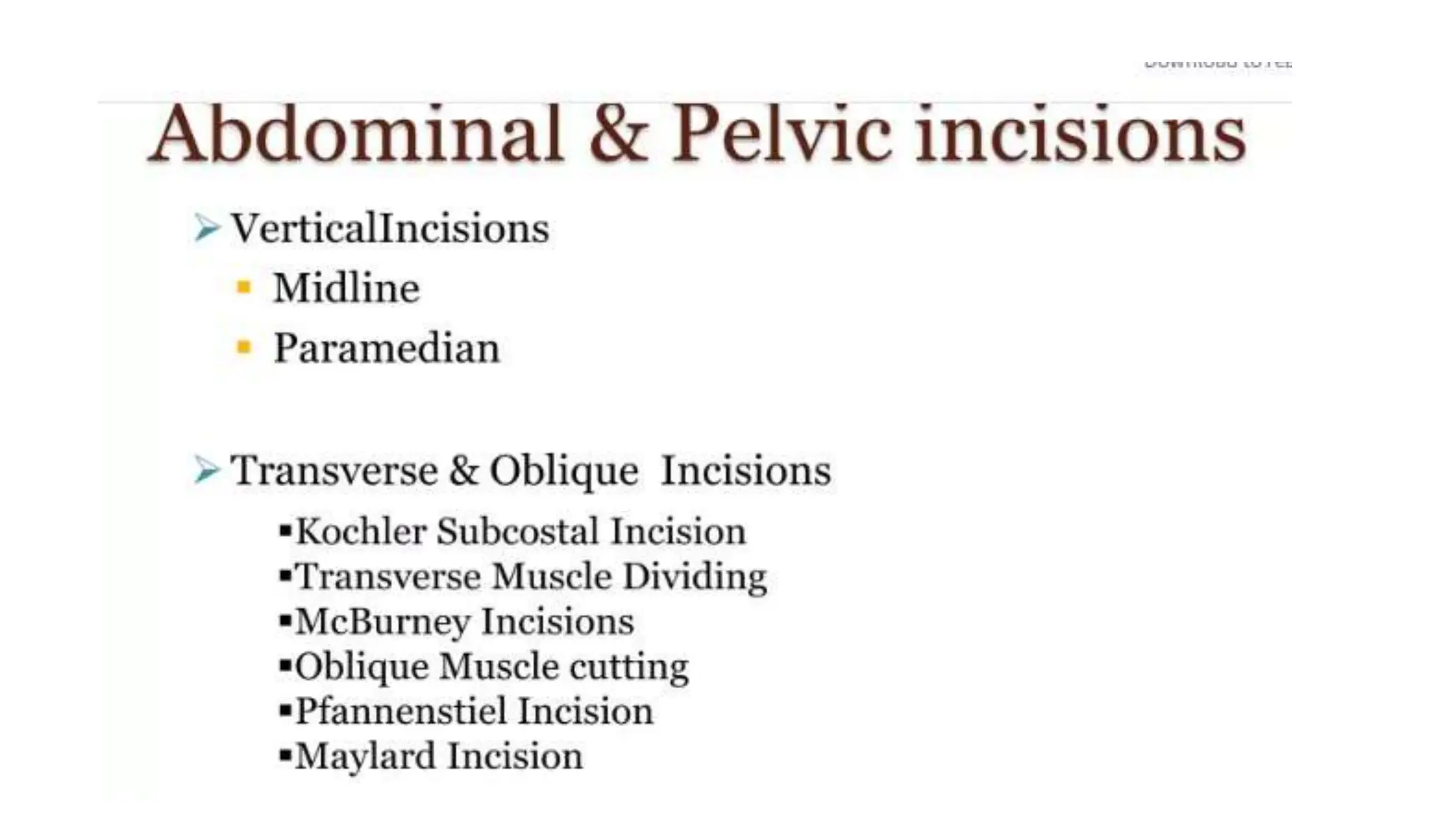
ANTERIOR ABDOMINAL WALL
Competency no. AN 44.1, 44.2, 44.3, 44.6, 44.7
By-Dr Shashi Munjal
AN 44.1 Describe: Regions & Quadrants of Abdomen
Lateral vertical, Transpyloric , Transtubercular and Subcostal planes
Linea Alba and Linea Semilunaris lines of Abdomen
AN 44.2 Describe anterior abdominal wall: 1. Fascia, 2. Nerves & 3.Blood vessels
AN 44.6 Describe attachments of Muscles of Anterior Abdominal wall
AN 44.3 Describe the formation of Rectus Sheath and its contents
AN 44.7 Enumerate common Abdominal Incisions
Specific learning objectives
• The Nine regions thus formed are:
• Epigastrium;
• Right and Left Hypochondrium;
• Central or Umbilical;
• Right and Left Lumbar;
• Hypogastrium or Suprapubic;
• Right and Left Iliac Fossa.
The abdomen can be divided into
Nine Arbitrary Regions by :
Two horizontal planes
Subcostal and Transtubercular planes
and Two vertical planes
Right &Left Mid Clavicular Planes
projected onto the surface of the body
LINEA ALBA AND LINEA SEMILUNARIS
• It is the
raphae
formed due
to the
interlacing
of the
abdominal
wall muscle
apponurosis
of right and
left side of
abdominal
wall
• It is the semilunar line
at the lateral margin of
rectus abdominis
muscle extending
from the tip of 9th
costal cartilage to
pubic tubercle.
The aponeuroses of
three flat muscles seem to end
in the fibrous raphe- linea alba.
Layers of Anterior Abdominal Wall
The anterior
abdominal wall is
made up of :
1. Skin
2. Superficial fascia
3. Muscles
4. Fascia transversalis
5. Extraperitoneal fat
6. Parietal peritoneum.
The Superficial Fascia of the anterior abdominal wall
Below the level of the umbilicus,
is divided into a superficial fatty layer
(Fascia of Camper)
and a deep membranous layer
(Fascia of Scarpa).
The various contents of the superficial fascia run between
these two layers.
The fatty layer is continuous with the superficial fascia of the adjoining part of
the body.
The membranous layer is continuous below with a similar membranous layer of
superficial fascia of the perineum known as Colles’ fascia.
Superficial Fascia
The cutaneous nerves of the anterior abdominal wall
Arteries of the anterior abdominal wall
The subcutaneous venous circulation
in: Normal subjects,
Superficial Lymphatics
Above the level of the umbilicus the
lymphatics run upwards to drain into the
axillary lymph nodes. Below the level of
the umbilicus they run downwards to
drain into the superficial inguinal lymph
nodes.
MUSCLES OF THE ANTEROLATERALABDOMINAL WALL
On either side of the midline, there are four large muscles.
These are the
External Oblique,
Internal Oblique,
Transversus abdominis
Rectus Abdominis.
Two small muscles, the Pyramidalis, conjoint tendon are also present.
External Oblique Muscle Origin
The muscle arises by eight fleshy slips from
the outer surfaces of the shaft of the lower
eight ribs. The fibres run downwards,
forwards and medially
Insertion
Most of the fibres of the muscle end in a
broad aponeurosis through which they are
inserted into the xiphoid process, linea alba,
Pubic symphysis,pubic crest& pectineal line
The lower fibres of the muscle are inserted directly into the anterior two-thirds of
the outer lip of the iliac crest. Between the ASIS & pubic tubercle the aponeurosis
has a free inferior border that is folded on itself to form the inguinal ligament.
Internal Oblique Muscle
Origin
From lateral 2/3 of inguinal Ligament,
anterior 2/3 of intermediate area of the
iliac crest and Thoracolumbar fascia.
Insertion
The uppermost fibres are inserted directly
into the lower three or four ribs and their
cartilages.
.
The greater part of the muscle ends in an aponeurosis through which it is inserted
into the seventh, eighth and ninth costal cartilages, the xiphoid process, linea alba,
pubic crest and the pectineal line of the pubis. It does not extend beyond the costal
margin.
Other Points of insertion of Internal oblique
In the upper 3/4th of the wall, the aponeurosis splits into an anterior lamina that
passes medially in front of the rectus abdominis; and a posterior lamina that lies
behind the rectus.
Below a level midway between the umbilicus and the pubic symphysis (lower 1/4th
of the wall) the aponeurosis remains a single layer. It passes in front of rectus
abdominis to reach linea alba.
Its lower free crescent shaped border is the Arcuate line
It is also modified to form:
• The conjoint tendon
• The cremaster muscle
Transversus Abdominis Muscle
Origin
The muscle has a fleshy origin from:
The lateral one-third of the inguinal ligament.
The anterior 2/3 of the inner lip of the iliac crest
The thoracolumbar fascia.
The fibres are directed horizontally forwards in
inner surfaces of the lower six costal cartilages.
Insertion
The fibres end in a broad aponeurosis which is
inserted into the xiphoid process, the linea alba,
pubic crest, and the pectineal line of the pubis.
The lowest fibres of the muscle
fuse with the lowest fibres of
internal oblique to form
Conjoint tendon.
Other Points of Insertion of Transversus abdominis
The aponeurosis of the transversus abdominis takes part in forming the rectus
sheath as follows:
Above the level of the arcuate line (upper 3/4th), the aponeurosis passes medially
behind the rectus abdominis muscle along with the posterior lamina of the internal
oblique aponeurosis.
The lower edge of this part of the aponeurosis helps to form the arcuate line along
with internal oblique muscle.
Below the level of the arcuate line the aponeurosis passes in front of the rectus
abdominis and helps to form the anterior wall of the rectus sheath.
Rectus Abdominis Muscle
OriginThe muscle arises by two tendinous
heads as follows.
1. Lateral head from the lateral part of the
pubic crest.
2. Medial head from the medial part of pubic
crest and anterior pubic ligament.
The fibres run vertically upwards.
Insertion On the front of the wall of the thorax, along a horizontal line passing
laterally from the xiphoid process, and cutting in that order, the 7th, 6th and 5th
costal cartilages.
Tendinous intersections: These are three transverse fibrous bands which divide
the muscle into smaller parts.One lies opposite the umbilicus Second opposite
the free end of the xiphoid process Third in between the two.
Rectus Sheath
The rectus sheath is the durable, resilient, fibrous compartment that covers the
rectus abdominis muscle as an enclosed sheath.
It is formed mainly by the aponeuroses of the three flat muscles of the abdominal
wall i.e. External Oblique, Internal Oblique and the Transversis abdominis muscle
Rectus sheath is firmly adherent to the tendinous intersections of the rectus muscle.
And has two walls, anterior and posterior.
Anterior Wall
It is complete, covering the muscle from end to end. Its composition is variable.
Above the costal margin Only External oblique
Between costal margin and arcuate line
External oblique and upper lamina of Internal Oblique
Below arcuate line External oblique, Internal Oblique &Transversus abdominis
Posterior Wall
It is incomplete,being deficient Above the costal margin
where the rectus muscle rests directly on the 5th, 6th and 7th costal cartilages.
and Below the arcuate line Only the Fascia Transversalis forms the Post.wall
Between costal margin and arcuate line External oblique, Lower lamina of
Internal Oblique and whole of theTransversus abdominis
Transverse sections through the rectus abdominis, and its sheath:
(a) Above the costal margin, A of inset, (b) Between costal margin and arcuate line,
B of inset and (c) Below arcuate line, C of inset
Contents
Muscles
Rectus abdominis
Arteries
Superior epigastric artery
Inferior epigastric artery
Veins
The superior epigastric venae comitantes
The inferior epigastric venae comitantes
Nerves
These are the terminal parts of the lower six thoracic nerves, including the lower
five intercostal nerves and the subcostal nerves.
INCISIONS
• Surgical incision
• is a cut made through the skin
• to facilitate an operation or procedure
The umbilicus
It is the normal scar in the anterior abdominal wall formed by the remnants of the
root of the Umbilical cord.
In the lymphatic and venous drainage, the level of the umbilicus is a watershed.
The skin around the umbilicus is supplied by segment T10 of the spinal cord.
The umbilicus is one of the important sites at which tributaries of the portal vein
anastomose with systemic veins (Portocaval anastomoses).
Umbilicus is the meeting point of the Four folds
(two lateral, head and tail) of embryonic plate.
This is also the meeting point of Three systems, namely
the Digestive (vitellointestinal duct), the Excretory (urachus),
and Vascular (umbilical vessels).
The subcutaneous venous circulation in: (a)
Normal subjects, (b) portal obstruction showing
caput medusae, (c) superior vena caval
obstruction, and (d) inferior vena caval
obstruction (arrows indicate the direction of
blood flow)
Nerve Supply of
External oblique muscle
Lower six thoracic nerves
Nerve Supply of
Internal Oblique muscle
&
Transversus abdominis
Lower six thoracic nerves
and the first lumbar nerve.

ANTERIOR ABDOMINAL WALL.pptxAnterior abdomen wall Rectus sheath

  • 1.
    ANTERIOR ABDOMINAL WALL Competencyno. AN 44.1, 44.2, 44.3, 44.6, 44.7 By-Dr Shashi Munjal
  • 2.
    AN 44.1 Describe:Regions & Quadrants of Abdomen Lateral vertical, Transpyloric , Transtubercular and Subcostal planes Linea Alba and Linea Semilunaris lines of Abdomen AN 44.2 Describe anterior abdominal wall: 1. Fascia, 2. Nerves & 3.Blood vessels AN 44.6 Describe attachments of Muscles of Anterior Abdominal wall AN 44.3 Describe the formation of Rectus Sheath and its contents AN 44.7 Enumerate common Abdominal Incisions Specific learning objectives
  • 3.
    • The Nineregions thus formed are: • Epigastrium; • Right and Left Hypochondrium; • Central or Umbilical; • Right and Left Lumbar; • Hypogastrium or Suprapubic; • Right and Left Iliac Fossa. The abdomen can be divided into Nine Arbitrary Regions by : Two horizontal planes Subcostal and Transtubercular planes and Two vertical planes Right &Left Mid Clavicular Planes projected onto the surface of the body
  • 5.
    LINEA ALBA ANDLINEA SEMILUNARIS • It is the raphae formed due to the interlacing of the abdominal wall muscle apponurosis of right and left side of abdominal wall • It is the semilunar line at the lateral margin of rectus abdominis muscle extending from the tip of 9th costal cartilage to pubic tubercle. The aponeuroses of three flat muscles seem to end in the fibrous raphe- linea alba.
  • 6.
    Layers of AnteriorAbdominal Wall The anterior abdominal wall is made up of : 1. Skin 2. Superficial fascia 3. Muscles 4. Fascia transversalis 5. Extraperitoneal fat 6. Parietal peritoneum.
  • 7.
    The Superficial Fasciaof the anterior abdominal wall Below the level of the umbilicus, is divided into a superficial fatty layer (Fascia of Camper) and a deep membranous layer (Fascia of Scarpa). The various contents of the superficial fascia run between these two layers. The fatty layer is continuous with the superficial fascia of the adjoining part of the body. The membranous layer is continuous below with a similar membranous layer of superficial fascia of the perineum known as Colles’ fascia. Superficial Fascia
  • 8.
    The cutaneous nervesof the anterior abdominal wall
  • 9.
    Arteries of theanterior abdominal wall
  • 10.
    The subcutaneous venouscirculation in: Normal subjects,
  • 11.
    Superficial Lymphatics Above thelevel of the umbilicus the lymphatics run upwards to drain into the axillary lymph nodes. Below the level of the umbilicus they run downwards to drain into the superficial inguinal lymph nodes.
  • 12.
    MUSCLES OF THEANTEROLATERALABDOMINAL WALL On either side of the midline, there are four large muscles. These are the External Oblique, Internal Oblique, Transversus abdominis Rectus Abdominis. Two small muscles, the Pyramidalis, conjoint tendon are also present.
  • 14.
    External Oblique MuscleOrigin The muscle arises by eight fleshy slips from the outer surfaces of the shaft of the lower eight ribs. The fibres run downwards, forwards and medially Insertion Most of the fibres of the muscle end in a broad aponeurosis through which they are inserted into the xiphoid process, linea alba, Pubic symphysis,pubic crest& pectineal line The lower fibres of the muscle are inserted directly into the anterior two-thirds of the outer lip of the iliac crest. Between the ASIS & pubic tubercle the aponeurosis has a free inferior border that is folded on itself to form the inguinal ligament.
  • 15.
    Internal Oblique Muscle Origin Fromlateral 2/3 of inguinal Ligament, anterior 2/3 of intermediate area of the iliac crest and Thoracolumbar fascia. Insertion The uppermost fibres are inserted directly into the lower three or four ribs and their cartilages. . The greater part of the muscle ends in an aponeurosis through which it is inserted into the seventh, eighth and ninth costal cartilages, the xiphoid process, linea alba, pubic crest and the pectineal line of the pubis. It does not extend beyond the costal margin.
  • 16.
    Other Points ofinsertion of Internal oblique In the upper 3/4th of the wall, the aponeurosis splits into an anterior lamina that passes medially in front of the rectus abdominis; and a posterior lamina that lies behind the rectus. Below a level midway between the umbilicus and the pubic symphysis (lower 1/4th of the wall) the aponeurosis remains a single layer. It passes in front of rectus abdominis to reach linea alba. Its lower free crescent shaped border is the Arcuate line It is also modified to form: • The conjoint tendon • The cremaster muscle
  • 17.
    Transversus Abdominis Muscle Origin Themuscle has a fleshy origin from: The lateral one-third of the inguinal ligament. The anterior 2/3 of the inner lip of the iliac crest The thoracolumbar fascia. The fibres are directed horizontally forwards in inner surfaces of the lower six costal cartilages. Insertion The fibres end in a broad aponeurosis which is inserted into the xiphoid process, the linea alba, pubic crest, and the pectineal line of the pubis. The lowest fibres of the muscle fuse with the lowest fibres of internal oblique to form Conjoint tendon.
  • 18.
    Other Points ofInsertion of Transversus abdominis The aponeurosis of the transversus abdominis takes part in forming the rectus sheath as follows: Above the level of the arcuate line (upper 3/4th), the aponeurosis passes medially behind the rectus abdominis muscle along with the posterior lamina of the internal oblique aponeurosis. The lower edge of this part of the aponeurosis helps to form the arcuate line along with internal oblique muscle. Below the level of the arcuate line the aponeurosis passes in front of the rectus abdominis and helps to form the anterior wall of the rectus sheath.
  • 20.
    Rectus Abdominis Muscle OriginThemuscle arises by two tendinous heads as follows. 1. Lateral head from the lateral part of the pubic crest. 2. Medial head from the medial part of pubic crest and anterior pubic ligament. The fibres run vertically upwards. Insertion On the front of the wall of the thorax, along a horizontal line passing laterally from the xiphoid process, and cutting in that order, the 7th, 6th and 5th costal cartilages. Tendinous intersections: These are three transverse fibrous bands which divide the muscle into smaller parts.One lies opposite the umbilicus Second opposite the free end of the xiphoid process Third in between the two.
  • 22.
    Rectus Sheath The rectussheath is the durable, resilient, fibrous compartment that covers the rectus abdominis muscle as an enclosed sheath. It is formed mainly by the aponeuroses of the three flat muscles of the abdominal wall i.e. External Oblique, Internal Oblique and the Transversis abdominis muscle
  • 23.
    Rectus sheath isfirmly adherent to the tendinous intersections of the rectus muscle. And has two walls, anterior and posterior. Anterior Wall It is complete, covering the muscle from end to end. Its composition is variable. Above the costal margin Only External oblique Between costal margin and arcuate line External oblique and upper lamina of Internal Oblique Below arcuate line External oblique, Internal Oblique &Transversus abdominis Posterior Wall It is incomplete,being deficient Above the costal margin where the rectus muscle rests directly on the 5th, 6th and 7th costal cartilages. and Below the arcuate line Only the Fascia Transversalis forms the Post.wall Between costal margin and arcuate line External oblique, Lower lamina of Internal Oblique and whole of theTransversus abdominis
  • 24.
    Transverse sections throughthe rectus abdominis, and its sheath: (a) Above the costal margin, A of inset, (b) Between costal margin and arcuate line, B of inset and (c) Below arcuate line, C of inset
  • 25.
    Contents Muscles Rectus abdominis Arteries Superior epigastricartery Inferior epigastric artery Veins The superior epigastric venae comitantes The inferior epigastric venae comitantes Nerves These are the terminal parts of the lower six thoracic nerves, including the lower five intercostal nerves and the subcostal nerves.
  • 26.
    INCISIONS • Surgical incision •is a cut made through the skin • to facilitate an operation or procedure
  • 29.
    The umbilicus It isthe normal scar in the anterior abdominal wall formed by the remnants of the root of the Umbilical cord. In the lymphatic and venous drainage, the level of the umbilicus is a watershed. The skin around the umbilicus is supplied by segment T10 of the spinal cord. The umbilicus is one of the important sites at which tributaries of the portal vein anastomose with systemic veins (Portocaval anastomoses). Umbilicus is the meeting point of the Four folds (two lateral, head and tail) of embryonic plate. This is also the meeting point of Three systems, namely the Digestive (vitellointestinal duct), the Excretory (urachus), and Vascular (umbilical vessels).
  • 30.
    The subcutaneous venouscirculation in: (a) Normal subjects, (b) portal obstruction showing caput medusae, (c) superior vena caval obstruction, and (d) inferior vena caval obstruction (arrows indicate the direction of blood flow)
  • 31.
    Nerve Supply of Externaloblique muscle Lower six thoracic nerves
  • 32.
    Nerve Supply of InternalOblique muscle & Transversus abdominis Lower six thoracic nerves and the first lumbar nerve.