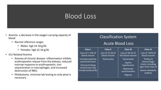
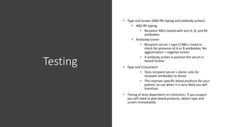
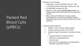
Anemia and Blood
Transfusions
Blood Loss
• Anemia- a decrease in the oxygen carrying capacity of
blood
• Normal reference ranges
• Males: hgb 14-18 g/dL
• Females: hgb 12-16 g/dL
• ICU-Related Anemia
• Anemia of chronic disease- inflammation inhibits
erythropoietin release from the kidneys, reduced
marrow response to erythropoietin, iron
sequestration in macrophages, and increased
destruction of RBCs
• Phlebotomy- minimize lab testing to only what is
necessary
Classification System
Acute Blood Loss
Class I
•Loss of < 15% of
blood volume
•Compensated by
vasoconstriction
•Clinical findings
minimal but may
include tachycardia
Class II
Loss of 15-30 of
blood volume
Tachycardia
Class III
Loss of 30-35 of
the blood volume
Tachycardia
Supine
hypotension
Depressed
consciousness
Oliguria
Class IV
Loss of >45% of
blood volume
Profound
hemorrhagic
shock, including
signs from class III
Severe metabolic
acidosis
Equations
Testing
• Type and Screen (ABO-Rh typing and antibody screen)
• ABO-Rh typing
• Recipient RBCs tested with anti-A, B, and Rh
antibodies
• Antibody screen
• Recipient serum + type O RBCs mixed to
check for presence of A or B antibodies. No
agglutination = negative screen
• If antibody screen is positive the serum is
tested further
• Type and Crossmatch
• Tests recipient serum + donor cells for
recipient antibodies to donor
• This reserves specific blood products for your
patient, so use when it is very likely you will
transfuse
• Timing of tests dependent on institution. If you suspect
you will need to give blood products, obtain type and
screen immediately.
Universal Donors & Recipients
Donor Recipient
O* universal O, A, B, AB
A A, AB
B B, AB
AB AB
Donor Recipient
O O
A A, O
B B, O
AB* universal O, A, B, AB
*In an emergency without a known blood type, the universal donor of red blood cells is O (–) because it is
devoid of A, B, and Rh antigens on its surface and won’t trigger a hemolytic transfusion reaction
Compatibility for PRBCs
Compatibility for FFP
Packed Red
Blood Cells
(pRBCs)
• Definition, Use, & Storage
• Single donor; volume 250-300 ml with Hct ~70%
• 1 unit pRBCs increases adult Hgb ~1 g/dl or Hct ~3%
• 10 ml/kg PRBC increases Hct 10%
• Always run with a bag of normal saline (do NOT use
lactated ringers due to theoretical clot formation from
calcium)
• Often stored with CPDA (lasts 35 days): Citrate
(anticoagulant)- also binds iCa, phosphate (buffer),
dextrose (energy source), adenosine (precursor to ATP
synthesis)
• Indications (ASA Guidelines)
1. Hgb/Hct < 6/24 in young, healthy patients
2. Usually unnecessary when H/H is 10/30
3. At Hgb 6-10 g/dl, the decision to transfuse is based
on:
• Ongoing indications of organ ischemia
• Potential or ongoing blood loss
• Volume status
• Risk factors for complications of inadequate O2
(example: myocardial ischemia)
Platelets
• Definition, Use, & Storage
• Platelet Concentrate (PC)
• Platelets from one donated unit, volume = 50-70
ml; increase plt ~5000- 10,000
• “6-pack” = 6 pooled PCs; rarely used anymore
• Apheresis Unit Platelets
• From a single donor; volume = 200-400 ml;
increase plt ~50,000
• Can give ABO-incompatible platelets, Rh tested only
• Stored at room temperature for 5 days
• Indications (ASA Guidelines)
1. Rarely when plt > 100,000
2. Usually when plt < 50,000 (spontaneous bleed at <
20K) in the presence of excessive bleeding
3. With platelet dysfunction (e.g. cardiopulmonary
bypass, plt inhibitors)
Fresh Frozen
Plasma
• Definition, Use, & Storage
• Fluid portion from whole blood
• Contains all coagulation factors (except platelets)
• 1 unit increases clotting factors 2-3%
• Use ABO-compatible; Rh-incompatible is OK
• Stored frozen; takes 30 min to thaw; use within 24 hrs
of thawing
• Indications (ASA Guidelines)
1. Urgent reversal of warfarin
2. Correction of known factor deficiency
3. Correction of excessive microvascular bleeding with
INR>2
4. During massive transfusion (before lab results
available)
5. Heparin resistance (i.e. antithrombin III deficiency)
in patients requiring heparinization
Cryoprecipitate
• Definition
• Fraction of plasma that precipitates when FFP is thawed
• Contains Factors VIII, XIII, I (fibrinogen), and vWF
• 1 unit contains ~5X more fibrinogen than 1 unit FFP
• Use within 4-6 hours after thawed if want to replace
Factor VIII
• Indications (ASA Guidelines)
1. Rarely when fibrinogen >150 mg/dl
2. When fibrinogen <100mg/dl with microvascular
bleeding
3. During massive transfusion when fibrinogen level not
available
4. Bleeding patients with von Willebrand Disease
5. Congenital fibrinogen deficiency
Transfusion-Related Infections
*Bacterial contamination is
most common with platelets
due to their storage in dextrose
at room temperature, pRBCs
are less common cause due to
their storage at 4˚C, but Yersinia
is the most likely organism
Transfusion Reactions
• *Whenever you suspect a transfusion reaction, STOP THE TRANSFUSION IMMEDIATELY, alert
team and blood bank
• Febrile Non-Hemolytic Reaction
• Due to recipient reaction to residual donor WBCs or platelets
• Benign; occurs with 0.5-1% of transfusions
• Treatment: Tylenol, Benadryl, slow transfusion, prevention by giving a patient
leukoreduced blood
• Anaphylactic Reaction
• Occurs within minutes; life-threatening
• Usually associated with IgA deficiency because they have IgA antibodies
• Signs/Symptoms: shock, angioedema, ARDS
• Treatment:
• Stop blood
• Give fluids, antihistamines, and epinephrine
• In a patient with known IgA deficiency, get washed blood (it reduces the amount of
plasma proteins and immunoglobins)
Transfusion
Reactions
• Acute Hemolytic Reaction
• Due to ABO incompatibility
• Symptoms: fever, chills, flank pain; watch for
unexplained tachycardia and hypotension, diffuse
oozing and brown urine; monitor for ARF and DIC
• Treatment:
• Stop Blood products
• Maintain alkaline UOP (bicarbonate,
mannitol, Lasix/crystalloid)
• Supportive care
• Delayed Hemolytic Reaction
• Due to antibodies (not anti-A or anti-B) to
antigens on donor RBCs
• More insidious, develops on day 2-21
TRALI vs. TACO
• Both can present with dyspnea, hypoxemia, and
pulmonary edema after blood transfusion
• TRALI (Transfusion-Related Acute Lung Injury)
Incidence: 1 in 1120, Mortality 5-10%
• Due to plasma-containing products (platelets
and FFP > pRBCs) - usually donor origin
antibodies to leukocytes
• Diagnosis of exclusion: first rule out sepsis,
volume overload, and cardiogenic pulmonary
edema
• Treatment: supportive care, similar to ARDS
(O2, mechanical ventilation, volume), NO
diuresis
• TACO- (Transfusion-Associated Circulatory Overload)
• Due to volume overload in the setting of rapid
and/or large volume blood transfusion
• Treatment: diuresis and supportive care
Massive Transfusion
• Massive Transfusion Definition: Acute administration of greater than 1 blood volume (~10
units) in 24 hours
• Each hospital has their own Massive Transfusion Guideline (MTG)- generally, a MTG
bucket will contain roughly 6 pRBCs, 4 FFP, and 1 unit of platelets.
Consequences
1. Hypothermia: Blood products are stored cold - use a fluid warmer!
2. Coagulopathy: Dilutional thrombocytopenia and coagulopathies
3. Citrate Toxicity
• Citrate is in CPDA storage solution as a Ca2+ chelator
• Massive transfusion can cause an acute hypocalcemia
• Binds magnesium as well causing hypomagnesemia
4. Acid-Base Abnormalities: At 21 days, stored blood has pH < 7.0, mostly due to CO2
production
5. Hyperkalemia: K+ moves out of pRBCs during storage
• If EKG changes occur, stop transfusion and treat hyperkalemia.
6. Impaired O2-Carrying Capacity: 2,3-DPG decreases in stored blood, causing a left-
shifted O2-Hb dissociation curve
References
• ASA Task Force on Perioperative Blood Transfusion and
Adjuvant Therapies. 2006. Practice guidelines for
perioperative Adjuvant Therapies. 2006. Practice
guidelines for perioperative blood transfusion and
adjuvant therapies. Anesthesiology, 105: 198-208.
• Stanford Anesthesia Cognitive Aid Group*. Emergency
Manual: Cognitive aids for perioperative clinical events.
See http://emergencymanual.stanford.edu for latest
version. Creative Commons BY-NC-ND. 2013 (creative
commons.org/licenses/by-nc-nd/3.0/legalcode). *Core
contributors in random order: Howard SK, Chu LK,
GoldhaberFiebert SN, Gaba DM, Harrison TK.
• Goodnough LT. 2003. Risks of blood transfusion. Crit Care
Med, 31: S678-86.
• Morgan GE, Mikhail MS, and Murray MJ. Clinical
Anesthesiology, 4th ed. New York: McGraw-Hill
Companies, Inc., 2006.
• Marino, P. Marino’s the ICU book, 4th Edition.
Philadelphia: Wolters Kluwer/Lippincott, Williams, &
Wilkins, 2014.

Anemia and Blood Transfusions

  • 1.
  • 2.
    Blood Loss • Anemia-a decrease in the oxygen carrying capacity of blood • Normal reference ranges • Males: hgb 14-18 g/dL • Females: hgb 12-16 g/dL • ICU-Related Anemia • Anemia of chronic disease- inflammation inhibits erythropoietin release from the kidneys, reduced marrow response to erythropoietin, iron sequestration in macrophages, and increased destruction of RBCs • Phlebotomy- minimize lab testing to only what is necessary Classification System Acute Blood Loss Class I •Loss of < 15% of blood volume •Compensated by vasoconstriction •Clinical findings minimal but may include tachycardia Class II Loss of 15-30 of blood volume Tachycardia Class III Loss of 30-35 of the blood volume Tachycardia Supine hypotension Depressed consciousness Oliguria Class IV Loss of >45% of blood volume Profound hemorrhagic shock, including signs from class III Severe metabolic acidosis
  • 3.
  • 4.
    Testing • Type andScreen (ABO-Rh typing and antibody screen) • ABO-Rh typing • Recipient RBCs tested with anti-A, B, and Rh antibodies • Antibody screen • Recipient serum + type O RBCs mixed to check for presence of A or B antibodies. No agglutination = negative screen • If antibody screen is positive the serum is tested further • Type and Crossmatch • Tests recipient serum + donor cells for recipient antibodies to donor • This reserves specific blood products for your patient, so use when it is very likely you will transfuse • Timing of tests dependent on institution. If you suspect you will need to give blood products, obtain type and screen immediately.
  • 5.
    Universal Donors &Recipients Donor Recipient O* universal O, A, B, AB A A, AB B B, AB AB AB Donor Recipient O O A A, O B B, O AB* universal O, A, B, AB *In an emergency without a known blood type, the universal donor of red blood cells is O (–) because it is devoid of A, B, and Rh antigens on its surface and won’t trigger a hemolytic transfusion reaction Compatibility for PRBCs Compatibility for FFP
  • 6.
    Packed Red Blood Cells (pRBCs) •Definition, Use, & Storage • Single donor; volume 250-300 ml with Hct ~70% • 1 unit pRBCs increases adult Hgb ~1 g/dl or Hct ~3% • 10 ml/kg PRBC increases Hct 10% • Always run with a bag of normal saline (do NOT use lactated ringers due to theoretical clot formation from calcium) • Often stored with CPDA (lasts 35 days): Citrate (anticoagulant)- also binds iCa, phosphate (buffer), dextrose (energy source), adenosine (precursor to ATP synthesis) • Indications (ASA Guidelines) 1. Hgb/Hct < 6/24 in young, healthy patients 2. Usually unnecessary when H/H is 10/30 3. At Hgb 6-10 g/dl, the decision to transfuse is based on: • Ongoing indications of organ ischemia • Potential or ongoing blood loss • Volume status • Risk factors for complications of inadequate O2 (example: myocardial ischemia)
  • 7.
    Platelets • Definition, Use,& Storage • Platelet Concentrate (PC) • Platelets from one donated unit, volume = 50-70 ml; increase plt ~5000- 10,000 • “6-pack” = 6 pooled PCs; rarely used anymore • Apheresis Unit Platelets • From a single donor; volume = 200-400 ml; increase plt ~50,000 • Can give ABO-incompatible platelets, Rh tested only • Stored at room temperature for 5 days • Indications (ASA Guidelines) 1. Rarely when plt > 100,000 2. Usually when plt < 50,000 (spontaneous bleed at < 20K) in the presence of excessive bleeding 3. With platelet dysfunction (e.g. cardiopulmonary bypass, plt inhibitors)
  • 8.
    Fresh Frozen Plasma • Definition,Use, & Storage • Fluid portion from whole blood • Contains all coagulation factors (except platelets) • 1 unit increases clotting factors 2-3% • Use ABO-compatible; Rh-incompatible is OK • Stored frozen; takes 30 min to thaw; use within 24 hrs of thawing • Indications (ASA Guidelines) 1. Urgent reversal of warfarin 2. Correction of known factor deficiency 3. Correction of excessive microvascular bleeding with INR>2 4. During massive transfusion (before lab results available) 5. Heparin resistance (i.e. antithrombin III deficiency) in patients requiring heparinization
  • 9.
    Cryoprecipitate • Definition • Fractionof plasma that precipitates when FFP is thawed • Contains Factors VIII, XIII, I (fibrinogen), and vWF • 1 unit contains ~5X more fibrinogen than 1 unit FFP • Use within 4-6 hours after thawed if want to replace Factor VIII • Indications (ASA Guidelines) 1. Rarely when fibrinogen >150 mg/dl 2. When fibrinogen <100mg/dl with microvascular bleeding 3. During massive transfusion when fibrinogen level not available 4. Bleeding patients with von Willebrand Disease 5. Congenital fibrinogen deficiency
  • 10.
    Transfusion-Related Infections *Bacterial contaminationis most common with platelets due to their storage in dextrose at room temperature, pRBCs are less common cause due to their storage at 4˚C, but Yersinia is the most likely organism
  • 11.
    Transfusion Reactions • *Wheneveryou suspect a transfusion reaction, STOP THE TRANSFUSION IMMEDIATELY, alert team and blood bank • Febrile Non-Hemolytic Reaction • Due to recipient reaction to residual donor WBCs or platelets • Benign; occurs with 0.5-1% of transfusions • Treatment: Tylenol, Benadryl, slow transfusion, prevention by giving a patient leukoreduced blood • Anaphylactic Reaction • Occurs within minutes; life-threatening • Usually associated with IgA deficiency because they have IgA antibodies • Signs/Symptoms: shock, angioedema, ARDS • Treatment: • Stop blood • Give fluids, antihistamines, and epinephrine • In a patient with known IgA deficiency, get washed blood (it reduces the amount of plasma proteins and immunoglobins)
  • 12.
    Transfusion Reactions • Acute HemolyticReaction • Due to ABO incompatibility • Symptoms: fever, chills, flank pain; watch for unexplained tachycardia and hypotension, diffuse oozing and brown urine; monitor for ARF and DIC • Treatment: • Stop Blood products • Maintain alkaline UOP (bicarbonate, mannitol, Lasix/crystalloid) • Supportive care • Delayed Hemolytic Reaction • Due to antibodies (not anti-A or anti-B) to antigens on donor RBCs • More insidious, develops on day 2-21
  • 13.
    TRALI vs. TACO •Both can present with dyspnea, hypoxemia, and pulmonary edema after blood transfusion • TRALI (Transfusion-Related Acute Lung Injury) Incidence: 1 in 1120, Mortality 5-10% • Due to plasma-containing products (platelets and FFP > pRBCs) - usually donor origin antibodies to leukocytes • Diagnosis of exclusion: first rule out sepsis, volume overload, and cardiogenic pulmonary edema • Treatment: supportive care, similar to ARDS (O2, mechanical ventilation, volume), NO diuresis • TACO- (Transfusion-Associated Circulatory Overload) • Due to volume overload in the setting of rapid and/or large volume blood transfusion • Treatment: diuresis and supportive care
  • 14.
    Massive Transfusion • MassiveTransfusion Definition: Acute administration of greater than 1 blood volume (~10 units) in 24 hours • Each hospital has their own Massive Transfusion Guideline (MTG)- generally, a MTG bucket will contain roughly 6 pRBCs, 4 FFP, and 1 unit of platelets. Consequences 1. Hypothermia: Blood products are stored cold - use a fluid warmer! 2. Coagulopathy: Dilutional thrombocytopenia and coagulopathies 3. Citrate Toxicity • Citrate is in CPDA storage solution as a Ca2+ chelator • Massive transfusion can cause an acute hypocalcemia • Binds magnesium as well causing hypomagnesemia 4. Acid-Base Abnormalities: At 21 days, stored blood has pH < 7.0, mostly due to CO2 production 5. Hyperkalemia: K+ moves out of pRBCs during storage • If EKG changes occur, stop transfusion and treat hyperkalemia. 6. Impaired O2-Carrying Capacity: 2,3-DPG decreases in stored blood, causing a left- shifted O2-Hb dissociation curve
  • 16.
    References • ASA TaskForce on Perioperative Blood Transfusion and Adjuvant Therapies. 2006. Practice guidelines for perioperative Adjuvant Therapies. 2006. Practice guidelines for perioperative blood transfusion and adjuvant therapies. Anesthesiology, 105: 198-208. • Stanford Anesthesia Cognitive Aid Group*. Emergency Manual: Cognitive aids for perioperative clinical events. See http://emergencymanual.stanford.edu for latest version. Creative Commons BY-NC-ND. 2013 (creative commons.org/licenses/by-nc-nd/3.0/legalcode). *Core contributors in random order: Howard SK, Chu LK, GoldhaberFiebert SN, Gaba DM, Harrison TK. • Goodnough LT. 2003. Risks of blood transfusion. Crit Care Med, 31: S678-86. • Morgan GE, Mikhail MS, and Murray MJ. Clinical Anesthesiology, 4th ed. New York: McGraw-Hill Companies, Inc., 2006. • Marino, P. Marino’s the ICU book, 4th Edition. Philadelphia: Wolters Kluwer/Lippincott, Williams, & Wilkins, 2014.