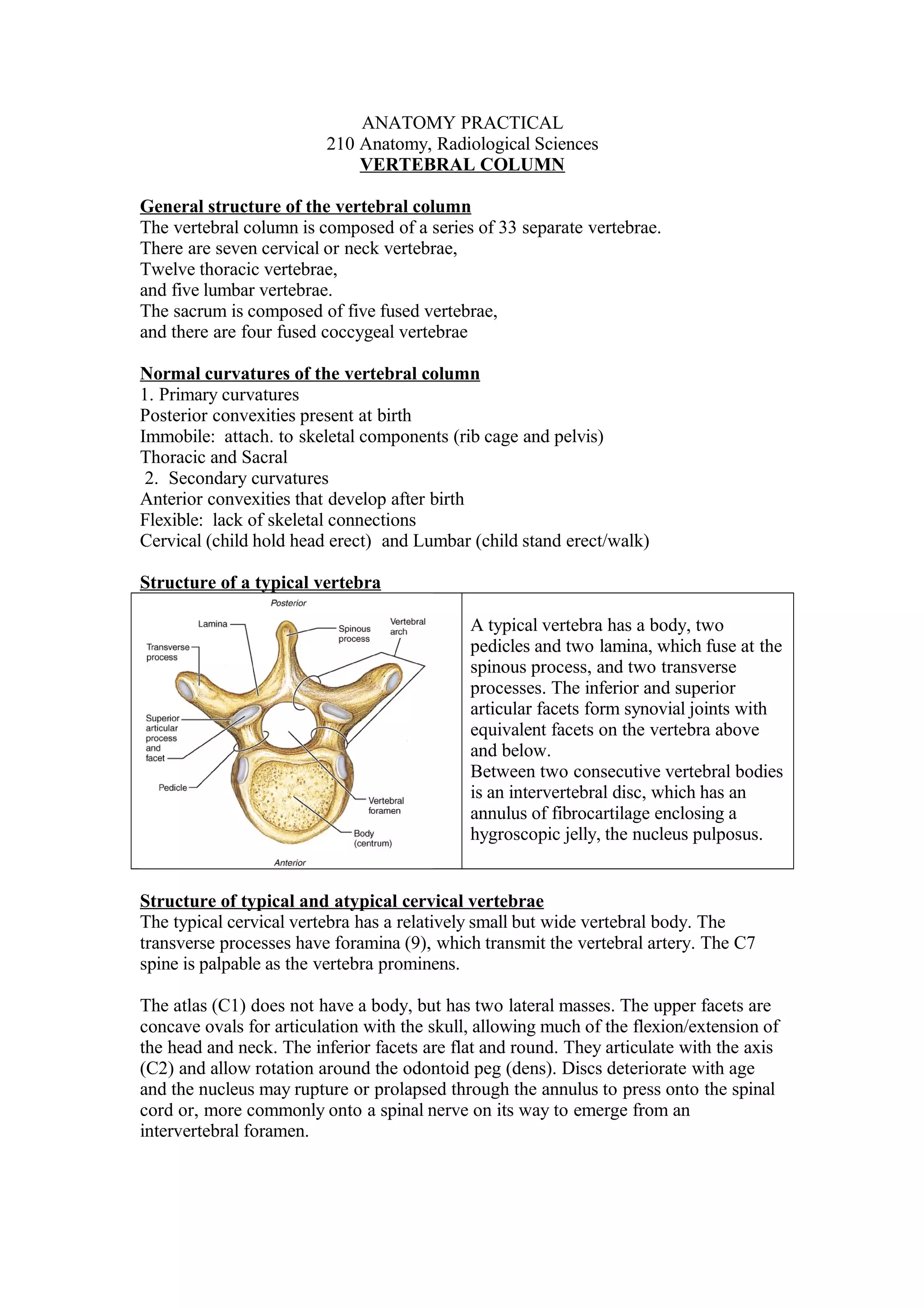
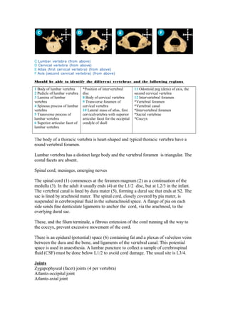
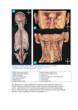
ANATOMY PRACTICAL
                         210 Anatomy, Radiological Sciences
                             VERTEBRAL COLUMN

General structure of the vertebral column
The vertebral column is composed of a series of 33 separate vertebrae.
There are seven cervical or neck vertebrae,
Twelve thoracic vertebrae,
and five lumbar vertebrae.
The sacrum is composed of five fused vertebrae,
and there are four fused coccygeal vertebrae

Normal curvatures of the vertebral column
1. Primary curvatures
Posterior convexities present at birth
Immobile: attach. to skeletal components (rib cage and pelvis)
Thoracic and Sacral
 2. Secondary curvatures
Anterior convexities that develop after birth
Flexible: lack of skeletal connections
Cervical (child hold head erect) and Lumbar (child stand erect/walk)

Structure of a typical vertebra

                                             A typical vertebra has a body, two
                                             pedicles and two lamina, which fuse at the
                                             spinous process, and two transverse
                                             processes. The inferior and superior
                                             articular facets form synovial joints with
                                             equivalent facets on the vertebra above
                                             and below.
                                             Between two consecutive vertebral bodies
                                             is an intervertebral disc, which has an
                                             annulus of fibrocartilage enclosing a
                                             hygroscopic jelly, the nucleus pulposus.


Structure of typical and atypical cervical vertebrae
The typical cervical vertebra has a relatively small but wide vertebral body. The
transverse processes have foramina (9), which transmit the vertebral artery. The C7
spine is palpable as the vertebra prominens.

The atlas (C1) does not have a body, but has two lateral masses. The upper facets are
concave ovals for articulation with the skull, allowing much of the flexion/extension of
the head and neck. The inferior facets are flat and round. They articulate with the axis
(C2) and allow rotation around the odontoid peg (dens). Discs deteriorate with age
and the nucleus may rupture or prolapsed through the annulus to press onto the spinal
cord or, more commonly onto a spinal nerve on its way to emerge from an
intervertebral foramen.
C Lumbar vertebra (from above)
D Cervical vertebra (from above)
E Atlas (first cervical vertebra) (from above)
F Axis (second cervical vertebra) (from above)

Should be able to identify the different vertebrae and the following regions

1 Body of lumbar vertebra       *Position of intervertebral         11 Odontoid peg (dens) of axis, the
2 Pedicle of lumbar vertebra    disc                                second cervical vertebra
3 Lamina of lumbar              8 Body of cervical vertebra         12 Intervertebral foramen
vertebra                        9 Transverse foramen of             *Vertebral foramen
4 Spinous process of lumbar     cervical vertebra                   *Vertebral canal
vertebra                        10 Lateral mass of atlas, first     *Intervertebral foramen
5 Transverse process of         cervicalvertebra with superior      *Sacral vertebrae
lumbar vertebra                 articular facet for the occipital   *Coccyx
6 Superior articular facet of   condyle of skull
lumbar vertebra


The body of a thoracic vertebra is heart-shaped and typical thoracic vertebra have a
round vertebral foramen.

Lumbar vertebra has a distinct large body and the vertebral foramen is triangular. The
costal facets are absent.

Spinal cord, meninges, emerging nerves

The spinal cord (1) commences at the foramen magnum (2) as a continuation of the
medulla (3). In the adult it usually ends (4) at the L1/2 disc, but at L2/3 in the infant.
The vertebral canal is lined by dura mater (5), forming a dural sac that ends at S2. The
sac is lined by arachnoid mater. The spinal cord, closely covered by pia mater, is
suspended in cerebrospinal fluid in the subarachnoid space. A flange of pia on each
side sends fine denticulate ligaments to anchor the cord, via the arachnoid, to the
overlying dural sac.

These, and the filum terminale, a fibrous extension of the cord running all the way to
the coccyx, prevent excessive movement of the cord.

There is an epidural (potential) space (6) containing fat and a plexus of valveless veins
between the dura and the bone, and ligaments of the vertebral canal. This potential
space is used in anaesthesia. A lumbar puncture to collect a sample of cerebrospinal
fluid (CSF) must be done below L1/2 to avoid cord damage. The usual site is L3/4.

Joints
Zygapophyseal (facet) joints (4 per vertebra)
Atlanto-occipital joint
Atlanto-axial joint
A Skull and vertebral column opened, with spinal cord in situ (from behind)
B Brainstem and cervical part of the spinal cord (from behind)
C Vertebral column, cervical region (from behind)


1 Spinal cord (spinal medulla)                8 Dorsal rootlets of spinal nerve
2 Margin of foramen magnum                    9 Ventral rootlets of spinal nerve
3 Medulla oblongata                           10 Spinal nerve
4 Lower end of spinal cord                    11 Dorsal root ganglion within dural sheath
5 Dura mater (reflected)                      12 Cauda equina
6 Epidural space                              13 Ventral rami
7 Posterior spinal arteries                   14 Intervertebral disc

The spinal cord is usually supplied by one anterior and two posterior arteries (7).
Dorsal rootlets (8), which are sensory, emerge from the cord and combine with the
motor, ventral rootlets (9) to form the mixed spinal nerves (10). The mixed spinal
nerves, still encased in pia, arachnoid and dura mater emerge from each intervertebral
foramen, where a swelling, the dorsal root ganglion (11).

210 vc-practical-rs

  • 1.
    ANATOMY PRACTICAL 210 Anatomy, Radiological Sciences VERTEBRAL COLUMN General structure of the vertebral column The vertebral column is composed of a series of 33 separate vertebrae. There are seven cervical or neck vertebrae, Twelve thoracic vertebrae, and five lumbar vertebrae. The sacrum is composed of five fused vertebrae, and there are four fused coccygeal vertebrae Normal curvatures of the vertebral column 1. Primary curvatures Posterior convexities present at birth Immobile: attach. to skeletal components (rib cage and pelvis) Thoracic and Sacral 2. Secondary curvatures Anterior convexities that develop after birth Flexible: lack of skeletal connections Cervical (child hold head erect) and Lumbar (child stand erect/walk) Structure of a typical vertebra A typical vertebra has a body, two pedicles and two lamina, which fuse at the spinous process, and two transverse processes. The inferior and superior articular facets form synovial joints with equivalent facets on the vertebra above and below. Between two consecutive vertebral bodies is an intervertebral disc, which has an annulus of fibrocartilage enclosing a hygroscopic jelly, the nucleus pulposus. Structure of typical and atypical cervical vertebrae The typical cervical vertebra has a relatively small but wide vertebral body. The transverse processes have foramina (9), which transmit the vertebral artery. The C7 spine is palpable as the vertebra prominens. The atlas (C1) does not have a body, but has two lateral masses. The upper facets are concave ovals for articulation with the skull, allowing much of the flexion/extension of the head and neck. The inferior facets are flat and round. They articulate with the axis (C2) and allow rotation around the odontoid peg (dens). Discs deteriorate with age and the nucleus may rupture or prolapsed through the annulus to press onto the spinal cord or, more commonly onto a spinal nerve on its way to emerge from an intervertebral foramen.
  • 2.
    C Lumbar vertebra(from above) D Cervical vertebra (from above) E Atlas (first cervical vertebra) (from above) F Axis (second cervical vertebra) (from above) Should be able to identify the different vertebrae and the following regions 1 Body of lumbar vertebra *Position of intervertebral 11 Odontoid peg (dens) of axis, the 2 Pedicle of lumbar vertebra disc second cervical vertebra 3 Lamina of lumbar 8 Body of cervical vertebra 12 Intervertebral foramen vertebra 9 Transverse foramen of *Vertebral foramen 4 Spinous process of lumbar cervical vertebra *Vertebral canal vertebra 10 Lateral mass of atlas, first *Intervertebral foramen 5 Transverse process of cervicalvertebra with superior *Sacral vertebrae lumbar vertebra articular facet for the occipital *Coccyx 6 Superior articular facet of condyle of skull lumbar vertebra The body of a thoracic vertebra is heart-shaped and typical thoracic vertebra have a round vertebral foramen. Lumbar vertebra has a distinct large body and the vertebral foramen is triangular. The costal facets are absent. Spinal cord, meninges, emerging nerves The spinal cord (1) commences at the foramen magnum (2) as a continuation of the medulla (3). In the adult it usually ends (4) at the L1/2 disc, but at L2/3 in the infant. The vertebral canal is lined by dura mater (5), forming a dural sac that ends at S2. The sac is lined by arachnoid mater. The spinal cord, closely covered by pia mater, is suspended in cerebrospinal fluid in the subarachnoid space. A flange of pia on each side sends fine denticulate ligaments to anchor the cord, via the arachnoid, to the overlying dural sac. These, and the filum terminale, a fibrous extension of the cord running all the way to the coccyx, prevent excessive movement of the cord. There is an epidural (potential) space (6) containing fat and a plexus of valveless veins between the dura and the bone, and ligaments of the vertebral canal. This potential space is used in anaesthesia. A lumbar puncture to collect a sample of cerebrospinal fluid (CSF) must be done below L1/2 to avoid cord damage. The usual site is L3/4. Joints Zygapophyseal (facet) joints (4 per vertebra) Atlanto-occipital joint Atlanto-axial joint
  • 3.
    A Skull andvertebral column opened, with spinal cord in situ (from behind) B Brainstem and cervical part of the spinal cord (from behind) C Vertebral column, cervical region (from behind) 1 Spinal cord (spinal medulla) 8 Dorsal rootlets of spinal nerve 2 Margin of foramen magnum 9 Ventral rootlets of spinal nerve 3 Medulla oblongata 10 Spinal nerve 4 Lower end of spinal cord 11 Dorsal root ganglion within dural sheath 5 Dura mater (reflected) 12 Cauda equina 6 Epidural space 13 Ventral rami 7 Posterior spinal arteries 14 Intervertebral disc The spinal cord is usually supplied by one anterior and two posterior arteries (7). Dorsal rootlets (8), which are sensory, emerge from the cord and combine with the motor, ventral rootlets (9) to form the mixed spinal nerves (10). The mixed spinal nerves, still encased in pia, arachnoid and dura mater emerge from each intervertebral foramen, where a swelling, the dorsal root ganglion (11).