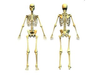
The document outlines the processes of bone growth and development, detailing the stages of ossification both prenatally and postnatally. It explains intramembranous and endochondral ossification, as well as hormone regulation affecting growth. Additionally, it describes how bones lengthen and thicken throughout development, highlighting the roles of various cells and hormones.

![(3) Intramembranous Ossification
(3) Intramembranous Ossification
[~Begins Week 8]
• Cells produce fibrous membranes surrounding cartilage.
• Ossification Center forms within fibrous membrane.
• Osteoid (Nutrient + Osteoblast Mixture) secreted from
membrane.
• Osteoblasts develop into Osteocytes.
• Osteoid mineralizes around blood vessels, forming spongy
bone trabeculae.
• Remaining external Mesenchymal Cells form periosteum.
• Outer trabeculae lament and form thicker compact bone.
• Cranial Bones + Clavicles formed.](https://image.slidesharecdn.com/anatunit4bonegrowthanddevelopmentnotes-250129134843-00cca1f7/85/ANAT_unit-4_bone-growth-and-development-notes-ppt-5-320.jpg)
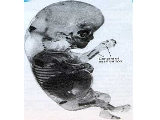
![(4) Endochondral Ossification
(4) Endochondral Ossification
[~Begins Week 10-12]
• Ossification Center forms within cartilage.
• Blood Vessels infiltrate, allowing for periosteum formation.
• Bone Collar forms around diaphysis of cartilage model.
• Osteoblasts within the Periosteum secrete osteoid, encasing
cartilage.
• Ossification Center calcifies, cartilage cells enlarge and die.
• Inner cavity invaded by periosteal bud (blood vessels, nerves,
osteoblasts).
• Osteoblasts surround dead cartilage cells, spongy bone trabeculae
formed.
• Innermost trabeculae devoured by osteoclasts, medullary cavity
formed.
• Outer trabeculae lament and form compact bone.](https://image.slidesharecdn.com/anatunit4bonegrowthanddevelopmentnotes-250129134843-00cca1f7/85/ANAT_unit-4_bone-growth-and-development-notes-ppt-8-320.jpg)
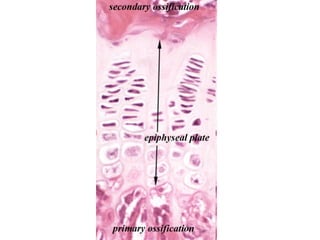
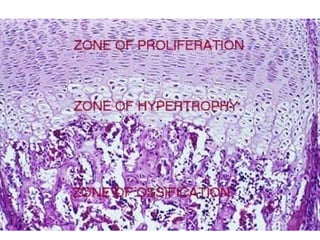
![(5) Endochondral Ossification:
(5) Endochondral Ossification:
The Epiphyses
The Epiphyses
[~Begins Week 27-29]
• Center (both inner and outer) of bone model
continues to ossify.
• Ends remain as cartilage, continue to undergo
mitosis Elongation.
• Ossification was “chasing” Cartilage
Formation to the ends of the bone.
• Epiphyses ossify…. Cartilage remains on
outer surface of epiphyses and at Epiphyseal
Plate.](https://image.slidesharecdn.com/anatunit4bonegrowthanddevelopmentnotes-250129134843-00cca1f7/85/ANAT_unit-4_bone-growth-and-development-notes-ppt-10-320.jpg)