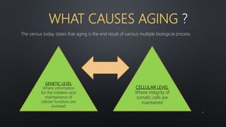
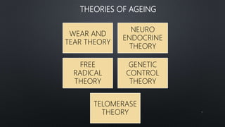
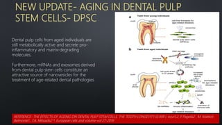
Aging is a continuous, detrimental, and innate phenomenon in an organism. It is a time-related process, which happens in a constant and steady manner right from birth and continues till death. Ageing is seen in teeth as well like other parts of body
Radiographic Assessment of the Prevalence of Pulp Stones in Malaysians
Kannan et al.
JOE — Volume 41, Number 3, March 2015
Pulp stones are discrete calcified bodies found in the dental pulp.
They have calcium phosphorous ratios similar to dentin and can be seen in healthy, diseased, or even unerupted teeth
Radiographically, pulp stones appear as radiopaque structures in the pulp space that frequently act as an impediment during endodontic treatment
Coronal and radicular pulp
Apical foramen
Accessory canal
Functions of dental pulp
Components of dental pulp
Functions of pulpal extracellular matrix
Organization of cells in the pulp
The principle cells of the pulp
The pathways of collagen synthesis
Matrix and ground substances
Vasculature and lymphatic supply
Innervation of Dentin- pulp complex
Disorders of the dental pulp
Advances in pulp vitality testing
PHYSICAL PROPERTIES
CHEMICAL PROPERTIES
STRUCTURE OF ENAMEL
DEVELOPMENT OF ENAMEL
EPITHELIAL ENAMEL ORGAN
AMELOGENESIS
LIFE CYCLE OF AMELOBLASTS
AGE CHANGES IN ENAMEL
DEFECTS OF AMELOGENESIS
CLINICAL IMPLICATIONS
It is a presentation in detail about the strongest structure of the oral cavity "ENAMEL". It is a simple topic but people find it difficult to learn about it. I hope my presentation is a simple method to learn about it. I would like to thank my professors for assign me this project and i learn't a lot from it and still learning my basics daily.
A Small effort to make this theory topic a little practical!!
The knowledge of this basic structure is actually the first building block towards dentistry.
Proper knowledge leads to correct diagnosis and henceforth desired treatment.
Difference between primary and permanent teethprincesoni3954
The presentation features the basic difference between primary and permanent dentition. The differences are tabulated under the headings of crown, roor and pulp.
Age changes in oral tissues/ /certified fixed orthodontic courses by Indian d...Indian dental academy
Welcome to Indian Dental Academy
The Indian Dental Academy is the Leader in continuing dental education , training dentists in all aspects of dentistry and offering a wide range of dental certified courses in different formats.
Indian dental academy has a unique training program & curriculum that provides students with exceptional clinical skills and enabling them to return to their office with high level confidence and start treating patients
State of the art comprehensive training-Faculty of world wide repute &Very affordable.
Age changes in oral tissues /certified fixed orthodontic courses by Indian de...Indian dental academy
The Indian Dental Academy is the Leader in continuing dental education , training dentists in all aspects of dentistry and offering a wide range of dental certified courses in different formats.
Indian dental academy provides dental crown & Bridge,rotary endodontics,fixed orthodontics,
Dental implants courses.for details pls visit www.indiandentalacademy.com ,or call
0091-9248678078
GERIATRIC CARE DENTISTRY - ORAL MANIFESTATIONS IN GERIATRIC PATIENTSdrpriyanka8
INTRODUCTION
CLASSIFICATION
AGE CHANGES AFFECTING STRUCTURES IN ORAL CAVITY
COMMON DISEASE AMONG GERIATRIC PATIENTS
MOST COMMON ORAL DISEASE WITH SUGGESTED TREATMENT
GOAL OF ORAL PHYSICIAN
COMMON SYSTEMIC DISEASE IN OLDER ADULT
CONCLUSION
REFERENCES
The wasting diseases of teeth, namely attrition, abrasion and dental erosion have taken their toll in the population around the world due to the changing lifestyles, increase in the stress levels and many others factors that were persistent earlier but have suddenly increased drastically. This presentation brings to light the new factors that have attributed to this condition as well as discusses the previous ones.
Prevalence of tooth loss
Tooth wear and causes
Tooth wear indices
Classification of tooth wear
Non carious cervical lesions include attrition, abrasion, abfraction, erosion and their combined lesion
developmental defects
resorption
trauma and fractures
How to Create Map Views in the Odoo 17 ERPCeline George
The map views are useful for providing a geographical representation of data. They allow users to visualize and analyze the data in a more intuitive manner.
Operation “Blue Star” is the only event in the history of Independent India where the state went into war with its own people. Even after about 40 years it is not clear if it was culmination of states anger over people of the region, a political game of power or start of dictatorial chapter in the democratic setup.
The people of Punjab felt alienated from main stream due to denial of their just demands during a long democratic struggle since independence. As it happen all over the word, it led to militant struggle with great loss of lives of military, police and civilian personnel. Killing of Indira Gandhi and massacre of innocent Sikhs in Delhi and other India cities was also associated with this movement.
2024.06.01 Introducing a competency framework for languag learning materials ...Sandy Millin
http://sandymillin.wordpress.com/iateflwebinar2024
Published classroom materials form the basis of syllabuses, drive teacher professional development, and have a potentially huge influence on learners, teachers and education systems. All teachers also create their own materials, whether a few sentences on a blackboard, a highly-structured fully-realised online course, or anything in between. Despite this, the knowledge and skills needed to create effective language learning materials are rarely part of teacher training, and are mostly learnt by trial and error.
Knowledge and skills frameworks, generally called competency frameworks, for ELT teachers, trainers and managers have existed for a few years now. However, until I created one for my MA dissertation, there wasn’t one drawing together what we need to know and do to be able to effectively produce language learning materials.
This webinar will introduce you to my framework, highlighting the key competencies I identified from my research. It will also show how anybody involved in language teaching (any language, not just English!), teacher training, managing schools or developing language learning materials can benefit from using the framework.
Model Attribute Check Company Auto PropertyCeline George
In Odoo, the multi-company feature allows you to manage multiple companies within a single Odoo database instance. Each company can have its own configurations while still sharing common resources such as products, customers, and suppliers.
Palestine last event orientationfvgnh .pptxRaedMohamed3
An EFL lesson about the current events in Palestine. It is intended to be for intermediate students who wish to increase their listening skills through a short lesson in power point.
The French Revolution, which began in 1789, was a period of radical social and political upheaval in France. It marked the decline of absolute monarchies, the rise of secular and democratic republics, and the eventual rise of Napoleon Bonaparte. This revolutionary period is crucial in understanding the transition from feudalism to modernity in Europe.
For more information, visit-www.vavaclasses.com
Read| The latest issue of The Challenger is here! We are thrilled to announce that our school paper has qualified for the NATIONAL SCHOOLS PRESS CONFERENCE (NSPC) 2024. Thank you for your unwavering support and trust. Dive into the stories that made us stand out!
How to Split Bills in the Odoo 17 POS ModuleCeline George
Bills have a main role in point of sale procedure. It will help to track sales, handling payments and giving receipts to customers. Bill splitting also has an important role in POS. For example, If some friends come together for dinner and if they want to divide the bill then it is possible by POS bill splitting. This slide will show how to split bills in odoo 17 POS.
Students, digital devices and success - Andreas Schleicher - 27 May 2024..pptxEduSkills OECD
Andreas Schleicher presents at the OECD webinar ‘Digital devices in schools: detrimental distraction or secret to success?’ on 27 May 2024. The presentation was based on findings from PISA 2022 results and the webinar helped launch the PISA in Focus ‘Managing screen time: How to protect and equip students against distraction’ https://www.oecd-ilibrary.org/education/managing-screen-time_7c225af4-en and the OECD Education Policy Perspective ‘Students, digital devices and success’ can be found here - https://oe.cd/il/5yV
Digital Tools and AI for Teaching Learning and Research
Age changes in tooth
1. AGE CHANGES IN ENAMEL,
DENTIN , PULP AND CEMENTUM
PRESENTED BY-
DR. AISHWARYA ARYA
MMCDSR
2. 1. Introduction
2. Various terminologies
3. What causes aging ?
4. Theories of ageing
5. Factors affecting ageing
6. Importance in dentistry
7. Importance in Conservative dentistry Endodontics
8. General factors of aging
9. Dental age estimation methods
10. Age related changes in oral tissues
11. Age changes in Enamel
12. Age changes in Dentin
13. Age changes in Cementum
14. Age changes in pulp
15. New Updates
16. Conclusion
17. References
CONTENTS
2
3. Aging is a continuous, detrimental, and innate phenomenon in an
organism. It is a time-related process, which happens in a constant and
steady manner right from birth and continues till death…
3
6. WHAT CAUSES AGING
The census today states that aging is the end result of various multiple biological process
GENETIC LEVEL
Where information
for the initiation and
maintainence of
cellular functions are
involved
CELLULAR LEVEL
Where integrity of
somatic cells are
maintained
6
7. THEORIES OF AGEING
WEAR AND
TEAR THEORY
NEURO
ENDOCRINE
THEORY
FREE
RADICAL
THEORY
GENETIC
CONTROL
THEORY
TELOMERASE
THEORY 7
8. WEAR AND TEAR THEORY
8
Given by August Weihmann (1882)
These theories (also called simple deterioration
theories)
Proposed that organs and tissues were damaged
by continuous use and abuse
When a person ages , body’s mechanism to
repair the damage caused by wear and tear is
reduced.
Hence , in old age people die of diseases which
they could have resisted when they were young
9. FREE RADICAL THEORY
First postulated by Harman in 1956, this theory
suggests that free radicals cause oxidative damage to
cells and their products, resulting in aging.
Free radicals are atoms or molecules that have one
free electron.
They are very reactive due to the tendency to pair.
Free radical activity is required to produce energy ,
maintain immunity, nerve transmission.
But free radicals also attack cell membranes producing
metabolic waste products –lipofuschins
9
10. GENETIC THEORY
Another theory proposed by
Leo Szilard in 1959
He puts forth the idea that
genes involved in DNA
metabolism and repair become
faulty with time.
This leads to accumulation of
DNA damage that manifests as
aging.
10
12. THE TELOMERASE THEORY OF
AGEING
•
Similarly, Watson in 1972
suggested that telomeres may be
involved in aging.
Age related changes do not occur
uniformly in individuals , but they
are under the influence of genetic
and environment factors
12
13. IMPORTANCE OF AGING IN DENTISTRY
FORENSIC
DENTISTRY
PROPER
EVALUATION
AND DIAGNOSIS
RESPONSE OF TOOTH
STRUCTURE TO
VARIOUS
ENIVRONMENTAL
FACTORS OVER A
PERIOD OF TIME
MORPHOLOGY OF
THE TOOTH
STRUCTURE
DIFFERENT
TREATMENT
PROTOCOLS
AGE RELATED
DISORDERS
13
14. RELEVANCE IN CONSERVATIVE DENTISTRY AND ENDOODNTICS
Etiology behind the pathology
Differentiating between normal and abnormal pathologies
Prognosis of the treatment
Restorative material used,
Endodontic outcomes-Interproximal root caries is difficult to restore, and restoration
failure as a result of continued decay is common
14
16. DENTALAGE ESTIMATION METHODS-ROLE IN
FORENSIC DENTISTRY
• Based on the evaluation of the sequence of teeth eruption in
oral cavity and the morphological changes on tooth structure
due to functions such as attrition, changes in color that are
indicators of aging.
VISUAL METHOD
• The development of each tooth can be assessed over long
periods of time using radiographs in a continuous pattern,
using different crown and root maturity stages of tooth
formation
RADIOGRAPHIC
METHODS
• Calculates the correlation between the reduction of the
coronal pulp cavity and the chronological age. The tooth-
coronal index (TCI) is calculated for each tooth and regressed
on the real age of the sample using a formula
CORONAL PULP
CAVITY INDEX
• They measured pulp length and width as well as root length
and width. Because of magnification and angulation on the
radiographs, different ratios between root and pulp were
measured
PULP TO TOOTH
RATIO METHOD
16
17. MORPHOLOGICAL METHODS
Morphological methods are based on assessment of ex-vivo teeth for age estimation of adults.
The samples of the extracted tooth can be sectioned or unsectioned and observed with the eye or with
microscope .
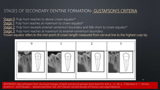
The first technique was published by Gustafson in 1950 AND noted the following changes:
1.Attrition of the incisal or occlusal surfaces due
to mastication [A]
1.Periodontitis [P]—the loss of periodontal attachment
Cementum apposition [C] at the root apex
Transparency of the root [T]
Root resorption [R] amount
1.Secondary dentin [S]—the amount of coronal secondary
dentine formation
17
18. Prenatal stage (up to 6 months) radiological methods cannot be accurate in dental
age estimation given that the dentin and enamel images are radiolucent.
BIOCHEMICAL METHODS
.
STACK S METHOD
Fetal age is linearly related to the square root of the weight of mineralized
tissue in the deciduous anterior teeth during the last trimester
At 6 months intrauterine life, the weight is 60 mg; in the newborn child,
18
19. Biochemical and histological methods are preferred in
adults.
The biochemical methods determine alterations in ion
levels with age.
The calcium/phosphorus ratio in peritubular dentine
increases significantly with age
The rate of racemization of D and L enantiomers of
aspartic acid residues in the collagen of dentin is time-
dependent
Aspartic acid racemization is demonstrated to be an
19
20. The d/l ratio has been shown to be highly correlated with age
The racemization rate of the cementum shows the fastest
reaction than enamel and dentine.
l-aspartic
acids
d-aspartic
acids
The levels of d-aspartic acid
in human enamel, dentine
and cementum
Increase
with age.
20
21. This technique is more appropriate
for postmortem situations.
The histological methods can detect
mineralization before being detected
in the radiographs.
Prenatal dental maturity can be
assessed by using dissection and
alizarin staining of fetal tooth germs.
HISTOLOGICAL METHOD
21
22. NEONATAL LINES
The neonatal lines are an optical phenomenon produced
due to alteration in the dimension, degree and
mineralization of enamel prisms caused due to the
sudden change from intrauterine to extrauterine
environment .
Neonatal lines can be used to differentiate between
live birth and still birth and are an important tool in
forensic cases and for pre epidemiological studies.
22
Role of Cross Striations
23. DIAGNOSTIC BIOMARKERS IN SALIVA RELATING TO AGE
Noninvasive approach for prediction of aging
processes as saliva biomarkers are related to age
features of teeth.
The saliva status during the different age periods
is related to age features of tooth enamel.
23
24. AGE RELATED CHANGES IN ORAL TISSUES
TISSUE CHANGES
1. TEETH
2.PERIODONTIUM
- BONE
- PDL
ORAL MUCOUS
MEMBRANE
FUNCTIONAL CHANGE
1. SALIVARY CHANGES
2. TASTE
3. MASTICATION
4. DEGLUTITION
24
26. MORPHOLOGICAL AND HISTOLOGICAL CHANGES IN ENAMEL
The outer surfaces of the enamel remain in equilibrium with the surrounding oral fluids.
When this equilibrium is not maintained, such as during an acid attack, H+ ions from the acid adhere
to different sites on the surface of the dental hard tissues and cause demineralization
In acidic environment, the concentrations of PO4 3– and OH– ions are lower, so PO4 3– and OH– ions
dissolve out of the tooth and into the surrounding fluids to maintain the equilibrium.
Throughout our lifetime, the teeth are not only constantly subjected to such acidic environments, but
also to mechanical forces, which intensify the wear of the tooth surfaces.
TOOTH WEAR IS A CHEMICAL-MECHANICAL PROCESS.
26
27. PHYSICAL CHANGES
Wearing away of occlusal and proximal
contacts due to overload of mastication
CLINICAL MANIFESTATIONS-
Appearance of small polished facets on cusp
tip and ridges and slight flattening of incisal
edges.
Reduction of cuspal height and flattening of
proximal contour
Shortening of the length of dental arch
Loss of vertical dimensions
Enamel becomes harder and brittle with age
CHEMICAL CHANGES:
• Increase in the Organic content while
some state there is decrease
• Increase in Nitrogen and Fluoride
• Increase in size of enamel crystals
27
28. AGE CHANGES IN ENAMEL
- CHANGES IN COLOUR-
BECOMES DARKER
-ATTRITION, EROSION, ABRASION
, ABFRACTION
- PRESENCE OF LONGITUDINAL
CRACKS
DECREASED - PERIKYMATA
- ENAMEL RODS
- PERMEABILITY TO FLUIDS
INCREASED – RESISTANCE TO DECAY.
- NITROGEN
- FLUORINE
28
29. COLOUR CHANGES IN ENAMEL
The nitrogen content of enamel appears to increase, which correlates with increased pigment,
leading to tooth darkening.
Refractive and light absorptive changes are related to enamel and dentinal structural changes.
29
30. DISCOLORATION AND STAINS
With ageing, the teeth become darker.
Extrinsic causes- staining brought about by tobaceo, tea, coffee, red
wine, chlorhexidene or discoloured restorations.
Intrinsic causes- staining by leakage of breakdown products of
haemoglobin into the dentinal tubules in non-vital teeth or contaminants
from root-filling materials or caries or resorption.
Clinical significance
The alterations to tooth colour and contour leads to modification in
treatment relating to esthetics
Use of adhesive restorations through bleaching, veneers and crowns to
the provision of prostheses.
Very subjective area as patients' perception of their oral self image varies
from individual to individual. 30
31. AGE CHANGES IN PERMEABILITY
LESS
PERMEABLE
31
Increase in the size of the crystal.
The crystal size increases due to ions
acquired by it from the oral fluids- saliva.
The pores between th crystals decreases
Permits the slow passage of water and
substances of small molecular size
through pores between the crystals.
The increase in size of the crystal
decreases the pores between them
causing a reduction in permeability
32. HARDNESS
An increase in brittleness,' The organic substance
between enamel prisms apparently becomes calcified,
causing brittleness and fractures
Some studies have shown that enamel surface hardness
presents a decreasing slope from the enamel surface
towards the dentino-enamel junction
Older teeth, which have already lost their surface layer
have lower hardness values.
Teeth from older patients (55 years or older) had
higher elastic modulus and surface hardness than
teeth from younger patients (between 18 and 30 years
old).
This increase in elastic modulus
Reduction in the interprismatic organic matrix 32
33. CONCENTRATION OF FLUORIDE
In enamel of young teeth the concentration is highest
towards the incisal edge and lowest towards the cervical
edge.
With increasing age, a great part of the surface fluoride is
lost
After the age of 30, there is a shift in the gradient of
fluoride concentration, which increases towards the cervical
edge of the teeth
Anterior teeth of elderly patients present relatively high
fluoride concentration on the cervical margin of the enamel,
Relatively low fluoride concentration on the remaining
enamel surface.
33
34. Root-surface caries lesions develops on exposed root dentine surfaces or exposed cementum as a result of
gingival recession and not on the actual cervical enamel itself
In addition, the lower fluoride concentration on the remaining enamel surface has also been associated
with a greater liability to demineralization (greater porosity within the lesion) than young teeth
Role of saliva as the age advances
34
36. ABRASION
• CAUSES
Pathological and physiological abrasion occurs in dental
hard tissues with age. These changes produce differences
in mineral exchange in different age groups
Definition — The pathologic wearing away of a substance or structure (such as a tooth), through some
unusual or abnormal mechanical process. (Glossary of Endodontics- Updated March 2020)
36
37. FEATURES OF ABRASION
Most common teeth involved are the premolars and the cuspids
Surface – extremely smooth and polished walls
On probing it stimulates pain 37
Saucer shaped defects.
Exposed root surface.
Associated with Gingival recession.
38. EROSION
38
A loss of tooth substance by a chemical process without
bacteria. (Glossary of Endodontics- Updated March 2020)
TYPES
i. Intrinsic
ii. Extrinsic
BROAD concavities with smooth surface
enamel.
Cupping of occlusal surface with dentinal
exposure.
In deciduous teeth –Loss of enamel surface
characteristics and pulp exposure .
40. ABFRACTION
40
The article was titled "Abfractions: A New
Classification of Hard Tissue Lesions of Teeth"
by John O. Grippo.
Definition — A V-shaped loss of hard tooth structure caused by biomechanical loading forces such as from flexure
and/or chemical fatigue degradation of enamel and/or dentin at some location distant from the actual point of
loading. (Glossary of Endodontics- Updated March 2020
41. ATTRITION
The physiologic wearing away of a substance or
structure, such as a tooth, in the course of normal
use or parafunctional habits. (Glossary of
Endodontics- Updated March 2020)
The initial signs of tooth wear are-
The flattening of the tooth surface
Loss of the outer layers of enamel, resulting in
smoother enamel surfaces without perikymata
41
CAUSES:
- MASTICATORY STRESS
- PARA-FUNCTIONAL HABITS.
-ABNORMAL OCCLUSION
42. CLINICAL PRESENTATION OF ATTRITION
• Loss of tooth anatomy.
• Sensitivity and pain.
•Tooth discoloration.
• Compromised periodontal support
leading to drifting of teeth.
• Altered occlusion due to decreasing
occlusal vertical dimension.
42
43. 43
Gustafson's criteria of attrition were measured by slicing the images sagitally in the centre of the
tooth and classified in accordance to the stages reported by Olze et al.
Stage 0: No attrition
Stage 1: Beginning attrition with loss of cusp tips
Stage 2: Attrition reaching into dentine
Stage 3: Attrition reaching into dentine with opening of pulp cavity
REFERENCE-Age estimation from structural changes of teeth and buccal alveolar bone level K.K. Koh a , J.S. Tan a , P
. Nambiar b, * , Norliza
Ibrahim b , Sunil Mutalik c , Muhammad Khan Asif. 2017 Elsevier Ltd and Faculty of Forensic and Legal Medicine
44. RELATIONSHIP BETWEEN ATTRITION AND DENTAL CARIES
• Physiological Attrition wears out the protective enamel increasing seeding of acid into the enamel matrix
therefore increasing demineralization and the chance of developing carious lesions.
• Pathological attrition which involves severe enamel wear, the dentine is exposed and this predisposes the tooth
to bacterial invasion by S. mutans which consequently leads to development and rapid progression of dental
caries .
44
48. .
CHANGES IN DENTIN TAKES PLACE TO ENSURE THAT THE FUNCTION OF PROTECTION IS TAKEN
OVER FROM ENAMEL AS THE ENAMEL WEARS WITH AGING.
TWO MOST IMPORTANT CHANGES
Increase in the thickness of dentin due
to continuous, gradual deposition of
secondary dentin
Increased sclerosis or obliteration
of dentinal tubules
The increase in the mineral
density is responsible for
increased hardness of dentin
with aging. 48
49. ODONTOBLASTIC CHANGES
Degeneration of odontoblasts with presence of vacuoles being visible prior to their atrophy .
Odontoblasts lining sclerotic dentine become reduced in number and disappear
Due to a cell mediated change.
49
50. Aged odontoblasts have a Decreased Secretory Function
Oodontoblasts reduce their secretory machinery acquire
a mature stage characterized by the presence of
autophagic vacuoles (AV).
Accumulation of lipofuscin deposits- LF
The size of the odontoblast layer changes from columnar
in young to cuboidal in older indivuals.
Nestin and Notch protein, which are expressed in young
odontoblasts , are absent in adult tissue
but are reexpressed during reparative dentin formation
50
51. REPARATIVE DENTIN
The hard tissue thus formed is best termed reparative
dentin, although the terms tertiary dentin, response, or
reactive dentin are also used.
Reparative dentin is characterized as having fewer and
more twisted tubules than normal dentin.
Tertiary dentin matrix secreted by odontoblast-like
cells in response to an appropriate stimulus after
the death of odontoblasts;
stimuli tend to be much stronger than those causing
reactionary dentin formation.( Glossary of
Endodontics- Updated March 2020)
51
52. ARRANGEMENT OF TUBULES IN REPARATIVE DENTIN
A- NORMAL DENTIN
B-E- OTHERTYPES OF REPARATIVE
DENTIN
B- REPARATIVE DENTIN CONTAINS
FEWER TUBULES THAN NORMAL TUBULES
C- IT INCLUDES CELLS WITHIN THE
MATRIX
D- SHOWS IRREGULARLY ARRANGED
TUBULES
E- COMBINATION OF DIFFERENT TYPES
52
53. Dead tracts In dried ground sections of
normal dentin shows the odontoblast
processes disintegrate, and the empty
tubules are filled with air.
Their degeneration is often observed in
the area of narrow pulpal horns because
of crowding of odontoblasts.
Where reparative dentin seals dentinal
tubules at their pulpal ends, dentinal
tubules fill with fluid or gaseous
substances.
DEAD TRACTS
Dentinal tubules seen in histologic sections to be devoid of cellular processes indicating odontoblastic destruction;
may contain debris and microorganisms. (Glossary of Endodontics- Updated March 2020)
53
55. SCLEROTIC DENTIN/ TRANSPARENT DENTIN
Dentin characterized by calcification of the dentinal tubules as a result of injury or normal aging; it appears
translucent in ground sections due to the difference in refractive indices of calcified dentinal tubules and adjacent
normal tubules when examined by transmitted light. (Glossary of Endodontics- Updated March 2020)
PREVELANT IN
OLDER INDIVUALS
An increase in the transluceney of older sclerotic dentine would tend to confine caries to the
dentinoenamel junction as opposed to the dead tracts in younger dentine which would allow caries
to progress towards the pulp more easily.
55
56. CLINICAL SIGNIFICANCE
Have a profound influence on surface treatment
techniques for adhesive materials.
When non-sclerotic dentine was treated with maleic acid
primer, patent tubules resulted into which resinous tags
could penetrate.
However, with increasing sclerosis of the dentine the
primer solution was less effective in opening patent
tubules
On application of one resin (CIcarfil Liner Bond System)
to normal dentine there was extensive resin tag
development into the opened tubules .
However when the same resin was applied to sclerotic
dentine there were few resin tags formation.
CASE 1
CASE 2
56
Reference- Progressive changes in the pulpo-dentinal complex and their clinical consequences F.M. Burke and D.Y.D. Samarawickrama Department of Conservative Dentistry,
St Bartholomew's and the Royal Eondon School of Medicine and Dentistry, London, UK. Gerodontology 1995; 12(2): 57-66
57. Use of an adhesive system which bonded chemically to
calcium, N (p-toly) giycine glycidyl methacrylate
resulted in reduced microleakage compared to an
adhesive which bonded to the organic portion of
dentine in aged teeth. The higher content of calcified
tissue in aged dentine may be responsible for this.
Cervical lesions which were more highly sclerosed
performed less well than less sclerosed lesions in terms of
loss of retention, marginal integrity and discolouration
CASE 4
CASE 3
57
Reference- Progressive changes in the pulpo-dentinal complex and their clinical consequences F.M. Burke and D.Y.D. Samarawickrama Department of Conservative Dentistry, St
Bartholomew's and the Royal Eondon School of Medicine and Dentistry, London, UK. Gerodontology 1995; 12(2): 57-66
58. PERITUBULAR DENTIN
This dentine lines the walls of dentinal
tubules.
Old dentine has deposition of a
phosphoric acid-resistant intratubular
material.
This dentine has a reduced permeability
when compared with younger dentin.
An increase in the amount of peritubular dentine
has been reported with attrition.
Clinical Significance
The pulps of young teeth with open dentinal tubules
would be more susceptible to toxic stimuli from dental
materials than older teeth with sclerosed canals
58
59. SECONDARY DENTIN
Secondary dentine forms pulpally throughout life
mainly on the floor and to a lesser extent, on the roof of
the pulp chamber .
In older patients, the dentine deposited on the pulpal
floor is thicker and more irregular than that on the
roof of the pulp chamber.
This irregular floor dentine also appeared to be more
radiolucent due few odontoblasts in this site having to
cover an increasing surface area with a consequent
irregular deposition of dentine.
59
Reference- Progressive changes in the pulpo-dentinal complex and their clinical consequences F.M. Burke and D.Y.D. Samarawickrama Department of Conservative Dentistry, St
Bartholomew's and the Royal Eondon School of Medicine and Dentistry, London, UK. Gerodontology 1995; 12(2): 57-66
60. GUSTAFSON'S CRITERIA
60
Stage 0: Pulp horn reaches to above crown equator*
Stage 1: Pulp horn reaches at maximum to crown equator*
Stage 2: Pulp horn exceeds enamel-cementum boundary and falls short to crown equator*
Stage 3: Pulp horn reaches at maximum to enamel-cementum boundary
*crown equator refers to the mid-point of crown length measured from cervical line to the highest cusp tip.
REFERENCE-Age estimation from structural changes of teeth and buccal alveolar bone level K.K. Koh a , J.S. Tan a , P
. Nambiar b, * , Norliza
Ibrahim b , Sunil Mutalik c , Muhammad Khan Asif. 2017 Elsevier Ltd and Faculty of Forensic and Legal Medicine
61. CLINICAL SIGNIFICANCE OF SECONDARY DENTIN FORMATION
61
While it is more difficult to expose the pulp during tooth preparation it is
also more difficult to locate the pulp chamber and canals when carrying
out root canal therapy
Increasing deposition of dentine in the pulp canal reduces the amount of
instrumentation necessary during root canal treatment
The increased amount of dentin at the apical foramen increases the
amount of apical slops.
Reference- Progressive changes in the pulpo-dentinal complex and their clinical consequences F.M. Burke and D.Y.D. Samarawickrama Department of Conservative Dentistry, St
Bartholomew's and the Royal Eondon School of Medicine and Dentistry, London, UK. Gerodontology 1995; 12(2): 57-66
62. PROGRESSION OF CARIES RELATING TO AGE
The carious process
often develops during
the first months after
tooth eruption but is
much less in adulthood
and old age
Leading to LESSER
progression of
caries
These processes are
mainly associated with
the mechanisms of ionic
exchange between the
oral cavity and hard
dental tissues.
A mature enamel has
small intercrystalline
spaces and, in this
connection, is poorly
adapted for ion
exchange.
62
63. CLINICAL CONSIDERATION IN DENTIN
DENTIN PERMEABILITY DENTIN SENITIVITY
REACTIONS TO DIFFERENT
CAVITY PREPARATION
63
65. CELLULAR CHANGES
VOLUME-The volume of pulp decreases with age,
which may be attributed to continuous deposition of
secondary dentin throughout the life.
CELLS- The number, nature, properties, and capabilities
of the cells change.
DENSITY- The decrease in pulp cell density is greater in root compared to crown and
at all ages . The pulp cell densities including odontoblasts is greater within the crown
compared to the root.The density of odontoblasts and the pulpal fibroblasts decreases
with age in general
NERVE SUPPLY-There is also considerable decrease in the nerve supply. This
compounded with increased thickness of dentin may be responsible for the decreased
pulpal response and its healing capability.
65
66. CYTOPLASMIC ORGANELLES- the cells are characterized by a
decrease in size and number of cytoplasmic organelles.
FIBROCYTES-The fibroblasts in the aging pulp exhibit less perinuclear
cytoplasm and possess long, thin cytoplasmic processes.
INTRACELLULAR ORGANELLES- The intracellular
organelles, mainly the mitochondria and endoplasmic
reticulum are reduced in number and size.
CHANGE IN THE SIZE OF THE PULP CAVITY - The continuous secondary
dentin deposition on the pulpal wall especially the floor of the pulp chamber leads
to its gradual reduction in size and its irregularity by age.
66
67. IF CELLS DIE THEN HOW DOES
FIBRES INCREASE IN THE PULP ?
67
68. The canal entrances also narrow and move centrally, whilst
in cross-section, the canals narrow concentrically.
Pulps continue to secrete dentine throughout life. In
molars, this is mainly on the floor and roof of the chamber,
converting it from a deep cavern to a flat disc .
This process also narrows the entrances and moves
them closer to the centre of the tooth.
68
69. VASCULAR CHANGES
Blood flow decreases with age.
This is due to decrease in the number of blood vessels
and due to formation of atherosclerotic plaques within
pulpal vessels.
In other cases, the outer diameter of vessel walls
becomes greater as collagen fibers increase in the
medial and adventitial layers.
Calcification in the walls of blood vessels is found
most often in the region near the apical foramen.
69
70. FIBROSIS OF PULP
In the aging pulp accumulations of both diffuse fibrillar
components as well as bundles of collagen fibers appear.
ARRANGEMENT CHANGES- Fiber bundles may appear
arranged longitudinally in bundles in the radicular pulp,
and in a random more diffuse arrangement in the
coronal area.
The increase in fibers in the pulp organ is gradual and is
generalized throughout the organ.
70
71. THEN WHY THE SIZE AND VOLUME OF PULP
DECREASES ?
Collagen increase Is noted in the medial and
adventitial layers of blood vessels as well.
The increase in collagen fibers may be more
apparent than actual, being attributable to the
decrease in the size of the pulp
This makes the fibers present occupy less space,
and hence they become more concentrated
without increasing in total volume
71
72. PULP STONES
Pulp stones or denticles are nodular, calcified masses
appearing in either or both coronal and root portions of
the pulp organ.
Calcifications in the coronal region are known as pulp
stones
whereas those in the radicular pulp are diffuse and may
lead to a complete calcific degeneration, a process
termed pulpal obliteration (Murray et al., 2002; Goldberg,
2014; Montoya et al., 2015).
All these events take place approximately in the same
period (20-39 years of age), and they are often followed
by a decrease in odontoblast cellularity
72
73. CALCIFICATION OF PULP
True denticles
Its structure is similar to dentin.
They exhibit dentinal tubules containing the processes
of the odontoblasts that exist on their surface. They are
rare and are usually located near the apical foramen.
False denticles
These denticles do not exhibit dentinal tubules but
appear as concentric layers of calcified tissue.
The surrounding pulp tissue may appear quite
normal. They are seen more frequently in the
coronal pulp.
73
74. Diffuse calcifications appear as irregular calcific deposits in the pulp
tissue, usually following collagenous fiber bundles or blood vessels .
Larger masses or fine calcified spicules.
DIFFUSE CALCIFICATIONS
74
75. TEENAGE-EXTENSIVE
BRANCHING OF PULP
NERVES AND RICH
ODONTOBLASIC
NETWORK
30 YEAR OLD- CENTRAL
PORTION OF THE PULP
CHAMBER- INTERIOR
VASCULAR CHAMBERS
ARE REDUCED
40-70 YEAR OLD-
CENTRALLY LOCATED
VESSSLES VISIBLE-
SUBODONTOBLASTIC
CAPILLARY PLEXUS
ABSENT
50 YEAR OLD-Only the
pulpal nerve is
demonstrable; the cuspal
branches and
subodontoblastic network
are absent
Reference- Seltzer and Bender's Dental Pulp, Second Edition
75
A
D
C
B
PULP CHAMBER SUBODONTOBLASTIC
CAPILLARY NERVES OF PULP
76. A- irregular secondary dentin formation is visible,
mostly on the lingual wall of the pulp chamber
B-The irregular secondary dentin fills the entire pulp
chamber of the crown and extends down to the
apical third of the root canal.
15-year-old subject.
Unmyelinated axons
form groups of
varying sizes
34-year-old-
Unmyelinated axons
are usually in small
groupings
Reference- Seltzer and Bender's Dental Pulp, Second Edition
76
SECONDARY DENTIN
UNMYELINATED AXONS
77. NEW UPDATE- AGING IN DENTAL PULP
STEM CELLS- DPSC
Dental pulp cells from aged individuals are
still metabolically active and secrete pro-
inflammatory and matrix-degrading
molecules.
Furthermore, mRNAs and exosomes derived
from dental pulp stem cells constitute an
attractive source of nanovesicles for the
treatment of age-related dental pathologies
77
REFERENCE- THE EFFECTS OF AGEING ON DENTAL PULP STEM CELLS, THE TOOTH LONGEVITY ELIXIR I. Iezzi1,2, P
. Pagella2 , M. Mattioli-
Belmonte1 , T.A. Mitsiadis2 *. European cells and volume-vol 27-2019
79. THICKNESS- the thickness of
cementum increases with age
Commonly seen in the apex of the root
reason- passive eruption occurring to
compensate the occlusal and proximal wear
due to attrition
Triples from the age 0f 10 to 70
WIDTH- increases- due to elimination of waste
products from cementum
CEMENTOCYTES-
Lowest proliferative stage
Gradually die due to reduced
accessibility to nutrition
HYERCEMENTOSIS- Abnormal thickening
of cementum which might be diffused or
circumscribed,
2 types- cementum hypertrophy
Cementum hyperplsia
EXCEMENTOSIS- Localized
hypercementosis observed in areas of
enamel drops that have been
developed over dentin.
CEMENTICLES- calcified bodies
present in the PDL and are also
referred to as coronal cementum
79
80. AGING IN ENDODONTICS- COHEN
• MEDICAL HISTORY
• OBJECTIVE SIGNS AND SYMTOMS
• ROLE OF SALIVA
• PREVALANCE OF ROOT CARIES ROOT CARIES
• PULP TESTING
• RADIOGRAPHS
• TREATMENT PLAN
• ASSOCIATION OF PERIODONTALLY COMPROMISED TOOTH WITH ENDODONTICS
• ACCESS PREPARATION
• SUCCESS AND FAILURE
80
82. CONCLUSION
The increased longevity of the dentition, made possible by expanding fields of
advanced restorative procedures and endodontics, implies that dentistry will see a
substantial growth in the number of older patients. The desire for root canal treatment
among aging patients has increased considerably in recent years. Older patients are
aware that treatment can be performed comfortably and that age is not a factor in
predicting success
82
“ Geriatric endodontics will gain importance as the aging
population recognises that COMPLETE DENTITION , AND NOT
COMPLETE DENTURE AS ITS DESTINY ” -Cohen
83. REFERENCES
REFERENCE-Age estimation from structural changes of teeth and buccal alveolar bone level K.K. Koh a , J.S. Tan a , P. Nambiar
b, * , Norliza Ibrahim b , Sunil Mutalik c , Muhammad Khan Asif. 2017 Elsevier Ltd and Faculty of Forensic and Legal Medicine
Seltzer and Bender's Dental Pulp, Second Edition
BOOKS
ARTICLES
ORBANS- ORAL HISTOLOGY AND EMBRYOLOGY- SN BHASKAR, GS KUMAR
Cohen – pathways of pulp- Kenneth M. Hargreaves, Stephen Cohen- 11th edition
Textbook of dental and oral histology with embryology- Satish Chandra
Oral anatomy , histology and embryology – international edition- B.K.B Berkovitz, G.R Holland , B.J.
moxham
Age Changes In Oral Tissues Dr Abhishek Khare Reader Dept. of Oral Pathology & Microbiology Career Post
Graduate Institute of Dental Science Lucknow Dr Amit thahriani Private Practicinor
Progressive changes in the pulpo-dentinal complex and their clinical consequences F.M. Burke and D.Y.D. Samarawickrama
Department of Conservative Dentistry, St Bartholomew's and the Royal Eondon School of Medicine and Dentistry, London, UK.
Glossary of Endodntics- Updated March 2020
Aging should not be confused with senescence. Senescence occurs after maturation, and it increases the amenability of an organism to death
according to these theories, humans age for the same reasons and because of the same processes that cause aging in automobiles and exterior paint.
Lipofuschin is considered to be wear and tear aging pigment found in the liver ,kidney heart retina,nerve cells.
One such widely accepted theory is the concept of free radicals. First postulated by Harman in 1956, this theory suggests that free radicals cause oxidative damage to cells and their products, resulting in aging. Another theory proposed by Leo Szilard in 1959 puts forth the idea that genes involved in DNA metabolism and repair become faulty with time. This leads to accumulation of DNA damage that manifests as aging. Similarly, Watson in 1972 suggested that telomeres may be involved in aging. Telomeres are small repetitive sequences seen at the ends of chromosomes. During every cell division, some of this telomere is lost. This theory was based on the fact that cells with short telomeres do not divide and become senescent. Although it can be replenished by the enzyme telomerase, over time, the length of telomere becomes short, and cell division no longer happens. The main drawback of these theories is that it is very difficult to prove whether each characteristic is a cause for aging, or is the result of aging. Lipofuscin, an intracellular pigment seen in many organisms, was initially thought to be a cause for aging. However, experiments have shown that lipofuscin is rather a product of aging process than being its cause
DESICCATION TO DRY OUT/ REMOVAL OF WATER
Fully formed teeth show aging changes. Thus, examination of dentition considering the tooth wear/attrition, tooth color and stains, periodontal status, etc. can provide valuable information on an individual’s development and age
Radiographic methods
Dental age may be estimated by two approaches: based on the time of emergence of the tooth in the oral cavity and the pattern of tooth development, in another word the dental maturity stages. Dental maturity is considered to be more reliable than the emergence of teeth into the oral cavity with a high heritability and low coefficient variation, and to be independent of environmental effects, nutritional and endocrine status [6, 7, 8]. The development of each tooth can be assessed over long periods of time using radiographs in a continuous pattern, using different crown and root maturity stages of tooth formation as criteria [27, 28]. Radiological methods are based on the evaluation of tooth development on the various radiographic images as intraoral periapical, panoramic radiographs, digital and advanced imaging technologies to assess the extent of tooth mineralization from the moment when radiopaque spots become visible prior to tooth calcification until the tooth apex is closed (Figure
Stack’s method he demonstrated that the dry weight of the mineralized tooth cusps gives an approximate age of the child by using gravimetric methods.
Cross striations are seen across enamel rods representing the daily incremental deposition of enamel can be used to find the exact age of the baby in days by counting the cross striations from the neonatal line.
the detection of neonatal line depends on various factors like axis of tooth section, thickness of the section and light source used
PERIKYMATA- TRANSVERSE GROOVES RUN THAT RUN OVER THE ENAMEL. EXT.MANIFESTATION OF STRIAE OF RETZIUS. LONGITUDINAL CRACKS-DECREASES WITH AGE DUE TO REDUCTION IN PORE SIZE MAY BE DUE TO ACQUISITION F IONS FROM SALIVA DO NOT INCREASE WITH AGE , THEY BECOME MORE OBVIOUS
PERMEABILITY AND WATER CONTENT:
an aging change is the greatly reduced permeability of older teeth to fluids.. Though there is insufficient evidence to show that enamel becomes harder with age, recent studies have suggested that the superficial layers of enamel become extremely hard with age and that both hardness and modulus of elasticity of enamel increases by about 12% by 55 years. This has been attributed to the uptake of fluoride by the surface layers. Though the hardness of enamel increases with age, it has been suggested that it tends to become more brittle in nature. The level of enamel permeability is different at different ages.
It is the highest in non-erupted teeth
it reduces in permanent teeth immediately after their eruption;
primary teeth are less permeable,
and permanent teeth of adults have the lowest permeability
It should be noted that the enamel permeability decreases with age due to the deposition of mineral substances in the enamel coming from the saliva.
Dental plaque plays a significant role in changing enamel permeability. It contributes the penetration of mineral components into the enamel by increasing its permeability
Despite the higher fluoride concentration on the cervical margin of enamel, root caries incidence among the elderly is still high. This is because
2-Increased sclerosis or obliteration of dentinal tubules during which minerals are added into the tubules with an associated decrease in the amount of dentinal fluid, reduced sensitivity and permeability.
the continuous formation of dentin and sclerosis are interlinked with functional demands placed on dentin. The stimuli for their formation may be either physiological or pathological. Therefore, these age changes and functional changes are considered together
Reparative dentin If by extensive abrasion, erosion, caries, or operative procedures the odontoblast processes are exposed or cut, the odontoblasts die or survive, depending on the intensity of the injury.
Those odontoblasts that die are replaced by the migration of undifferentiated cells arising in deeper regions of the pulp to the dentin interface.
The newly differentiated odontoblasts then begin deposition of reparative dentin.
The odontoblast processes are exposed or cut, the odontoblasts die or survive, depending on the intensity of the injury.
If they survive the dentin that is produced is known as reactionary or regenerated dentin.
In ground sections, such groups of tubules may entrap air and appear black in transmitted and white in reflected light.
Dentin areas characterized by degenerated odontoblast processes give rise to dead tracts.
These areas demonstrate decreased sensitivity and appear to a greater extent in older teeth.
Dead tracts are probably the initial step in the formation of sclerotic dentin. They appear black in transmitted and white in reflected light .
Stimuli may not only induce additional formation of reparative dentin but also lead to protective changes in the existing dentin.
Apatite crystals are initially only sporadic in a dentinal tubule, but gradually the tubule becomes filled with a fine meshwork of crystals .
Gradually, the tubular lumen is obliterated with mineral, which appears very much like the peritubular dentin
Transparent or sclerotic dentin can be observed in the teeth of elderly people, especially in the roots .
This may be regarded as an age change.
The sclerosis of radicular dentin begins at the apex in the dentin adjacent to the cementodentinal junction and progresses inward and coronally with advancing age. Due to the formation of sclerotic dentin in the roots, the roots become more brittle and may fracture during extraction. Sclerotic dentin may also be found under slowly progressing caries. Sclerosis reduces the permeability of the dentin and may help prolong pulp vitality. Mineral density is greater in this area of dentin, as shown both by radiography and permeability studies. The hardness of sclerotic dentin varied, those formed as a result of aging were harder than those found below carious lesions.
Caries, attrition, abrasion, erosion, or cavity preparation, sufficient stimuli are generated to cause collagen fibers and apatite crystals to begin appearing in the dentinal tubules.
In such cases, blocking of the tubules may be considered a defensive reaction of the dentin
The altered response of scleroscd dentine may have an influence on the usage of dentine adhesive systems and alteration in tooth preparation; application or composition of adhesive systems will have to be explored further.
and is more mineralised than intertubular dentine
Dentine deposition on the chamber roof and floor converts it to a flat disc which is easily traversed without realising by a poorly orientated bur.
Vascular changes occur in the aging pulp organ as they do in any organ. The basement membrane was noted to be discontinuous in old pulp vessels and appeared more thick and homogeneous. Cytoplasm of endothelial cells showed certain remarkable changes like an increase in the pinocytic and micropinocytic vesicles,
It has been postulated that remnants of epithelial root sheath have become enclosed in the pulp as a result of local disturbance at the root apex during development. These epithelial remnants induce the cells of the pulp to differentiate into odontoblasts that form dentin masses of the true pulp stones.
FALSE- Remnants of necrotic and calcified cells and calcification of thrombi in blood vessels may serve as nidus for the deposition of concentric layers in typical calcospherite form giving rise to small nodules, these nodules increase in size by incremental growth in their surface. A theory has been advanced that the development of the true denticle is caused by the inclusion of remnants of the epithelial root sheath within the pulp. These epithelial remnants induce the cells of the pulp to differentiate into odontoblasts, which then form the dentin masses called true pulp
The pulp organ may appear quite normal in its coronal portion without signs of inflammation or other pathologic changes but may exhibit these calcifications in the roots
The location of canal orifices is difficult
Iatrogenic errors
Reduces the size of pulp chamber
MORE DEPOSITION OF CALICIFIED CANALS
MORE AMOUNT OF SECONDARY DENTIN FORMATION