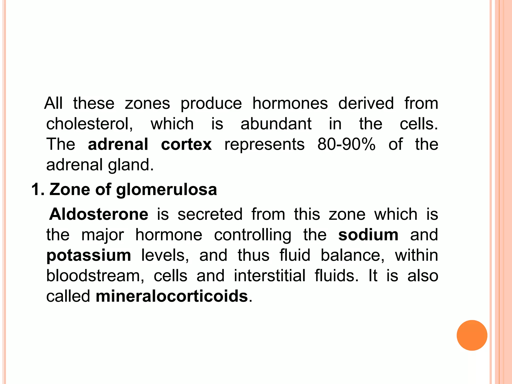
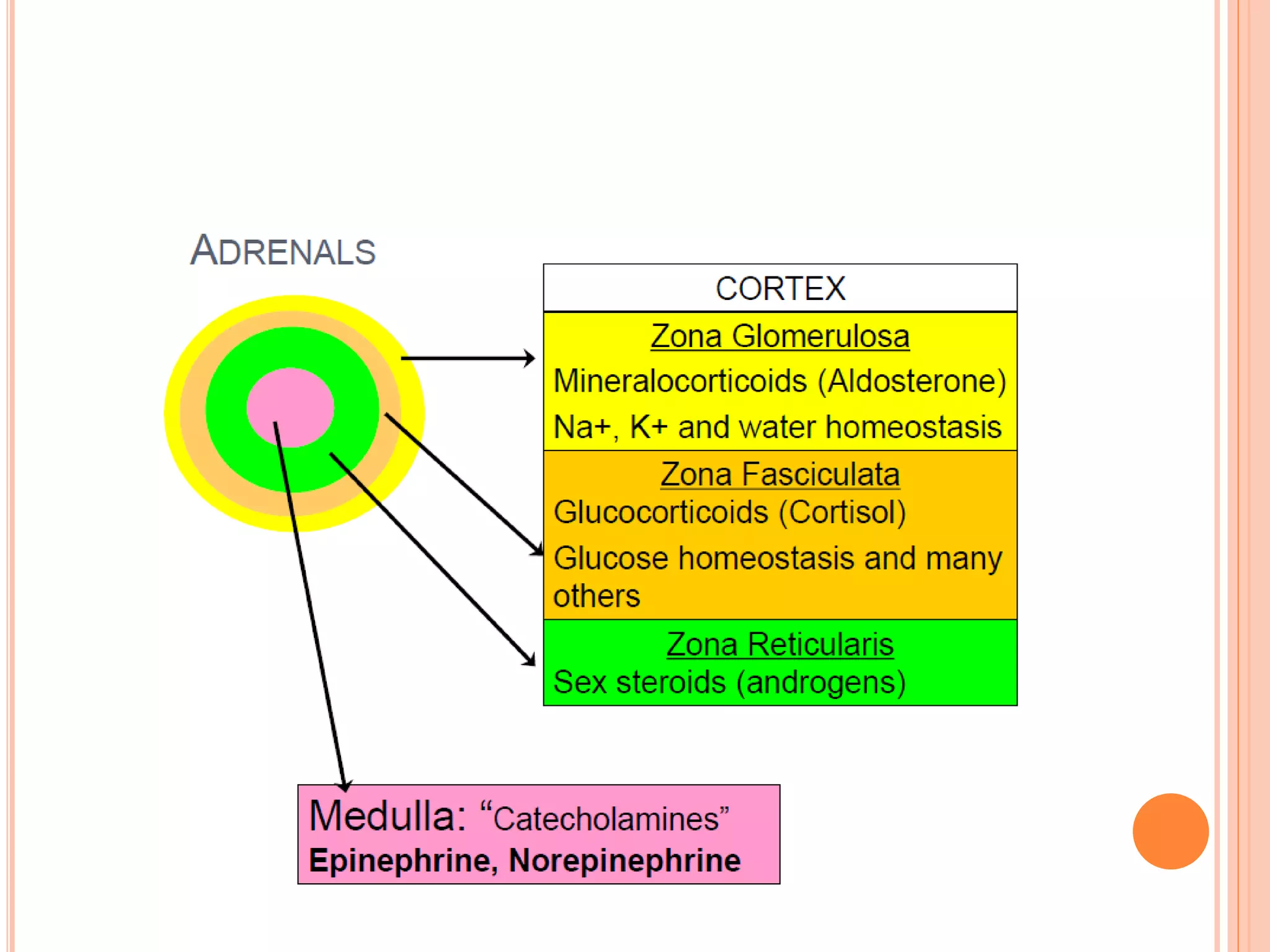
ADRENALS - CHEMISTRY AND
BIOSYNTHESIS OF ADRENAL
MEDULLARY AND ADRENAL CORTICAL
HORMONES
Dr. ACHLA JAIN
GUEST LECTURER
SOS IN LIFE SCIENCES
PT. RAVISHANKAR SHUKLA UNIVERSITY,
RAIPUR
ADRENAL GLANDS
The adrenals are orange-colored glands that sit on top of
the kidneys near the spine, just underneath the last rib
and extending down about an inch.
The glands consist of two layers;
the cortex and medulla.
The adrenal cortex is composed of three zones.
From the outer to inner, the layers are;
1. Zona glomerulosa
2. Zona fasciculata
3. Zona reticularis
All these zones produce hormones derived from
cholesterol, which is abundant in the cells.
The adrenal cortex represents 80-90% of the
adrenal gland.
1. Zone of glomerulosa
Aldosterone is secreted from this zone which is
the major hormone controlling the sodium and
potassium levels, and thus fluid balance, within
bloodstream, cells and interstitial fluids. It is also
called mineralocorticoids.
2. Zone of fasciculata
Coritsol (hydrocortisone) is produced, affects glucose,
amino acid and fat metabolism, which is called
glucocorticoids. Cells of this zone are arranged into
fascicles separated by venous sinuses.
3. Zone of reticularis
Dehydroepiandrosterone (DHEA)-precursor for
androgen is synthesized.
1. SYNTHESIS OF STEROID HORMONES
i. Cholesterol is first acted upon by desmolase and a 6-carbon
unit is cleaved off, forming the 21 carbon steroid,
pregnenolone. It is a common precursor for all the steroid
hormones. ACTH stimulates this step. This is the rate limiting
step for synthesis of all steroid hormones.
ii. Progesterone is the first steroid hormone
formed from pregnenolone in two steps. The beta hydroxyl
group is converted to a keto group by a 3-beta-ol-
dehydrogenase and the Δ5 double bond shifted to Δ4.
iii. Progesterone is further converted into glucocorticoids,
mineralocorticoids and sex steroids .
iv. The major adrenal glucocorticoids are cortisol,
cortisone and corticosterone in that order. The
major mineralocorticoid is aldosterone, but 11- deoxy-
corticosterone and corticosterone also have significant
mineralocorticoid activity.
v. These reactions are effected by hydroxylation.
These specific hydroxylases are monooxygenases.
All these enzymes are NADPH dependent.
vi. ACTH stimulates the synthesis of all steroid
hormones by activating desmolase so that the
availability of pregnenolone is increased.
2. SECRETION OF ADRENAL HORMONES
i. Secretion of all adrenocortical hormones is under the control
of ACTH.
ii. The diurnal variation of secretion of cortisol (highest
values early in the morning and minimum at night) parallels
the pulsatile release of ACTH from anterior pituitary under
the influence of CRF.
iii. Cortisol exerts the negative feedback effect on ACTH
secretion.
iv. ACTH increases the secretion of aldosterone.
v. The level of aldosterone is affected by position, highest
values in upright posture and lowest while lying down.
vi. All steroid hormones act through intracellular
messengers and increase the rate of transcription.
3. TRANSPORT AND METABOLISM
i. Approximately 70% of cortisol in blood is bound to an
alpha-1-globulin called cortisol binding globulin
(CBG) or transcortin.
ii. About 20% is bound to albumin and the rest is free,
which is the biologically active fraction. The half-life of
cortisol is about 2 hours.
iii.The steroid hormones are metabolized and inactivated
by the liver. The major processes are reduction and
conjugation.
iv. The C21 steroids are reduced to their tetrahydro
derivatives, which are excreted as their glucuronides or
sulfates in urine.
THE ADRENAL MEDULLA
The functional unit of the adrenal medulla is the chromaffin
cell, which functions as a neuroendocrine cell. In response
to stimulation, chromaffin cells secrete the hormones
epinephrine (adrenaline) and norepinephrine
(noradrenalin) directly into the blood.
The medulla is involved in extreme stress, epinephrine
and norepinephrine both work with cortisol from the adrenal
cortex. Epinephrine and norepinephrine are important
mainly in crisis situations. The adrenal medulla is primarily
involved in the production of catecholamines; epinephrine
and norepinephrine.
In fetal life, the adrenal medulla plays a role in
the autonomic nervous system. The medulla acts
as a sympathetic ganglion with the
postganglionic cells lacking axons. Through
sympathetic preganglionic fiber stimulation, the
medullary cells secrete catecholamines. The
adrenal medulla represents only 10-20% of the
adrenal gland.
 noradrenaline 20% 1.2 - 3.4 nmol/l
- 5 - 6 fold increase exerts biological action
 adrenaline 80% 0.1 - 0.8 nmol/l
2 fold increase can elicit physiological response
- These hormones prepare the body to deal with short- term
stress.
CATECHOLAMINES BIOSYNTHESIS
1. Tyrosine is precursor for the synthesis of
catecholamines.
2. The catecholamine are produced in response to
fight, fright and flight (3F). These include
emergencies like shock, cold, fatigue, emotional
condition like anger.
BIOCHEMICAL FUNCTION OF CATECHOLAMINE
1. Effect on carbohydrate metabolism: Both of them can
increase glycogenolysis and gluconeogenesis and decrease
glycogenesis.
 Catecholamine promote the release of glucose from liver and
 Decrease its utilization by muscle; Epinepherine inhibits
insulin secretion but promote glucagon secretion.
2. Effect on lipid metabolism: Both of them enhance the
breakdown of TAG in adipose tissue. This cause increase in
the free fatty acid in the circulation which are effectively
utilized by the heart and muscle as fuel source.
3. Effect on physiological function: Catecholamines
increase cardiac output, blood pressure and oxygen
consumption. They cause smooth muscle relaxation in
bronchi, GIT and blood vessels supplying skeletal muscle.
Adrenals   chemistry and biosynthesis of adrenal medullary and adrenal cortical hormones

Adrenals chemistry and biosynthesis of adrenal medullary and adrenal cortical hormones

  • 1.
    ADRENALS - CHEMISTRYAND BIOSYNTHESIS OF ADRENAL MEDULLARY AND ADRENAL CORTICAL HORMONES Dr. ACHLA JAIN GUEST LECTURER SOS IN LIFE SCIENCES PT. RAVISHANKAR SHUKLA UNIVERSITY, RAIPUR
  • 2.
    ADRENAL GLANDS The adrenalsare orange-colored glands that sit on top of the kidneys near the spine, just underneath the last rib and extending down about an inch.
  • 3.
    The glands consistof two layers; the cortex and medulla. The adrenal cortex is composed of three zones. From the outer to inner, the layers are; 1. Zona glomerulosa 2. Zona fasciculata 3. Zona reticularis
  • 4.
    All these zonesproduce hormones derived from cholesterol, which is abundant in the cells. The adrenal cortex represents 80-90% of the adrenal gland. 1. Zone of glomerulosa Aldosterone is secreted from this zone which is the major hormone controlling the sodium and potassium levels, and thus fluid balance, within bloodstream, cells and interstitial fluids. It is also called mineralocorticoids.
  • 5.
    2. Zone offasciculata Coritsol (hydrocortisone) is produced, affects glucose, amino acid and fat metabolism, which is called glucocorticoids. Cells of this zone are arranged into fascicles separated by venous sinuses. 3. Zone of reticularis Dehydroepiandrosterone (DHEA)-precursor for androgen is synthesized.
  • 7.
    1. SYNTHESIS OFSTEROID HORMONES i. Cholesterol is first acted upon by desmolase and a 6-carbon unit is cleaved off, forming the 21 carbon steroid, pregnenolone. It is a common precursor for all the steroid hormones. ACTH stimulates this step. This is the rate limiting step for synthesis of all steroid hormones. ii. Progesterone is the first steroid hormone formed from pregnenolone in two steps. The beta hydroxyl group is converted to a keto group by a 3-beta-ol- dehydrogenase and the Δ5 double bond shifted to Δ4. iii. Progesterone is further converted into glucocorticoids, mineralocorticoids and sex steroids .
  • 11.
    iv. The majoradrenal glucocorticoids are cortisol, cortisone and corticosterone in that order. The major mineralocorticoid is aldosterone, but 11- deoxy- corticosterone and corticosterone also have significant mineralocorticoid activity. v. These reactions are effected by hydroxylation. These specific hydroxylases are monooxygenases. All these enzymes are NADPH dependent. vi. ACTH stimulates the synthesis of all steroid hormones by activating desmolase so that the availability of pregnenolone is increased.
  • 13.
    2. SECRETION OFADRENAL HORMONES i. Secretion of all adrenocortical hormones is under the control of ACTH. ii. The diurnal variation of secretion of cortisol (highest values early in the morning and minimum at night) parallels the pulsatile release of ACTH from anterior pituitary under the influence of CRF. iii. Cortisol exerts the negative feedback effect on ACTH secretion. iv. ACTH increases the secretion of aldosterone. v. The level of aldosterone is affected by position, highest values in upright posture and lowest while lying down. vi. All steroid hormones act through intracellular messengers and increase the rate of transcription.
  • 14.
    3. TRANSPORT ANDMETABOLISM i. Approximately 70% of cortisol in blood is bound to an alpha-1-globulin called cortisol binding globulin (CBG) or transcortin. ii. About 20% is bound to albumin and the rest is free, which is the biologically active fraction. The half-life of cortisol is about 2 hours. iii.The steroid hormones are metabolized and inactivated by the liver. The major processes are reduction and conjugation. iv. The C21 steroids are reduced to their tetrahydro derivatives, which are excreted as their glucuronides or sulfates in urine.
  • 15.
    THE ADRENAL MEDULLA Thefunctional unit of the adrenal medulla is the chromaffin cell, which functions as a neuroendocrine cell. In response to stimulation, chromaffin cells secrete the hormones epinephrine (adrenaline) and norepinephrine (noradrenalin) directly into the blood. The medulla is involved in extreme stress, epinephrine and norepinephrine both work with cortisol from the adrenal cortex. Epinephrine and norepinephrine are important mainly in crisis situations. The adrenal medulla is primarily involved in the production of catecholamines; epinephrine and norepinephrine.
  • 16.
    In fetal life,the adrenal medulla plays a role in the autonomic nervous system. The medulla acts as a sympathetic ganglion with the postganglionic cells lacking axons. Through sympathetic preganglionic fiber stimulation, the medullary cells secrete catecholamines. The adrenal medulla represents only 10-20% of the adrenal gland.
  • 17.
     noradrenaline 20%1.2 - 3.4 nmol/l - 5 - 6 fold increase exerts biological action  adrenaline 80% 0.1 - 0.8 nmol/l 2 fold increase can elicit physiological response - These hormones prepare the body to deal with short- term stress.
  • 18.
    CATECHOLAMINES BIOSYNTHESIS 1. Tyrosineis precursor for the synthesis of catecholamines. 2. The catecholamine are produced in response to fight, fright and flight (3F). These include emergencies like shock, cold, fatigue, emotional condition like anger.
  • 20.
    BIOCHEMICAL FUNCTION OFCATECHOLAMINE 1. Effect on carbohydrate metabolism: Both of them can increase glycogenolysis and gluconeogenesis and decrease glycogenesis.  Catecholamine promote the release of glucose from liver and  Decrease its utilization by muscle; Epinepherine inhibits insulin secretion but promote glucagon secretion. 2. Effect on lipid metabolism: Both of them enhance the breakdown of TAG in adipose tissue. This cause increase in the free fatty acid in the circulation which are effectively utilized by the heart and muscle as fuel source. 3. Effect on physiological function: Catecholamines increase cardiac output, blood pressure and oxygen consumption. They cause smooth muscle relaxation in bronchi, GIT and blood vessels supplying skeletal muscle.