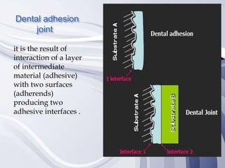
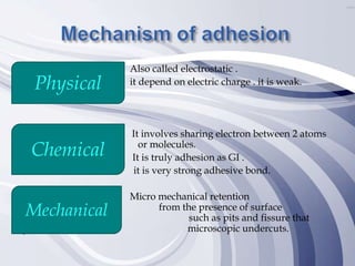
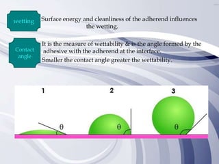
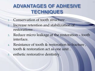
Adhesion in
Dentistry
 Introduction.
 Mechanism of adhesion.
 Factors affecting adhesion.
 Chemical Adhesion
 Micro-Mechanical adhesion
- Adhesion to enamel.
- Adhesion to dentin.
 Advantages of adhesion.
 Effect of treatment timing of the enamel and
dentin surfaces on shear bond strength.
 Definition :-
oAdhesion is derived from the Latin word
ADHEARERE which is composed of AD=
to & hearere = STICK, IT is the mutual
attraction between unlike molecules that
causes them to cling to one another.
oAdhesive /adherent is the material which
added to produce adhesion.
oAdherend is the surface to which adhesive
applied.
Dental adhesion
joint
it is the result of
interaction of a layer
of intermediate
material (adhesive)
with two surfaces
(adherends)
producing two
adhesive interfaces .

Adhesion started in 1950 with Dr. Oscar
Hugger ,then in 1955with Dr. Michael
Buonocore who is regarded as The Father Of
Adhesive Dentistry as he identified the
micromechanical adhesion to enamel via its
acid etching .
Also called electrostatic .
it depend on electric charge . it is weak.
It involves sharing electron between 2 atoms
or molecules.
It is truly adhesion as GI .
it is very strong adhesive bond.
Micro mechanical retention
results from the presence of surface
irregularities, such as pits and fissure that
give rise to microscopic undercuts.
Physical
Chemical
Mechanical
Surface energy Surface tension wetting
viscosity cleanliness
Surface energy
Surface tension
Surface energy and cleanliness of the adherend influences
the wetting.
It is the measure of wettability & is the angle formed by the
adhesive with the adherend at the interface.
Smaller the contact angle greater the wettability.
wetting
Contact
angle
GICs have good adhesive relations with tooth substrates, uniquely chemically
bonding to dentine and, to a lesser extend, to enamel. During initial dissolution,
both the glass particles and the hydroxyapatite structure are affected, and thus as
the acid is buffered the matrix reforms, chemically welded together at the
interface into a calcium phosphate polyalkenoate bond.
Chemical Adhesion
Micro Mechanical Adhesion
 Adhesion to enamel is easy and very simple than
adhesion to dentin.
 Hence it is composed of 96% inorganic content mainly
hydroxyapatite .
 Acid etching removes about 10 µm of enamel surface
and creates a microporous layer from 5-50 µm deep
which is infiltrated by the bonding agent creating the
adhesive Joint.
 Clinically:
Selective demineralization :-
How ????
Chemical
Acid
Physical
laser
Mechanical
Micro
abrasion
1. Kind of acid used. PHOSPHORIC ACID Most
commonly used.
2. Acid concentration. 30%: 40% average 37%
3. Etching time . 15 : 20 sec.
4. Form of etchant . gel /semi gel /solution
semi gel is preferably – better control.
1) Remove smear layer .
2) It converts smooth enamel to irregular surface
with microspores.
3) Increases surface area .
4) Increases the surface energy .
When adhesive resin infiltrates the etched enamel two types of resin
tags are subsequently formed ;
1- Macro-tags at the prism peripheries with the itched interprismatic
substance.
2- Micro-tags at the core of enamel with the intraprism etched material.
Micro-tags contribute more to the bonding strength due to their greater
quantity and their larger surface area.
Bonding to dentin, on the other hand, presents a much greater
challenge. Severe factors account for this difference between
enamel and dentin bonding.
1. Heterogeneous composition :- 70% inorganic
materials & high percentage of water and organic material .
2. Complex histological structure :- it is composed of
 Dentinal tubules .
 Intertubular dentin .
 Peritubular dentin .
the dentinal tubules diverge from the DEJ to pulp ,so no. &
diameter of it increases at pulpal dentin .
3. Intrapulpal fluid pressure :- Movement of fluid from
the pulp to the DEJ is a result of a slight but constant pulpal
pressure. Pulpal pressure has a magnitude of 25-30 mm of Hg .
4. Dentin is a dynamic tissue :- presence of more than one
type of dentin present challenges in dentin bonding .
 Normal dentin .
 Secondary dentin .
 Tertiary dentin .
5. Smear layer :- formed after cutting of tooth structure with
thickness of 2:5 microns which contain :
o Chips.
o Hydroxyapatite crystals .
o Saliva .
o Collagen .
o Blood .
o microorganism .
It fills the orifices of dentinal tubules forming smear plugs , so causing
of a weak joint , but it decreases dentinal permeability and
hypersensitivity .
Clinically:-
 Removal of smear layer to allow bonding
directly with dentin .
 Creation of microporous .
 Demineralization of peritubular dentin which
lead to increase diameter of opening of
dentinal tubules .
 Rinsing for removal of acid remnants
 the same time of etching .
 Dryness is a very sensitive step .
Two technique :-
Dry bond technique
•Using air tip .
Wet bond technique
•Using cotton pellet or minisponge .
 Primers are bonding-promoters monomer molecules
in organic solvents such as ethanol or acetone .
 Primers contain solvents to displace the water and
carry the monomers into the microporosities in the
collagen network. During application of the primer,
most of the solvent evaporates quickly.
 Requirements for an ideal primer
1. Should be hydrophilic .
2. Should contain hydrophobic part .
3. Should have low viscosity for better diffusion.
4. Should posses minimum film thickness for
better wettability.
5. Should be biocompatible .
6. Should posses high bond strength to both E.D.
7. Should have good shelf life .
8. Should minimize microleakage to
nanoleakage .
When the adhesive resin infiltrates the primed dentin it
fills up the inter-fibrillar spaces left between the collagen
fibrils leading to hybridization with dentin .
It also joins with the demineralized peritubular dentin
walls, the adhesive also enters the open dentinal tubules
forming resin tags in the bonding process .
- It was found that inter-tubular hybridization is more
important in the adhesive joint than the resin tags due to
its large surface area in the superficial dentin, but when
getting closer to the pulp the diameter of the dentinal
tubues increases so resin tags are more important there.
 Classification of adhesives is mandatory to provide an
overview of the current field. The classification that best
characterize adhesive systems was proposed by Van
Meerbeek et al.
 In 2003, as it considers the interaction mode with the
substrate and the number of clinical application steps.
Nowadays, two main adhesion strategies are identified:
etch-and-rinse adhesive systems, which remove the smear
layer and self-etching adhesive systems, which dissolve
and incorporate the smear layer.
 A new group of materials called universal or multi-mode
adhesive systems has recently been marketed. The main
feature of these systems is that they can be applied
according to different adhesion strategies
Category
Technique
(generation)
Etch Priming Adhesive
Etch-and-rinse
(ER)
Three steps (4th
generation)
Phosphoric acid
32–40%
Primer Adhesive resin
Two steps (5th
generation)
Phosphoric acid
32–40%
Primer + adhesive resin
Self-etch (SE)
Two steps (6th
generation)
Acidic monomers + primer Adhesive resin
One step (7th
generation)
Acidic monomers + primer
+ adhesive resin
Universal (U)
ER 2 steps
Phosphoric acid
32–40%
(E+D)
Acidic monomers + primer
+ adhesive resin (E+D)
SE 1 step
Acidic monomers + primer
+ adhesive resin(E+D)
Selective etching
enamel + SE
dentin
Phosphoric acid
32–40% (E)
Acidic monomers + primer
+ adhesive resin(E+D)
1. Conservation of tooth structure .
2. Increase retention and stabilization of
restorations .
3. Reduce micro leakage at the restoration – tooth
interface.
4. Resistance of tooth & restoration to fracture ,
tooth & restoration act as one unit .
5. esthetic restorative dentistry.
A study examined the effect of different enamel and dentin conditioning
times on the shear bond strength of a resin composite using etchand-rinse
and self-etch adhesive systems.
Shear bond strengths were determined following treatment of flat ground
human enamel and dentin surfaces (4000 grit) with 11 adhesive systems: 1)
AdheSE One Viva Pen–(ASE), 2) Adper Prompt LPop–(PLP), 3) Adper Single
Bond Plus–(SBP), 4) Clearfil SE Bond–(CSE), 5) Clearfil S3 Bond–(CS3), 6)
OptiBond All-In-One–(OBA), 7) OptiBond Solo Plus–(OBS), 8) Peak SE–
(PSE), 9) Xeno IV–(X4), 10) Xeno V–(X5) and 11) XP Bond–(XPB) using
recommended treatment times and an extended treatment time of 60 seconds
(n=10/group).
Composite (Z100) to enamel and dentin bond strengths (24 hours) were
determined using Ultradent fixtures and debonded with a crosshead speed
of 1 mm/minute. The data were analyzed with a three-way Analysis of
Variance (ANOVA) and Fisher’s LSD post hoc test.
 The highest shear bond strengths (MPa) to enamel were achieved by
the three etch-and-rinse systems at both the recommended treatment
time (SBP–40.5 ± 6.1; XPB–38.7 ± 3.7; OBS– 35.2 ± 6.2) and the extended
treatment time (SBP–44.5 ± 8.1; XPB–40.9 ± 5.7; OBS–35.0 ± 4.5).
 Extending the enamel treatment time did not produce a significant
change (p>0.05) in bond strength for the 11 adhesive systems tested.
OBS generated the high- est (46.2 ± 7.9) bond strengths to dentin at the
recommended treatment time. At the extended treatment time X4 (42.2 ±
11.7), PSE (42.1 ± 9.7) and OBS (41.4 ± 8.0) produced the highest bond
strengths to dentin.
- The bond strength change between recommend and extended
treatment times was significant PSE, but the other 10 systems did not
exhibit any significant change.

- Materials Science » "Adhesives – Applications and
Properties", book edited by Anna Rudawska, ISBN
978-953-51-2784-0, Print ISBN 978-953-51-2783-3,
Published: November 23, 2016 under CC BY 3.0 license
- Operative dentistry Book of Cairo university 2011
2012 edition – Chapter 9 (Introduction to adhesion )
- www.en.wikipedia.org/wiki/Glass_ionomer_cement
- Adhesive Bond Strengths to Enamel and Dentin Using
Recommended and Extended Treatment Times NS
Kimmes • WW Barkmeier RL Erickson • MA Latta
©Operative Dentistry, 2010, 35-1, 112-119.
Adhesion Presentation.pptx

Adhesion Presentation.pptx

  • 1.
  • 3.
     Introduction.  Mechanismof adhesion.  Factors affecting adhesion.  Chemical Adhesion  Micro-Mechanical adhesion - Adhesion to enamel. - Adhesion to dentin.  Advantages of adhesion.  Effect of treatment timing of the enamel and dentin surfaces on shear bond strength.
  • 4.
     Definition :- oAdhesionis derived from the Latin word ADHEARERE which is composed of AD= to & hearere = STICK, IT is the mutual attraction between unlike molecules that causes them to cling to one another. oAdhesive /adherent is the material which added to produce adhesion. oAdherend is the surface to which adhesive applied.
  • 5.
    Dental adhesion joint it isthe result of interaction of a layer of intermediate material (adhesive) with two surfaces (adherends) producing two adhesive interfaces .
  • 6.
     Adhesion started in1950 with Dr. Oscar Hugger ,then in 1955with Dr. Michael Buonocore who is regarded as The Father Of Adhesive Dentistry as he identified the micromechanical adhesion to enamel via its acid etching .
  • 7.
    Also called electrostatic. it depend on electric charge . it is weak. It involves sharing electron between 2 atoms or molecules. It is truly adhesion as GI . it is very strong adhesive bond. Micro mechanical retention results from the presence of surface irregularities, such as pits and fissure that give rise to microscopic undercuts. Physical Chemical Mechanical
  • 8.
    Surface energy Surfacetension wetting viscosity cleanliness
  • 9.
  • 10.
    Surface energy andcleanliness of the adherend influences the wetting. It is the measure of wettability & is the angle formed by the adhesive with the adherend at the interface. Smaller the contact angle greater the wettability. wetting Contact angle
  • 13.
    GICs have goodadhesive relations with tooth substrates, uniquely chemically bonding to dentine and, to a lesser extend, to enamel. During initial dissolution, both the glass particles and the hydroxyapatite structure are affected, and thus as the acid is buffered the matrix reforms, chemically welded together at the interface into a calcium phosphate polyalkenoate bond. Chemical Adhesion
  • 14.
  • 15.
     Adhesion toenamel is easy and very simple than adhesion to dentin.  Hence it is composed of 96% inorganic content mainly hydroxyapatite .  Acid etching removes about 10 µm of enamel surface and creates a microporous layer from 5-50 µm deep which is infiltrated by the bonding agent creating the adhesive Joint.
  • 16.
     Clinically: Selective demineralization:- How ???? Chemical Acid Physical laser Mechanical Micro abrasion
  • 19.
    1. Kind ofacid used. PHOSPHORIC ACID Most commonly used. 2. Acid concentration. 30%: 40% average 37% 3. Etching time . 15 : 20 sec. 4. Form of etchant . gel /semi gel /solution semi gel is preferably – better control.
  • 21.
    1) Remove smearlayer . 2) It converts smooth enamel to irregular surface with microspores. 3) Increases surface area . 4) Increases the surface energy .
  • 22.
    When adhesive resininfiltrates the etched enamel two types of resin tags are subsequently formed ; 1- Macro-tags at the prism peripheries with the itched interprismatic substance. 2- Micro-tags at the core of enamel with the intraprism etched material. Micro-tags contribute more to the bonding strength due to their greater quantity and their larger surface area.
  • 23.
    Bonding to dentin,on the other hand, presents a much greater challenge. Severe factors account for this difference between enamel and dentin bonding.
  • 26.
    1. Heterogeneous composition:- 70% inorganic materials & high percentage of water and organic material . 2. Complex histological structure :- it is composed of  Dentinal tubules .  Intertubular dentin .  Peritubular dentin . the dentinal tubules diverge from the DEJ to pulp ,so no. & diameter of it increases at pulpal dentin . 3. Intrapulpal fluid pressure :- Movement of fluid from the pulp to the DEJ is a result of a slight but constant pulpal pressure. Pulpal pressure has a magnitude of 25-30 mm of Hg . 4. Dentin is a dynamic tissue :- presence of more than one type of dentin present challenges in dentin bonding .  Normal dentin .  Secondary dentin .  Tertiary dentin .
  • 27.
    5. Smear layer:- formed after cutting of tooth structure with thickness of 2:5 microns which contain : o Chips. o Hydroxyapatite crystals . o Saliva . o Collagen . o Blood . o microorganism . It fills the orifices of dentinal tubules forming smear plugs , so causing of a weak joint , but it decreases dentinal permeability and hypersensitivity .
  • 29.
  • 30.
     Removal ofsmear layer to allow bonding directly with dentin .  Creation of microporous .  Demineralization of peritubular dentin which lead to increase diameter of opening of dentinal tubules .
  • 31.
     Rinsing forremoval of acid remnants  the same time of etching .  Dryness is a very sensitive step . Two technique :- Dry bond technique •Using air tip . Wet bond technique •Using cotton pellet or minisponge .
  • 33.
     Primers arebonding-promoters monomer molecules in organic solvents such as ethanol or acetone .  Primers contain solvents to displace the water and carry the monomers into the microporosities in the collagen network. During application of the primer, most of the solvent evaporates quickly.
  • 34.
     Requirements foran ideal primer 1. Should be hydrophilic . 2. Should contain hydrophobic part . 3. Should have low viscosity for better diffusion. 4. Should posses minimum film thickness for better wettability. 5. Should be biocompatible . 6. Should posses high bond strength to both E.D. 7. Should have good shelf life . 8. Should minimize microleakage to nanoleakage .
  • 35.
    When the adhesiveresin infiltrates the primed dentin it fills up the inter-fibrillar spaces left between the collagen fibrils leading to hybridization with dentin . It also joins with the demineralized peritubular dentin walls, the adhesive also enters the open dentinal tubules forming resin tags in the bonding process . - It was found that inter-tubular hybridization is more important in the adhesive joint than the resin tags due to its large surface area in the superficial dentin, but when getting closer to the pulp the diameter of the dentinal tubues increases so resin tags are more important there.
  • 37.
     Classification ofadhesives is mandatory to provide an overview of the current field. The classification that best characterize adhesive systems was proposed by Van Meerbeek et al.  In 2003, as it considers the interaction mode with the substrate and the number of clinical application steps. Nowadays, two main adhesion strategies are identified: etch-and-rinse adhesive systems, which remove the smear layer and self-etching adhesive systems, which dissolve and incorporate the smear layer.  A new group of materials called universal or multi-mode adhesive systems has recently been marketed. The main feature of these systems is that they can be applied according to different adhesion strategies
  • 38.
    Category Technique (generation) Etch Priming Adhesive Etch-and-rinse (ER) Threesteps (4th generation) Phosphoric acid 32–40% Primer Adhesive resin Two steps (5th generation) Phosphoric acid 32–40% Primer + adhesive resin Self-etch (SE) Two steps (6th generation) Acidic monomers + primer Adhesive resin One step (7th generation) Acidic monomers + primer + adhesive resin Universal (U) ER 2 steps Phosphoric acid 32–40% (E+D) Acidic monomers + primer + adhesive resin (E+D) SE 1 step Acidic monomers + primer + adhesive resin(E+D) Selective etching enamel + SE dentin Phosphoric acid 32–40% (E) Acidic monomers + primer + adhesive resin(E+D)
  • 40.
    1. Conservation oftooth structure . 2. Increase retention and stabilization of restorations . 3. Reduce micro leakage at the restoration – tooth interface. 4. Resistance of tooth & restoration to fracture , tooth & restoration act as one unit . 5. esthetic restorative dentistry.
  • 41.
    A study examinedthe effect of different enamel and dentin conditioning times on the shear bond strength of a resin composite using etchand-rinse and self-etch adhesive systems. Shear bond strengths were determined following treatment of flat ground human enamel and dentin surfaces (4000 grit) with 11 adhesive systems: 1) AdheSE One Viva Pen–(ASE), 2) Adper Prompt LPop–(PLP), 3) Adper Single Bond Plus–(SBP), 4) Clearfil SE Bond–(CSE), 5) Clearfil S3 Bond–(CS3), 6) OptiBond All-In-One–(OBA), 7) OptiBond Solo Plus–(OBS), 8) Peak SE– (PSE), 9) Xeno IV–(X4), 10) Xeno V–(X5) and 11) XP Bond–(XPB) using recommended treatment times and an extended treatment time of 60 seconds (n=10/group). Composite (Z100) to enamel and dentin bond strengths (24 hours) were determined using Ultradent fixtures and debonded with a crosshead speed of 1 mm/minute. The data were analyzed with a three-way Analysis of Variance (ANOVA) and Fisher’s LSD post hoc test.
  • 42.
     The highestshear bond strengths (MPa) to enamel were achieved by the three etch-and-rinse systems at both the recommended treatment time (SBP–40.5 ± 6.1; XPB–38.7 ± 3.7; OBS– 35.2 ± 6.2) and the extended treatment time (SBP–44.5 ± 8.1; XPB–40.9 ± 5.7; OBS–35.0 ± 4.5).  Extending the enamel treatment time did not produce a significant change (p>0.05) in bond strength for the 11 adhesive systems tested. OBS generated the high- est (46.2 ± 7.9) bond strengths to dentin at the recommended treatment time. At the extended treatment time X4 (42.2 ± 11.7), PSE (42.1 ± 9.7) and OBS (41.4 ± 8.0) produced the highest bond strengths to dentin. - The bond strength change between recommend and extended treatment times was significant PSE, but the other 10 systems did not exhibit any significant change.
  • 43.
     - Materials Science» "Adhesives – Applications and Properties", book edited by Anna Rudawska, ISBN 978-953-51-2784-0, Print ISBN 978-953-51-2783-3, Published: November 23, 2016 under CC BY 3.0 license - Operative dentistry Book of Cairo university 2011 2012 edition – Chapter 9 (Introduction to adhesion ) - www.en.wikipedia.org/wiki/Glass_ionomer_cement - Adhesive Bond Strengths to Enamel and Dentin Using Recommended and Extended Treatment Times NS Kimmes • WW Barkmeier RL Erickson • MA Latta ©Operative Dentistry, 2010, 35-1, 112-119.