Download to read offline

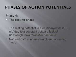
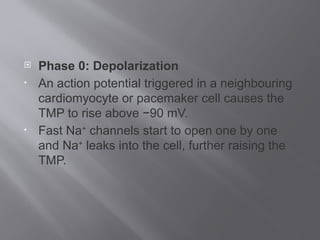
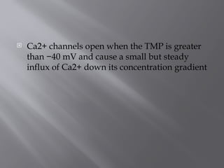
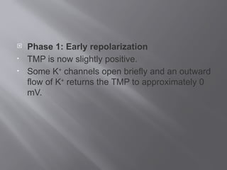
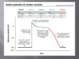


The document discusses action potentials in cardiomyocytes, focusing on the electrical stimulation generated by ion fluxes across cell membranes. It outlines the phases of action potential, including resting potential, depolarization, early repolarization, plateau phase, and repolarization, detailing the roles of various ions like Na+, Ca2+, and K+ during these phases. The process is crucial for maintaining ionic gradients and preparing the cell for subsequent depolarization cycles.


![Hypothalamus short ppt by Dr. Neha [PT].pptx](https://cdn.slidesharecdn.com/ss_thumbnails/hypothalamusbydr-260124145759-b9f94a93-thumbnail.jpg?width=640&height=640&fit=bounds)


