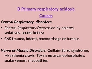
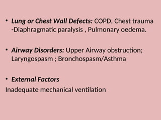
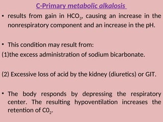
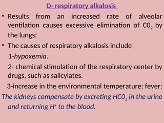
Acid-Base balance
&
Acid-Base disturbances
By
Dr: Sanaa nassar
An acid
is a substance that can yield a hydrogen ion (H+
) when
dissolved in water.
A base
is a substance that can yield hydroxyl ions (OH")
when dissolved in water. .
ACID-BASE BALANCE
Maintenance of H+
 The normal concentration of H+
in the extracellular body fluid
ranges from 36 to 44 nmol/L (pH 7.34 to pH 7.44).
 through metabolism, the body produces much greater
quantities of H+
.
 Any H+
value outside this range will cause alterations in the
rates of chemical reactions within the cell and affect the many
metabolic processes of the body and lead to alterations in
consciousness, tetany, coma, and death.
The reference value for arterial blood pH is 7.40 and
is equivalent to an H+
concentration of 40 nmol/L.
pH is the negative log of the H+
, so increase in H+
concentration decreases the pH, while a decrease in
H+
concentration increases the pH.
systems that regulate The arterial pH by production
and retention of acids and bases include: buffers,
the respiratory center and lungs, and the kidneys.
1
-
Buffer Systems: Regulation of H
+
• The first line of defense against changes in H+
concentration is the
buffer systems present in all body fluids. it is a system that resists
changes in pH.
• All buffers consist of a weak acid, and its salt , such as carbonic acid
(H2C03), bicarbonate (NaHC03), for the bicarbonate-carbonic acid
buffer system.
• The bicarbonate buffering system is referred to as an open system
(can be changed), and the dissolved CO2 (dC02) , which is controlled by
the lungs, is the respiratory component
the bicarbonate-carbonic acid system has low
buffering capacity, but it is an important buffer
because:
(1) H2C03 dissociates into C02 and H20, allowing C02
to be eliminated by the lungs and disposing of
H+
as water.
(2)changes in C02 modify the (respiratory) rate.
(3) HC03 concentration can be altered by the
kidneys.
Other buffer systems
 The phosphate buffer system
Plasma protein,
The lungs and kidneys
• The lungs regulate pH through retention or elimination
of CO2, by changing the rate and volume of ventilation.
• The kidneys regulate pH by
1- excreting acid, primarily in the ammonium ion
2- reabsorbing HC03
-
from the glomerular filtrate.
In the lungs
 The lungs, provide the first defense to changes in acid-base status.
 Inspired 02 diffuses from the alveoli into the blood and bind to
hemoglobin, forming oxyhemoglobin (02Hb).
 The H+
carried on the (reduced) hemoglobin in the venous blood is
released to recombine HC03 forming H2CO3, which dissociates into
H20 and C02.
 The C02 diffuses into the alveoli and is eliminated through
ventilation.
 When the lungs do not remove C02 , it accumulates in the blood,
causing an increase in H+
concentration.
 If CO2 removal is faster than production (hyperventilation), the H+
concentration will be decreased. Consequently, ventilation affects
the pH of the blood.
The kidneys
 The kidneys main role is to reabsorb HCO3 ( mainly in the
proximal tubules) from the glomerular filtrate.
 sodium (Na+
) in the glomerular filtrate is exchanged for H+
in the tubular cell. The H+
combines with HCO3 in the
filtrate to form H2CO3, which is converted into H2O and C02
by carbonic anhydrase.
 The CO2 easily diffuses into the tubule and reacts with H2O
to reform H2CO3 and then HCO3, which is reabsorbed into
the blood along with sodium.
Acid-Base Disorders: Acidosis and Alkalosis
acidemia,
a blood pH is less than the reference range (<7.34), it
reflects excess acid or H+
concentration.
alkalemia
A pH greater than the reference range (>7.44), it reflects
excess base.
primary respiratory acidosis or alkalosis
Any disorder caused by ventilatory dysfunction (a change
in the PCO2, the respiratory component) .
metabolic acidosis or alkalosis
A disorder resulting from a change in the bicarbonate level
(a renal or metabolic function).
Mixed respiratory and nonrespiratory disorders
arise from more than one pathologic process and represent a
serious of medical conditions as compensation for the
primary disorder is failing.
compensation
• Is the body trials to restore acid-base homeostasis
whenever an imbalance occurs, because the body's cellular
and metabolic activities are pH dependent,
• Compensation may successfully return the ratio to the
normal, but the primary abnormality is not corrected.
• the body accomplishes this by altering the factor not primarily
affected by the pathologic process.
Example:
• if the imbalance is of metabolic origin, the body compensates by
altering ventilation.
• For disturbances of the respiratory component, the kidneys
compensate by selectively excreting or reabsorbing anions and
cations.
• The lungs can compensate immediately, but the response is short
term and incomplete, while the kidneys are slower to respond (2-4
days), but the response is long term and complete.
• Fully compensated implies that the pH has returned to the normal
range, while partially compensated implies that the pH is
approaching normal.
A- (metabolic) acidosis
mechanisms
1- direct administration of an acid-producing substance, or by
excessive formation of organic acids as seen with diabetic
ketoacidosis and starvation.
2- reduced excretion of acids, as in renal tubular acidosis.
3- excessive loss of bicarbonate from diarrhea .
• The body compensates through hyperventilation, which is
an increase in the rate or depth of breathing. By "blowing
off CO2, the base-to-acid ratio will return toward normal.
metabolic acidosis
Causes
1- Diabetic ketoacidosis
2- Alcoholic ketoacidosis
3- Starvation ketoacidosis
4- Toxins : Ethylene glycol, Methanol, salicylates
5- Renal Causes : Renal Failure, Renal tubular acidosis
6- GIT Causes: Severe diarrhoea
B-Primary respiratory acidosis
Mechanism
A- Results from a decrease in alveolar
ventilation (hypoventilation), causing a
decreased elimination of C02 by the lungs.
B- Over-production of Carbon Dioxide.
C- Increased Intake of Carbon Dioxide
B-Primary respiratory acidosis
Causes
Central Respiratory disorders:
• Central Respiratory Depression by opiates,
sedatives, anaesthetics)
• CNS trauma, infarct, haemorrhage or tumour
Nerve or Muscle Disorders: Guillain-Barre syndrome,
Myasthenia gravis, Toxins eg organophosphates,
snake venom, myopathies
• Lung or Chest Wall Defects: COPD, Chest trauma
-Diaphragmatic paralysis , Pulmonary oedema.
• Airway Disorders: Upper Airway obstruction;
Laryngospasm ; Bronchospasm/Asthma
• External Factors
Inadequate mechanical ventilation
C-Primary metabolic alkalosis
• results from gain in HCO3, causing an increase in the
nonrespiratory component and an increase in the pH.
• This condition may result from:
(1)the excess administration of sodium bicarbonate.
(2) Excessive loss of acid by the kidney (diuretics) or GIT.
• The body responds by depressing the respiratory
center. The resulting hypoventilation increases the
retention of C02.
D- respiratory alkalosis
• Results from an increased rate of alveolar
ventilation causes excessive elimination of C02 by
the lungs:
• The causes of respiratory alkalosis include
1-hypoxemia.
2- chemical stimulation of the respiratory center by
drugs, such as salicylates.
3-increase in the environmental temperature; fever;
The kidneys compensate by excreting HC03 in the urine
and returning H+
to the blood.
causes
1. Central Causes: Head Injury; Stroke, drugs, Various
endogenous compounds (eg progesterone during
pregnancy, cytokines during sepsis)
2. Hypoxaemia: Respiratory stimulation via peripheral
chemoreceptors
3. Pulmonary Causes: Pneumonia, Asthma, Pulmonary
edema
4. Iatrogenic (act directly on ventilation) Excessive
controlled ventilation

acid base,PH,buffer edited by sanaa .ppt

  • 1.
  • 2.
    An acid is asubstance that can yield a hydrogen ion (H+ ) when dissolved in water. A base is a substance that can yield hydroxyl ions (OH") when dissolved in water. .
  • 3.
    ACID-BASE BALANCE Maintenance ofH+  The normal concentration of H+ in the extracellular body fluid ranges from 36 to 44 nmol/L (pH 7.34 to pH 7.44).  through metabolism, the body produces much greater quantities of H+ .  Any H+ value outside this range will cause alterations in the rates of chemical reactions within the cell and affect the many metabolic processes of the body and lead to alterations in consciousness, tetany, coma, and death.
  • 4.
    The reference valuefor arterial blood pH is 7.40 and is equivalent to an H+ concentration of 40 nmol/L. pH is the negative log of the H+ , so increase in H+ concentration decreases the pH, while a decrease in H+ concentration increases the pH. systems that regulate The arterial pH by production and retention of acids and bases include: buffers, the respiratory center and lungs, and the kidneys.
  • 5.
    1 - Buffer Systems: Regulationof H + • The first line of defense against changes in H+ concentration is the buffer systems present in all body fluids. it is a system that resists changes in pH. • All buffers consist of a weak acid, and its salt , such as carbonic acid (H2C03), bicarbonate (NaHC03), for the bicarbonate-carbonic acid buffer system. • The bicarbonate buffering system is referred to as an open system (can be changed), and the dissolved CO2 (dC02) , which is controlled by the lungs, is the respiratory component
  • 6.
    the bicarbonate-carbonic acidsystem has low buffering capacity, but it is an important buffer because: (1) H2C03 dissociates into C02 and H20, allowing C02 to be eliminated by the lungs and disposing of H+ as water. (2)changes in C02 modify the (respiratory) rate. (3) HC03 concentration can be altered by the kidneys.
  • 7.
    Other buffer systems The phosphate buffer system Plasma protein,
  • 8.
    The lungs andkidneys • The lungs regulate pH through retention or elimination of CO2, by changing the rate and volume of ventilation. • The kidneys regulate pH by 1- excreting acid, primarily in the ammonium ion 2- reabsorbing HC03 - from the glomerular filtrate.
  • 9.
    In the lungs The lungs, provide the first defense to changes in acid-base status.  Inspired 02 diffuses from the alveoli into the blood and bind to hemoglobin, forming oxyhemoglobin (02Hb).  The H+ carried on the (reduced) hemoglobin in the venous blood is released to recombine HC03 forming H2CO3, which dissociates into H20 and C02.  The C02 diffuses into the alveoli and is eliminated through ventilation.  When the lungs do not remove C02 , it accumulates in the blood, causing an increase in H+ concentration.  If CO2 removal is faster than production (hyperventilation), the H+ concentration will be decreased. Consequently, ventilation affects the pH of the blood.
  • 10.
    The kidneys  Thekidneys main role is to reabsorb HCO3 ( mainly in the proximal tubules) from the glomerular filtrate.  sodium (Na+ ) in the glomerular filtrate is exchanged for H+ in the tubular cell. The H+ combines with HCO3 in the filtrate to form H2CO3, which is converted into H2O and C02 by carbonic anhydrase.  The CO2 easily diffuses into the tubule and reacts with H2O to reform H2CO3 and then HCO3, which is reabsorbed into the blood along with sodium.
  • 11.
    Acid-Base Disorders: Acidosisand Alkalosis acidemia, a blood pH is less than the reference range (<7.34), it reflects excess acid or H+ concentration. alkalemia A pH greater than the reference range (>7.44), it reflects excess base. primary respiratory acidosis or alkalosis Any disorder caused by ventilatory dysfunction (a change in the PCO2, the respiratory component) . metabolic acidosis or alkalosis A disorder resulting from a change in the bicarbonate level (a renal or metabolic function).
  • 12.
    Mixed respiratory andnonrespiratory disorders arise from more than one pathologic process and represent a serious of medical conditions as compensation for the primary disorder is failing. compensation • Is the body trials to restore acid-base homeostasis whenever an imbalance occurs, because the body's cellular and metabolic activities are pH dependent, • Compensation may successfully return the ratio to the normal, but the primary abnormality is not corrected.
  • 13.
    • the bodyaccomplishes this by altering the factor not primarily affected by the pathologic process. Example: • if the imbalance is of metabolic origin, the body compensates by altering ventilation. • For disturbances of the respiratory component, the kidneys compensate by selectively excreting or reabsorbing anions and cations. • The lungs can compensate immediately, but the response is short term and incomplete, while the kidneys are slower to respond (2-4 days), but the response is long term and complete. • Fully compensated implies that the pH has returned to the normal range, while partially compensated implies that the pH is approaching normal.
  • 14.
    A- (metabolic) acidosis mechanisms 1-direct administration of an acid-producing substance, or by excessive formation of organic acids as seen with diabetic ketoacidosis and starvation. 2- reduced excretion of acids, as in renal tubular acidosis. 3- excessive loss of bicarbonate from diarrhea . • The body compensates through hyperventilation, which is an increase in the rate or depth of breathing. By "blowing off CO2, the base-to-acid ratio will return toward normal.
  • 15.
    metabolic acidosis Causes 1- Diabeticketoacidosis 2- Alcoholic ketoacidosis 3- Starvation ketoacidosis 4- Toxins : Ethylene glycol, Methanol, salicylates 5- Renal Causes : Renal Failure, Renal tubular acidosis 6- GIT Causes: Severe diarrhoea
  • 16.
    B-Primary respiratory acidosis Mechanism A-Results from a decrease in alveolar ventilation (hypoventilation), causing a decreased elimination of C02 by the lungs. B- Over-production of Carbon Dioxide. C- Increased Intake of Carbon Dioxide
  • 17.
    B-Primary respiratory acidosis Causes CentralRespiratory disorders: • Central Respiratory Depression by opiates, sedatives, anaesthetics) • CNS trauma, infarct, haemorrhage or tumour Nerve or Muscle Disorders: Guillain-Barre syndrome, Myasthenia gravis, Toxins eg organophosphates, snake venom, myopathies
  • 18.
    • Lung orChest Wall Defects: COPD, Chest trauma -Diaphragmatic paralysis , Pulmonary oedema. • Airway Disorders: Upper Airway obstruction; Laryngospasm ; Bronchospasm/Asthma • External Factors Inadequate mechanical ventilation
  • 19.
    C-Primary metabolic alkalosis •results from gain in HCO3, causing an increase in the nonrespiratory component and an increase in the pH. • This condition may result from: (1)the excess administration of sodium bicarbonate. (2) Excessive loss of acid by the kidney (diuretics) or GIT. • The body responds by depressing the respiratory center. The resulting hypoventilation increases the retention of C02.
  • 20.
    D- respiratory alkalosis •Results from an increased rate of alveolar ventilation causes excessive elimination of C02 by the lungs: • The causes of respiratory alkalosis include 1-hypoxemia. 2- chemical stimulation of the respiratory center by drugs, such as salicylates. 3-increase in the environmental temperature; fever; The kidneys compensate by excreting HC03 in the urine and returning H+ to the blood.
  • 21.
    causes 1. Central Causes:Head Injury; Stroke, drugs, Various endogenous compounds (eg progesterone during pregnancy, cytokines during sepsis) 2. Hypoxaemia: Respiratory stimulation via peripheral chemoreceptors 3. Pulmonary Causes: Pneumonia, Asthma, Pulmonary edema 4. Iatrogenic (act directly on ventilation) Excessive controlled ventilation