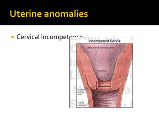
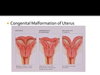
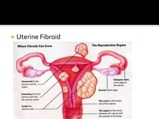
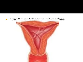
Presented by
Sinmayee devi ,
Associate professor
l.j.m con , bhubaneswar
Odisha
 Abortion: It is the expulsion or extraction
from its mother of an embryo or fetus
weighing 500 gm or less when it is not
capable of independent survival (WHO).
 The 500gm of fetal development is attained
approximately at 22 weeks of gestation.
 Expelled fetus- Abortus
ABORTION
Spontaneous
Induced
Spontaneous Abortion
Missed Threatened Incomplete Inevitable Complete Septic
Induced
Legal
Illegal
 Spontaneous: If Abortion takes place on its
own, without use of any medical or
mechanical means , it is known as
Spontaneous Abortion or miscarriage.
 Induced Abortion: If it takes place with the
use of medical or mechanical means in an
attempt to empty the uterus than it is known
as InducedAbortion.
Pregnancy
Miscarriage
10-20%
Illegal abortions
10%
ABORTION
<16th
week(75%)
<8th
week(75%)
8-16 weeks
(25%)>16th week
(25%)
Abortion
Fetal Causes
Maternal
Causes
Environmental
Causes
• Trisomy 16-most common
(30%)
AutosomalTrisomy
(50%)
• Presence of 3 or more haploid
number of chromosomes.
Polyploidy
22%
• Commonest- Monosomy 45 XMonosomy
(20%)
• Translocation, deletion,
inversion
Structural Chromosomal
Rearrangements(2-4%)
• Mosaic, DoubleTrisomy
Others(4%)
 Uterine anomolies
 Medical conditions
 Immunological causes
 Endocrinologic causes
 Cervical Incompetence
 Congenital Malformation of Uterus
 Uterine Fibroid
 Intra UterineAdhesions or Synechiae
 Cyanotic heart disease,
 hemoglobinopathies .
 Luteal Phase Defect (LPD)
 Deficient progesterone.
 Thyroid abnormalities (overt)
 Diabetes mellitus (poorly controlled) are
associated with increased fetal loss.

 Autoimmune Diseases-Anti nuclear
antibodies.Anti phospholipid antibodies-
lupus anticoagulants
 Alloimmune Diseases- Lack of maternal
blocking antibodies
 Infections
 Smoking
 Alcohol
 Radiation
 Toxins
 In spite of the numerous factors mentioned,
it is indeed difficult , in the majority, to
pinpoint the exact cause of abortion.
However, risk of abortion increases with
increased maternal age. Number of previous
abortions and the etiology are also
important.

• First death of ovum ->expulsion
• Ovum with villi, decidual coverings
expelled intact
<8weeks
• Fetus expels out leaving behind
placenta and membranes.
8-14
weeks
• Mini labour>14 weeks
 It is a clinical entity where the process of
abortion has started but has not progressed
to a state from which recovery is impossible.

 Bleeding perVaginam: light and bright red in
colour. On rare occasion the bleeding may be
brisk and sharp, specially in the late 2nd
trimester, suggestive of low implantation of
placenta.The bleeding usually stops
spontaneously .
 Pain: Bleeding is usually painless but there
may be mild backache or dull pain in lower
abdomen. Pain appears usually following
haemorrhage.
 Speculum examination reveals – bleeding, if
any, escapes through the external os. Any
local lesion in the cervix may co-exist .
 Digital examination reveals the closed
external os. The uterine size corresponds to
the period of amenorrhea.The uterus and
cervix feel soft. Pelvic examination is avoided
when ultrasonography is available .
 Routine Investigations
 Ultrasonography (TVS) findings may be :
A well formed gestation ring with central echoes
from the embryo indicating healthy fetus.
Observation of fetal cardiac motion With this
there is 98% chance of continuation of pregnancy.
 Serum Progesterone –Value of 25ng/ml or
more generally indicates a viable pregnancy
in about 95% cases .
 Serial serum chorionic gonadotrophin (hCG)
level is helpful to assess the fetal well being.
 Rest: until bleeding stops.
 Drugs: Sedation and relief of pain may be
ensured by phenobarbitone 30mg or
diazepam 5mg tablet twice daily.
 General Measures:
preserve the vulval pads and anything expelled
out per vaginam , for inspection .
To report if bleeding and/or pain becomes
aggravated.
Routine note of pulse, temperature and vaginal
 Advice on Discharge:
 The patient should limit her activities for
atleast 2 weeks and avoid heavy work.
 Coitus is contraindicated during this period
 She should be re- examined after one month
to assess the growth of the fetus.
 The prognosis is very unpredictable.
 In isolated spontaneous threatened abortion,
the following events may occur:
 In about 2/3rd , the pregnancy continues
beyond 28 weeks .
 In the rest, it terminates either in inevitable or
missed abortion. If the pregnancy continues ,
there is increased frequency of preterm
labour, placenta previa, intra uterine growth
retardation of the fetus and fetal anomalies.
 It is the clinical type of abortion where the
changes have progressed to a state from
where continuation of pregnancy is
impossible.
 The patient , having the features of
threatened abortion, develops the following
manifestations.
1. Increased vaginal bleeding .
2. Aggravation of pain in the lower abdomen
which may be colicky in nature.
3. The general condition of the patient is
proportionate to the visible blood loss
4. Internal examination reveals dilated internal
os of the cervix through which the products
 On occasion, the features may develop
quickly without prior clinical evidence of
threatened abortion.
 In the second trimester, however , it may
start with rupture of the membranes or
intermittent lower abdominal pain (mini
labour).
 To take appropriate measures to look after
the general condition .
 To accelerate the process of expulsion.
 To maintain strict asepsis as in conduction of
labor.
 Excessive Bleeding should promptly be
controlled by administering Oxytocin if the
cervix is dilated and the size of the uterus is
less than 12 weeks.
 The shock is corrected by intravenous fluid
therapy and blood transfusion .
 Before 12 weeks :
 1) Dilatation and evacuation followed by
curettage of the uterine cavity by blunt
curette under general anesthesia .
 2) Alternatively , suction evacuation followed
by curettage is done.
 The uterine contraction is accelerated by
oxytocin drip (10units in 500ml of normal
saline) 40-60 drops per minute. If the fetus is
expelled and the placenta is retained, it is
removed by ovum forceps, if lying separated.
If the placenta is not separated, digital
separation followed by its evacuation is to be
done under general anesthesia.
 If the bleeding is profuse with the cervix
closed (suggestive of low implantation of
placenta)- evacuation of the uterus may have
 When the products of conception are
expelled in mass, it is called complete
abortion.
 There is history of expulsion of a fleshy mass
per vaginam followed by :
 Abdominal pain.
 Vaginal bleeding becomes trace or absent.
 Uterus is smaller than the period of
amenorrhhea and a little firmer.
 Cervical os is closed.
 Bleeding is trace .
 Examination of the expelled fleshy mass is
found intact.
 The effect of blood loss, if any, should be
assessed and treated .
 If there is doubt about complete expulsion of
the products, uterine curettage should be
done.
 Transvaginal sonography is useful to prevent
unnecessary surgical procedure.
 When the entire products of conception are
not expelled , instead a part of it is left inside
the uterine cavity, it is called incomplete
abortion.
 This is the commonest type met amongst
women , hospitalized for abortion
complications.
 History of expulsion of a fleshy mass per
vaginam followed by:
 Continuation of pain lower abdomen , colicky
in nature, although in diminished magnitude.
 Persistence of bleeding of varying
magnitude.
 Uterus smaller than the period of
amenorrhea
 Patulous cervical os often admitting tip of the
finger
 Varying amount of bleeding
 On examination , the expelled mass is found
incomplete.
 The products left behind may lead to – a) Profuse
bleeding , b) sepsis ,c) placenta polyp and d) rarely
choriocarcinoma
 Patient may be in a state of shock due to blood loss.
She should be resuscitated before any active
treatment is undertaken.
 Early Abortion: Dilatation and evacuation under
general anesthesia is to be done.
 Late Abortion:The uterus is evacuated under GA and
the products are removed by ovum forceps or by blunt
curette.
 In late cases, dilatation and curettage operation is to
be done to remove the bits of tissues left behind.The
removed materials are subjected to a histological
examination.
 When the fetus is dead and retained inside
the uterus for a variable period, it is called
missed abortion or silent miscarriage or early
fetal demise.
 The patient usually presents with features of
threatened abortion followed by:
 Persistence of brownish vaginal discharge .
 Subsidence of pregnancy symptoms
 Retrogression of breast changes .
 Cessation of uterine growth which in fact becomes
smaller in size.
 Non audibility of the fetal heart sound even with
Doppler cardioscope if it had been audible before.
 Cervix feels firm.
 Immunological test for pregnancy becomes negative.
 Real time ultrasonography reveals an empty sac early
in the pregnancy or the absence of fetal motion or
fetal heart movement later in the pregnancy.
 Psychological Upset. Associated with it, there
may be malaise.
 Infection
 Uterus is less than 12 weeks:Vaginal
evacuation can be carried out without delay .
This can be effectively done by suction
evacuation or slow dilatation of the cervix by
laminaria tent followed by dilatation and
evacuation (D & E) of the uterus under
general anesthesia.
 The risk of damage to the uterine walls and
brisk haemorrhage during the operation
should be kept in mind.
 Induction is done by following methods:
 Oxytocin –
To start with 10-20 units of oxytocin in 500ml of
normal saline at 30 drops per minute .
If fails, escalating dose of oxytocin to the
maximum of 200 mIU/min. ,may be used with
monitoring.
 Prostaglandins are more effective than
oxytocin in such cases .The methods used
are-
 Prostaglandin E1 analogue (misoprostol)
200µg tablet is inserted into the posterior
vaginal fornix every 4 hrs. for a maximum of 5
such.
 I.M. administration of 15 methyl PGF2α 250µg
at three hourly intervals for a maximum of 10
such.
 Any abortion associated with clinical
evidences of infection of the uterus and its
contents, is called septic abortion.
 Although clinical criteria vary, abortion is
usually considered septic when there are:
1. Rise of temperature of at least 100.4F (38C)
for 24 hrs or more
2. Offensive or purulent vaginal discharge
3. Other evidences of pelvic infection such as
lower abdominal pain and tenderness.
 About 10% of abortions requiring admission
to hospital are septic.
 The majority of septic abortion are associated
with incomplete abortion.
 While in the majority of cases the infection
occurs following illegal induced abortion but
infection can occur even after spontaneous
abortion.
 Proper antiseptic and asepsis are not taken
 Incomplete evacuation
 Inadvertent injury to the genital organs and
adjacent structures , particularly the gut.
 Anaerobic - Bacteriodes group (fragilis),
anaerobic streptococci , Cl welchii, and
tetanus bacillus
 Aerobic - Escherichia coli , Klebsiella ,
Staphylococcus, Pseudomonas and
haemolytic Streptococcus.
 Mixed infection is more common.
 Pyrexia . Associated with chills and
rigors
 Pain abdomen of varying degrees is
almost a constant feature.
 A rising pulse rate of 100-120/min or
more is a significant finding than even
pyrexia. It indicates spread of infection
beyond the uterus.
 Internal examination reveals offensive
purulent vaginal discharge or a tender
uterus usually with patulous os
 Grade 1 :The infection is localised in the uterus
 Grade 2 :The infection spreads beyond the
uterus to the parametrium , tubes and ovaries or
pelvic peritoneum
 Grade 3 : Generalised peritonitis and /or
endotoxic shock or jaundice or acute renal
failure.
 Grade 1 is the commonest and is usually
associated with spontaneous abortion . Grade 3
is almost always associated with illegal induced
 Cervical or high vaginal swab is taken prior to
internal examination for-
1. culture in aerobic and anaerobic media to
find out the dominant micro organisms
2. sensitivity of the micro organisms to
antibiotics
3. smear for Gram stain
 Blood for haemoglobin estimation, total and
differential count of white cells, ABO and Rh
grouping
 Ultrasonography pelvis and abdomen to
detect intrauterine retained products of
conception , foreign body- intrauterine or
intra-abdominal , free fluid in the peritoneal
cavity or in the pouch of Douglas
 Blood - Culture
 Early : haemorrhage , injury, spread of
infection
 Remote: chronic pelvic pain, backache,
dyspareunia, ectopic pregnancy, scondary
infertility, depression.
 Hospitalization and isolation.
 To take high vaginal or cervical swab for
culture , drug sensitivity test and Gram Stain.
 Vaginal examination is done to note the state
of the abortion process and extension of the
infection. If the products are found loosely
lying in the cervix, it is removed by an ovum
forceps.
 Overall assessment & clinical grading.
 Investigation protocols
1. To control sepsis
2. To remove the source of infection
3. To give supportive therapy to bring back the
normal homeostatic and cellular
metabolism.
4. To assess the response of treatment.
 For Gram Positive aerobes-
 Aqueous penicillinG-5 milllion I.V. every 6
hours or
 Ampicillin 0.5-1 Gm. I.V. every 6 hrs.
 For Gram Negative aerobes-
 Gentamicin 1.5mg/kg I.V. every 8hrs
 Cefatriaxone 1G , I.V. every 12 hours
 For Anaerobes- Metonidazole 500mg I.V.
every 8 hrs , or clindamycin 600mg I.V. every
6 hrs
 Antibiotic regimens have to be modified
according to the culture and sensitivity report
as obtained later on.
 Prophylactic Anti gas - gangrene serum of
8000 units and 3000 units of antitetanus
serum i.m. are given if there is a history of
interference.
 Analgesic and sedatives , as required , are to
be prescribed.
 BloodTransfusion is given to improve anaemia
and body resistance.
 Evacuation of the uterus: As abortion is often
incomplete, evacuation should be performed at
a convenient time within 24 hrs following
antibiotic therapy . Excessive bleeding is, of
course, an urgent indication for evacuation .
Early emptying not only minimises the risk of
haemorrhage but also removes the nidus of
infection.The procedure should be gentle to
avoid injury to the uterus.
 Clinical Monitoring
 Surgery:
 Evacuation of the uterus- Should be withheld for
atleast 48hrs when the infection is controlled
and is localised , the only exception being
excessive bleeding.
 Posterior colpotomy-When the infection is
localised in the pouch of Douglas pelvic abscess
is formed. It is evidenced by spiky rise of
termperature, rectal tenesmus (frequent loose
stool mixed with mucus ) and boggy mass felt
through the posterior fornix. Posterior
colpotomy and drainage of the pus relieve the
 Supportive therapy is directed to
generalised peritonitis by gastric suction and
i.v. saline infusion.
 Management for endotoxic shock or renal
failure , if present, is to be conducted. Patient
may need intensive care unit management.
 Injury to the uterus.
 Suspected injury to bowel
 Presence of foreign body in the abdomen as
evidenced by the sonography or X-ray or felt
through the fornix on bimanual examination.
 Unresponsive peritonitis suggestive of
collection of pus .
 Septic Shock or oliguria not responding to the
conservative treatment.
 Uterus too big to be safely evacuated per
 Laprotomy should be done by experienced
surgeon with a skilled anaesthetist . Removal
of the uterus should be done irrespective of
parity . Adnexa is to b e removed or preserved
according to the pathology found.Thorough
inspection of the gut and omentum for
evidence of any injury is mandatory . Even
when nothing is found on laportomy, simple
drainage of the pus is effective.
 Recurrent miscarriage is defined as a
sequence of three or more consecutive
spontaneous abortion before 20 weeks
.Some however, consider two or more as a
standard.
 It may be primary or secondary (having
previous viable birth).
 Treated with natural micronized
progesterone 100mg daily as vaginal
suppository. Started 2 days after ovulation. If
the period is missed and pregnancy is
confirmed, progesterone supplementation is
continued till 10-12 weeks gestation.
 HCG therapy is thought to improve the
pregnancy outcome in LPD . It stimulates
corpus luteum to produce progesterone.
However benefits of both hormonal
 Treated with low dose Aspirin (50mg/day), or
low dose aspirin and heparin (5000 units SC
twice daily) upto 30 weeks or Prednisone (40-
50mg/day). I.V. immunoglobulin is also used.
 CerclageOperation
 Cervical cerclage (tracheloplasty), also
known as a cervical stitch, is used for the
treatment of cervical incompetence (or
insufficiency).
 Two types of operation are in current use
during pregnancy each claiming an equal
success rate of about 80% .The operations
are named after Shirodkar (1955) and Mc
Donald (1957).
 A non- absorbable encircling suture is placed
around the cervix at the level of internal os. It
operates by interfering with the uterine
polarity, preventing the internal os and the
adjacent lower segment from being "taken
up"
 In a proven case the operation should be
done around 14 weeks of pregnancy or at
least 2 wks earlier than the lower period of
previous wastage, as early as the 10th week.
 The patient is put under light general
anesthesia and placed in lithotomy position
with good exposure of the cervix by a
posterior vaginal speculum.The lips of the
cervix are pulled down by sponge holding
forceps or allis tissue forceps.
 A transverse incision is given anteriorly below
the base of the bladder on the vaginal wall
and the bladder is pushed up to expose the
level of the internal os . A vertical incision is
given posteriorly on the cervico-vaginal
 The non absorbable suture material-No. 4
braided nylon or Mersilence (Dacron) is
passed submucously with the help of an
aneurysm needle or cervical needle so as to
bring the suture ends through the posterior
incision.
 The ends of the suture are tied up posteriorly
by a reef knot. the bulging membranes , if
present , must be reduced beforehand in to
the uterine cavity .The anterior and posterior
incisions are repaired by interrupted stitches
 The non absorbable suture material is placed
as a purse string suture as high as possible at
the junction of the rugose vaginal epithelium
and the smooth vaginal part of the cervix
below the level of the bladder.
 The suture starts at the anterior wall of the
cervix.Taking successive deep bites (4-5 sites)
it is carried around the lateral and posterior
walls back to the anterior wall again where
the two ends of the suture are tied.
 The operation is simple having less blood
loss, and has got a good success rate.There is
less formation of cervical scar and hence less
chance of cervical dystocia during labour.
 Post Operative:The patient should be in bed
for at least 2-3 days . Isoxsuprine (tocolytics)
10ms tablet is given thrice daily to avoid
uterine irritability.
 Removal of Stitches:The stitch should be
removed at 38th week or earlier if labour pain
starts or features of abortion appear.If the
stitch is not cut in time, uterine rupture or
cervical tear may occur. If the stitch is cut
prior to the onset of labour, it is preferable to
cut it in operation theatre as there is
increased chance of cord prolapse specially in
the cases with floating head.

Abortion sin

  • 1.
    Presented by Sinmayee devi, Associate professor l.j.m con , bhubaneswar Odisha
  • 3.
     Abortion: Itis the expulsion or extraction from its mother of an embryo or fetus weighing 500 gm or less when it is not capable of independent survival (WHO).  The 500gm of fetal development is attained approximately at 22 weeks of gestation.  Expelled fetus- Abortus
  • 4.
  • 5.
    Spontaneous Abortion Missed ThreatenedIncomplete Inevitable Complete Septic
  • 7.
  • 8.
     Spontaneous: IfAbortion takes place on its own, without use of any medical or mechanical means , it is known as Spontaneous Abortion or miscarriage.  Induced Abortion: If it takes place with the use of medical or mechanical means in an attempt to empty the uterus than it is known as InducedAbortion.
  • 9.
  • 10.
  • 11.
  • 12.
    • Trisomy 16-mostcommon (30%) AutosomalTrisomy (50%) • Presence of 3 or more haploid number of chromosomes. Polyploidy 22% • Commonest- Monosomy 45 XMonosomy (20%) • Translocation, deletion, inversion Structural Chromosomal Rearrangements(2-4%) • Mosaic, DoubleTrisomy Others(4%)
  • 13.
     Uterine anomolies Medical conditions  Immunological causes  Endocrinologic causes
  • 14.
  • 15.
  • 16.
  • 17.
  • 18.
     Cyanotic heartdisease,  hemoglobinopathies .
  • 19.
     Luteal PhaseDefect (LPD)  Deficient progesterone.  Thyroid abnormalities (overt)  Diabetes mellitus (poorly controlled) are associated with increased fetal loss. 
  • 20.
     Autoimmune Diseases-Antinuclear antibodies.Anti phospholipid antibodies- lupus anticoagulants  Alloimmune Diseases- Lack of maternal blocking antibodies
  • 21.
     Infections  Smoking Alcohol  Radiation  Toxins
  • 22.
     In spiteof the numerous factors mentioned, it is indeed difficult , in the majority, to pinpoint the exact cause of abortion. However, risk of abortion increases with increased maternal age. Number of previous abortions and the etiology are also important. 
  • 23.
    • First deathof ovum ->expulsion • Ovum with villi, decidual coverings expelled intact <8weeks • Fetus expels out leaving behind placenta and membranes. 8-14 weeks • Mini labour>14 weeks
  • 24.
     It isa clinical entity where the process of abortion has started but has not progressed to a state from which recovery is impossible. 
  • 25.
     Bleeding perVaginam:light and bright red in colour. On rare occasion the bleeding may be brisk and sharp, specially in the late 2nd trimester, suggestive of low implantation of placenta.The bleeding usually stops spontaneously .  Pain: Bleeding is usually painless but there may be mild backache or dull pain in lower abdomen. Pain appears usually following haemorrhage.
  • 26.
     Speculum examinationreveals – bleeding, if any, escapes through the external os. Any local lesion in the cervix may co-exist .  Digital examination reveals the closed external os. The uterine size corresponds to the period of amenorrhea.The uterus and cervix feel soft. Pelvic examination is avoided when ultrasonography is available .
  • 27.
     Routine Investigations Ultrasonography (TVS) findings may be : A well formed gestation ring with central echoes from the embryo indicating healthy fetus. Observation of fetal cardiac motion With this there is 98% chance of continuation of pregnancy.
  • 28.
     Serum Progesterone–Value of 25ng/ml or more generally indicates a viable pregnancy in about 95% cases .  Serial serum chorionic gonadotrophin (hCG) level is helpful to assess the fetal well being.
  • 29.
     Rest: untilbleeding stops.  Drugs: Sedation and relief of pain may be ensured by phenobarbitone 30mg or diazepam 5mg tablet twice daily.  General Measures: preserve the vulval pads and anything expelled out per vaginam , for inspection . To report if bleeding and/or pain becomes aggravated. Routine note of pulse, temperature and vaginal
  • 30.
     Advice onDischarge:  The patient should limit her activities for atleast 2 weeks and avoid heavy work.  Coitus is contraindicated during this period  She should be re- examined after one month to assess the growth of the fetus.
  • 31.
     The prognosisis very unpredictable.  In isolated spontaneous threatened abortion, the following events may occur:  In about 2/3rd , the pregnancy continues beyond 28 weeks .  In the rest, it terminates either in inevitable or missed abortion. If the pregnancy continues , there is increased frequency of preterm labour, placenta previa, intra uterine growth retardation of the fetus and fetal anomalies.
  • 32.
     It isthe clinical type of abortion where the changes have progressed to a state from where continuation of pregnancy is impossible.
  • 33.
     The patient, having the features of threatened abortion, develops the following manifestations. 1. Increased vaginal bleeding . 2. Aggravation of pain in the lower abdomen which may be colicky in nature. 3. The general condition of the patient is proportionate to the visible blood loss 4. Internal examination reveals dilated internal os of the cervix through which the products
  • 34.
     On occasion,the features may develop quickly without prior clinical evidence of threatened abortion.  In the second trimester, however , it may start with rupture of the membranes or intermittent lower abdominal pain (mini labour).
  • 35.
     To takeappropriate measures to look after the general condition .  To accelerate the process of expulsion.  To maintain strict asepsis as in conduction of labor.
  • 36.
     Excessive Bleedingshould promptly be controlled by administering Oxytocin if the cervix is dilated and the size of the uterus is less than 12 weeks.  The shock is corrected by intravenous fluid therapy and blood transfusion .
  • 37.
     Before 12weeks :  1) Dilatation and evacuation followed by curettage of the uterine cavity by blunt curette under general anesthesia .  2) Alternatively , suction evacuation followed by curettage is done.
  • 38.
     The uterinecontraction is accelerated by oxytocin drip (10units in 500ml of normal saline) 40-60 drops per minute. If the fetus is expelled and the placenta is retained, it is removed by ovum forceps, if lying separated. If the placenta is not separated, digital separation followed by its evacuation is to be done under general anesthesia.  If the bleeding is profuse with the cervix closed (suggestive of low implantation of placenta)- evacuation of the uterus may have
  • 39.
     When theproducts of conception are expelled in mass, it is called complete abortion.
  • 40.
     There ishistory of expulsion of a fleshy mass per vaginam followed by :  Abdominal pain.  Vaginal bleeding becomes trace or absent.
  • 41.
     Uterus issmaller than the period of amenorrhhea and a little firmer.  Cervical os is closed.  Bleeding is trace .  Examination of the expelled fleshy mass is found intact.
  • 42.
     The effectof blood loss, if any, should be assessed and treated .  If there is doubt about complete expulsion of the products, uterine curettage should be done.  Transvaginal sonography is useful to prevent unnecessary surgical procedure.
  • 43.
     When theentire products of conception are not expelled , instead a part of it is left inside the uterine cavity, it is called incomplete abortion.  This is the commonest type met amongst women , hospitalized for abortion complications.
  • 44.
     History ofexpulsion of a fleshy mass per vaginam followed by:  Continuation of pain lower abdomen , colicky in nature, although in diminished magnitude.  Persistence of bleeding of varying magnitude.
  • 45.
     Uterus smallerthan the period of amenorrhea  Patulous cervical os often admitting tip of the finger  Varying amount of bleeding  On examination , the expelled mass is found incomplete.
  • 46.
     The productsleft behind may lead to – a) Profuse bleeding , b) sepsis ,c) placenta polyp and d) rarely choriocarcinoma  Patient may be in a state of shock due to blood loss. She should be resuscitated before any active treatment is undertaken.  Early Abortion: Dilatation and evacuation under general anesthesia is to be done.  Late Abortion:The uterus is evacuated under GA and the products are removed by ovum forceps or by blunt curette.  In late cases, dilatation and curettage operation is to be done to remove the bits of tissues left behind.The removed materials are subjected to a histological examination.
  • 47.
     When thefetus is dead and retained inside the uterus for a variable period, it is called missed abortion or silent miscarriage or early fetal demise.
  • 48.
     The patientusually presents with features of threatened abortion followed by:  Persistence of brownish vaginal discharge .  Subsidence of pregnancy symptoms  Retrogression of breast changes .  Cessation of uterine growth which in fact becomes smaller in size.  Non audibility of the fetal heart sound even with Doppler cardioscope if it had been audible before.  Cervix feels firm.  Immunological test for pregnancy becomes negative.  Real time ultrasonography reveals an empty sac early in the pregnancy or the absence of fetal motion or fetal heart movement later in the pregnancy.
  • 49.
     Psychological Upset.Associated with it, there may be malaise.  Infection
  • 50.
     Uterus isless than 12 weeks:Vaginal evacuation can be carried out without delay . This can be effectively done by suction evacuation or slow dilatation of the cervix by laminaria tent followed by dilatation and evacuation (D & E) of the uterus under general anesthesia.  The risk of damage to the uterine walls and brisk haemorrhage during the operation should be kept in mind.
  • 51.
     Induction isdone by following methods:  Oxytocin – To start with 10-20 units of oxytocin in 500ml of normal saline at 30 drops per minute . If fails, escalating dose of oxytocin to the maximum of 200 mIU/min. ,may be used with monitoring.
  • 52.
     Prostaglandins aremore effective than oxytocin in such cases .The methods used are-  Prostaglandin E1 analogue (misoprostol) 200µg tablet is inserted into the posterior vaginal fornix every 4 hrs. for a maximum of 5 such.  I.M. administration of 15 methyl PGF2α 250µg at three hourly intervals for a maximum of 10 such.
  • 53.
     Any abortionassociated with clinical evidences of infection of the uterus and its contents, is called septic abortion.  Although clinical criteria vary, abortion is usually considered septic when there are: 1. Rise of temperature of at least 100.4F (38C) for 24 hrs or more 2. Offensive or purulent vaginal discharge 3. Other evidences of pelvic infection such as lower abdominal pain and tenderness.
  • 54.
     About 10%of abortions requiring admission to hospital are septic.  The majority of septic abortion are associated with incomplete abortion.  While in the majority of cases the infection occurs following illegal induced abortion but infection can occur even after spontaneous abortion.
  • 55.
     Proper antisepticand asepsis are not taken  Incomplete evacuation  Inadvertent injury to the genital organs and adjacent structures , particularly the gut.
  • 56.
     Anaerobic -Bacteriodes group (fragilis), anaerobic streptococci , Cl welchii, and tetanus bacillus  Aerobic - Escherichia coli , Klebsiella , Staphylococcus, Pseudomonas and haemolytic Streptococcus.  Mixed infection is more common.
  • 57.
     Pyrexia .Associated with chills and rigors  Pain abdomen of varying degrees is almost a constant feature.  A rising pulse rate of 100-120/min or more is a significant finding than even pyrexia. It indicates spread of infection beyond the uterus.  Internal examination reveals offensive purulent vaginal discharge or a tender uterus usually with patulous os
  • 58.
     Grade 1:The infection is localised in the uterus  Grade 2 :The infection spreads beyond the uterus to the parametrium , tubes and ovaries or pelvic peritoneum  Grade 3 : Generalised peritonitis and /or endotoxic shock or jaundice or acute renal failure.  Grade 1 is the commonest and is usually associated with spontaneous abortion . Grade 3 is almost always associated with illegal induced
  • 59.
     Cervical orhigh vaginal swab is taken prior to internal examination for- 1. culture in aerobic and anaerobic media to find out the dominant micro organisms 2. sensitivity of the micro organisms to antibiotics 3. smear for Gram stain  Blood for haemoglobin estimation, total and differential count of white cells, ABO and Rh grouping
  • 60.
     Ultrasonography pelvisand abdomen to detect intrauterine retained products of conception , foreign body- intrauterine or intra-abdominal , free fluid in the peritoneal cavity or in the pouch of Douglas  Blood - Culture
  • 61.
     Early :haemorrhage , injury, spread of infection  Remote: chronic pelvic pain, backache, dyspareunia, ectopic pregnancy, scondary infertility, depression.
  • 62.
     Hospitalization andisolation.  To take high vaginal or cervical swab for culture , drug sensitivity test and Gram Stain.  Vaginal examination is done to note the state of the abortion process and extension of the infection. If the products are found loosely lying in the cervix, it is removed by an ovum forceps.  Overall assessment & clinical grading.  Investigation protocols
  • 63.
    1. To controlsepsis 2. To remove the source of infection 3. To give supportive therapy to bring back the normal homeostatic and cellular metabolism. 4. To assess the response of treatment.
  • 64.
     For GramPositive aerobes-  Aqueous penicillinG-5 milllion I.V. every 6 hours or  Ampicillin 0.5-1 Gm. I.V. every 6 hrs.  For Gram Negative aerobes-  Gentamicin 1.5mg/kg I.V. every 8hrs  Cefatriaxone 1G , I.V. every 12 hours
  • 65.
     For Anaerobes-Metonidazole 500mg I.V. every 8 hrs , or clindamycin 600mg I.V. every 6 hrs  Antibiotic regimens have to be modified according to the culture and sensitivity report as obtained later on.
  • 66.
     Prophylactic Antigas - gangrene serum of 8000 units and 3000 units of antitetanus serum i.m. are given if there is a history of interference.  Analgesic and sedatives , as required , are to be prescribed.
  • 67.
     BloodTransfusion isgiven to improve anaemia and body resistance.  Evacuation of the uterus: As abortion is often incomplete, evacuation should be performed at a convenient time within 24 hrs following antibiotic therapy . Excessive bleeding is, of course, an urgent indication for evacuation . Early emptying not only minimises the risk of haemorrhage but also removes the nidus of infection.The procedure should be gentle to avoid injury to the uterus.
  • 68.
     Clinical Monitoring Surgery:  Evacuation of the uterus- Should be withheld for atleast 48hrs when the infection is controlled and is localised , the only exception being excessive bleeding.  Posterior colpotomy-When the infection is localised in the pouch of Douglas pelvic abscess is formed. It is evidenced by spiky rise of termperature, rectal tenesmus (frequent loose stool mixed with mucus ) and boggy mass felt through the posterior fornix. Posterior colpotomy and drainage of the pus relieve the
  • 69.
     Supportive therapyis directed to generalised peritonitis by gastric suction and i.v. saline infusion.  Management for endotoxic shock or renal failure , if present, is to be conducted. Patient may need intensive care unit management.
  • 70.
     Injury tothe uterus.  Suspected injury to bowel  Presence of foreign body in the abdomen as evidenced by the sonography or X-ray or felt through the fornix on bimanual examination.  Unresponsive peritonitis suggestive of collection of pus .  Septic Shock or oliguria not responding to the conservative treatment.  Uterus too big to be safely evacuated per
  • 71.
     Laprotomy shouldbe done by experienced surgeon with a skilled anaesthetist . Removal of the uterus should be done irrespective of parity . Adnexa is to b e removed or preserved according to the pathology found.Thorough inspection of the gut and omentum for evidence of any injury is mandatory . Even when nothing is found on laportomy, simple drainage of the pus is effective.
  • 72.
     Recurrent miscarriageis defined as a sequence of three or more consecutive spontaneous abortion before 20 weeks .Some however, consider two or more as a standard.  It may be primary or secondary (having previous viable birth).
  • 73.
     Treated withnatural micronized progesterone 100mg daily as vaginal suppository. Started 2 days after ovulation. If the period is missed and pregnancy is confirmed, progesterone supplementation is continued till 10-12 weeks gestation.  HCG therapy is thought to improve the pregnancy outcome in LPD . It stimulates corpus luteum to produce progesterone. However benefits of both hormonal
  • 74.
     Treated withlow dose Aspirin (50mg/day), or low dose aspirin and heparin (5000 units SC twice daily) upto 30 weeks or Prednisone (40- 50mg/day). I.V. immunoglobulin is also used.
  • 75.
     CerclageOperation  Cervicalcerclage (tracheloplasty), also known as a cervical stitch, is used for the treatment of cervical incompetence (or insufficiency).  Two types of operation are in current use during pregnancy each claiming an equal success rate of about 80% .The operations are named after Shirodkar (1955) and Mc Donald (1957).
  • 76.
     A non-absorbable encircling suture is placed around the cervix at the level of internal os. It operates by interfering with the uterine polarity, preventing the internal os and the adjacent lower segment from being "taken up"
  • 77.
     In aproven case the operation should be done around 14 weeks of pregnancy or at least 2 wks earlier than the lower period of previous wastage, as early as the 10th week.
  • 80.
     The patientis put under light general anesthesia and placed in lithotomy position with good exposure of the cervix by a posterior vaginal speculum.The lips of the cervix are pulled down by sponge holding forceps or allis tissue forceps.  A transverse incision is given anteriorly below the base of the bladder on the vaginal wall and the bladder is pushed up to expose the level of the internal os . A vertical incision is given posteriorly on the cervico-vaginal
  • 81.
     The nonabsorbable suture material-No. 4 braided nylon or Mersilence (Dacron) is passed submucously with the help of an aneurysm needle or cervical needle so as to bring the suture ends through the posterior incision.  The ends of the suture are tied up posteriorly by a reef knot. the bulging membranes , if present , must be reduced beforehand in to the uterine cavity .The anterior and posterior incisions are repaired by interrupted stitches
  • 82.
     The nonabsorbable suture material is placed as a purse string suture as high as possible at the junction of the rugose vaginal epithelium and the smooth vaginal part of the cervix below the level of the bladder.  The suture starts at the anterior wall of the cervix.Taking successive deep bites (4-5 sites) it is carried around the lateral and posterior walls back to the anterior wall again where the two ends of the suture are tied.
  • 83.
     The operationis simple having less blood loss, and has got a good success rate.There is less formation of cervical scar and hence less chance of cervical dystocia during labour.
  • 84.
     Post Operative:Thepatient should be in bed for at least 2-3 days . Isoxsuprine (tocolytics) 10ms tablet is given thrice daily to avoid uterine irritability.
  • 85.
     Removal ofStitches:The stitch should be removed at 38th week or earlier if labour pain starts or features of abortion appear.If the stitch is not cut in time, uterine rupture or cervical tear may occur. If the stitch is cut prior to the onset of labour, it is preferable to cut it in operation theatre as there is increased chance of cord prolapse specially in the cases with floating head.