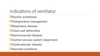
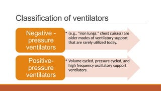
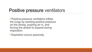
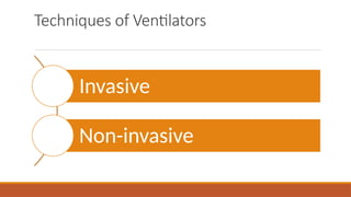
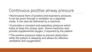
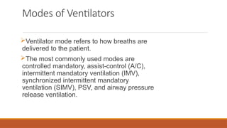
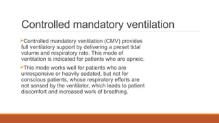
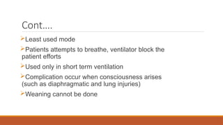
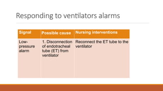
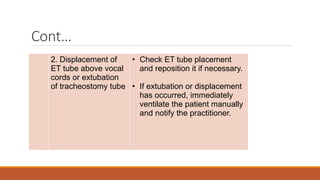
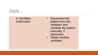
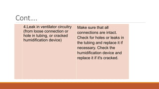
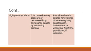
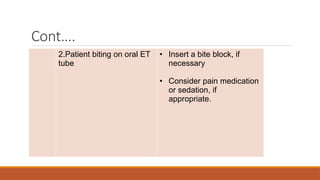
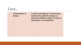
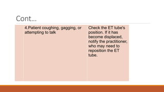
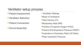
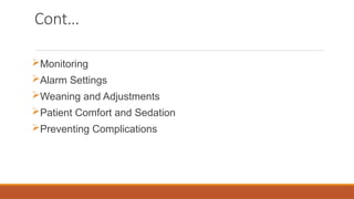
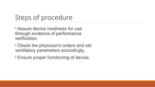
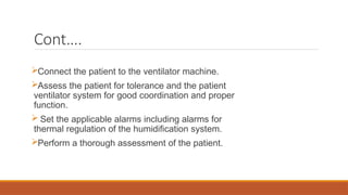
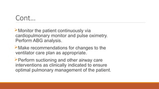
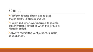
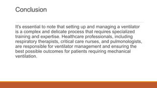
VENTILATOR SETUP
PRESENTED BY: KAJAL
M.SC. (N) 1ST
YEAR
What is ventilation
It is a method of controlling the
environment with the air, and
can be defined as movement of
air between the environment
and lung via inhalation and
exhalation.
What is ventilator
A ventilator is a medical device that
assists patients in breathing when they
are unable to do so on their own or need
support due to respiratory insufficiency.
Purposes of ventilator
 To control over the airway and over
the work of breathing.
 Replaces the work of exhausted or
temporarily inadequate respiratory
muscle.
Cont….
 The physiologic benefits of mechanical
ventilation are reversal of gas exchange
abnormalities, alteration of pressure- volume
relationships in the respiratory system, and
reduction in the work of breathing.
Indications of ventilator
Routine anesthesia
Postoperative management
Respiratory disease
Chest wall deformities
Neuromuscular disease
Central nervous system impairment
Cardiovascular disease
Neonatal conditions
Classification of ventilators
• (e.g., "iron lungs," chest cuirass) are
older modes of ventilatory support
that are rarely utilized today.
Negative -
pressure
ventilators
• Volume cycled, pressure cycled, and
high frequency oscillatory support
ventilators.
Positive-
pressure
ventilators
Positive pressure ventilators
Positive-pressure ventilators inflate
the lungs by exerting positive pressure
on the airway, pushing air in, and
forcing the alveoli to expand during
inspiration.
Expiration occurs passively.
Cont….
Three types of positive-pressure ventilators
are classified by the method of ending the
inspiratory phase of respiration.
Volume cycled ventilators
Pressure cycled ventilators
High frequency oscillatory support
ventilators
Volume cycled ventilators
Deliver a preset volume of air with each
inspiration.
From breath to breath, the volume of air
delivered by the ventilator is relatively constant,
ensuring consistent, adequate breaths despite
varying airway pressures.
Pressure cycled ventilators
Delivers a flow of air
(inspiration) until it reaches a
preset pressure, and then cycles
off, and expiration occurs.
High frequency oscillatory support ventilators
These types of ventilators deliver very high
respiratory rates (180 to 900
breaths/minute) that are accompanied by
very low tidal volumes and high airway
pressures (hence the name high-frequency
oscillatory support).
Techniques of Ventilators
Invasive
Non-invasive
Invasive
Life-saving intervention for patients with
respiratory failure.
Mechanical ventilation is indicated to support
the patient with respiratory failure when
adequate gas exchange cannot otherwise be
maintained.
Often chosen over noninvasive methods when
altered mental status or hemodynamic instability
accompany acute respiratory failure.
Non-invasive
Positive pressure ventilation can be given
via facemasks covering the nose and
mouth, nasal masks, or other nasal devices.
The most comfortable mode for the patient
is pressure-controlled ventilation with
pressure support.
Bilevel positive airway pressure
Offers independent control of inspiratory and
expiratory pressures while providing pressure
support ventilation.
Each inspiration can be initiated either by the
patient or by the machine if it is programmed with a
backup rate.
Most often used for patients who require
ventilatory assistance at night, such as those with
severe COPD or CSA.
Continuous positive airway pressure
Noninvasive form of positive end-expiratory pressure.
It can be given through a ventilator as a separate
mode. It can also be delivered by a machine.
It provides a constant end-expiratory pressure which
helps to keep the airway open. Some machines also
provide supplemental oxygen, if required by the patient.
The positive pressure helps to prevent obstruction
while the patient is sleeping and allows for effective
ventilation and oxygenation.
Modes of Ventilators
Ventilator mode refers to how breaths are
delivered to the patient.
The most commonly used modes are
controlled mandatory, assist-control (A/C),
intermittent mandatory ventilation (IMV),
synchronized intermittent mandatory
ventilation (SIMV), PSV, and airway pressure
release ventilation.
Controlled mandatory ventilation
Controlled mandatory ventilation (CMV) provides
full ventilatory support by delivering a preset tidal
volume and respiratory rate. This mode of
ventilation is indicated for patients who are apneic.
This mode works well for patients who are
unresponsive or heavily sedated, but not for
conscious patients, whose respiratory efforts are
not sensed by the ventilator, which leads to patient
discomfort and increased work of breathing.
Cont….
Least used mode
Patients attempts to breathe, ventilator block the
patient efforts
Used only in short term ventilation
Complication occur when consciousness arises
(such as diaphragmatic and lung injuries)
Weaning cannot be done
Assist control ventilation
Similar to CMV in that the ventilator will deliver
preset tidal volumes at a preset rate of respirations.
However, if the patient initiates a breath between the
machine's breaths, the ventilator delivers at the preset
volume (assisted breath). Therefore, every breath is
the preset volume.
Commonly used mode of ventilation in the intensive
care units.
Intermittent mandatory ventilation
Provides a combination of mechanically
assisted breaths and spontaneous breaths.
Mechanical breaths are delivered at preset
intervals and a preselected tidal volume,
regardless of the patient's efforts. Although the
patient can increase the respiratory rate by
initiating inspiration between ventilator-delivered
breaths, these spontaneous breaths are limited
to the tidal volume generated by the patient.
Cont….
IMV allows patients to use their
own muscles for ventilation to help
prevent muscle atrophy. It lowers
mean airway pressure, which can
assist in preventing barotrauma.
However, "fighting the ventilator” or
"bucking the ventilator may be
increased.
Synchronized intermittent mandatory
ventilation
Also delivers a preset tidal
volume and number of breaths per
minute.
Between ventilator-delivered
breaths, the patient can breathe
spontaneously with no assistance
from the ventilator on those extra
breaths.
Cont..
Because the ventilator senses patient
breathing efforts and does not initiate a breath
in opposition to the patient’s efforts fighting the
ventilator is reduced.
 As the patient ability to breathe spontaneously
increases, the preset number of ventilator
breaths is decreased and the patient does more
of the work of breathing.
Pressure-Controlled ventilation
During Pressure-Controlled ventilation,
the ventilator generates the preset
pressure during a preset inspiratory
time at the preset respiratory rate. The
pressure is constant during the
inspiratory time and the flow is
decelerating.
Pressure support ventilation (PSV)
Pressure support ventilation (PSV) applies a
pressure plateau to the airway throughout the
patient-triggered inspiration to decrease
resistance within the tracheal tube and ventilator
tubing.
Ventilators alarms
Ventilators offer audible and visual alarms
to alert the caregivers to changes in key
patient and ventilator functions and
settings. These alarms prompt a timely
response, safeguarding the patient and
proper functioning of the ventilator.
Responding to ventilators alarms
Signal Possible cause Nursing interventions
Low-
pressure
alarm
1. Disconnection
of endotracheal
tube (ET) from
ventilator
Reconnect the ET tube to the
ventilator
Cont…
2. Displacement of
ET tube above vocal
cords or extubation
of tracheostomy tube
• Check ET tube placement
and reposition it if necessary.
• If extubation or displacement
has occurred, immediately
ventilate the patient manually
and notify the practitioner.
Cont…
3. Ventilator
malfunction
• Disconnect the
patient from the
ventilator and
ventilate the patient
manually, if
necessary.
• Obtain another
ventilator.
Cont….
4.Leak in ventilator circuitry
(from loose connection or
hole in tubing, or cracked
humidification device)
Make sure that all
connections are intact.
Check for holes or leaks in
the tubing and replace it if
necessary. Check the
humidification device and
replace it if it's cracked.
Cont…
High-pressure alarm 1.Increased airway
pressure or
decreased lung
compliance caused
by worsening
disease
Auscultate breath
sounds for evidence
of increasing lung
consolidation,
barotrauma, or
wheezing. Notify the
practitioner, if
indicated.
Cont….
2.Patient biting on oral ET
tube
• Insert a bite block, if
necessary
• Consider pain medication
or sedation, if
appropriate.
Cont…
3.Secretions in
airway
Look for secretions in the airway;
suction the patient's airway or
have the patient cough to remove
secretions, as necessary
Cont…
4.Patient coughing, gagging, or
attempting to talk
Check the ET tube's
position. If it has
become displaced,
notify the practitioner,
who may need to
reposition the ET
tube.
Ventilator setup process
Patient Assessment
Ventilator Selection
Patient Connection
Circuit Assembly
Ventilator Settings
Mode of Ventilation
Tidal Volume (VT)
Respiratory Rate (RR)
Fraction of Inspired Oxygen (FiO2)
Positive End-Expiratory Pressure (PEEP)
Inspiratory to Expiratory Ratio (I:E Ratio)
Peak inspiratory Pressure
Cont…
Monitoring
Alarm Settings
Weaning and Adjustments
Patient Comfort and Sedation
Preventing Complications
Adjusting the Ventilator
The ventilator is adjusted so that the patient is comfortable and
breathes synchronously (i.e., "in sync") with the machine.
Minimal alteration of the normal cardiovascular and pulmonary
dynamics is desired. If the volume ventilator is adjusted
appropriately, the patient's arterial blood gas values will be
satisfactory and there will be little or no cardiovascular
compromise.
Equipments
Mannual resuscitator bag
and appropriate size mask
Cardiopulmonary monitor
and supplies
Pulse oximeter
Suction quipment
Intubation equipment
Stethoscope
Steps of procedure
Assure device readiness for use
through evidence of performance
verification.
Check the physician’s orders and set
ventilatory parameters accordingly.
Ensure proper functioning of device.
Cont….
Connect the patient to the ventilator machine.
Assess the patient for tolerance and the patient
ventilator system for good coordination and proper
function.
 Set the applicable alarms including alarms for
thermal regulation of the humidification system.
Perform a thorough assessment of the patient.
Cont…
Monitor the patient continuously via
cardiopulmonary monitor and pulse oximetry.
Perform ABG analysis.
Make recommendations for changes to the
ventilator care plan as appropriate.
Perform suctioning and other airway care
interventions as clinically indicated to ensure
optimal pulmonary management of the patient.
Cont…
Perform routine circuit and related
equipment changes as per unit
Policy and whenever required to restore
integrity of the circuit or when the circuit is
visually soiled.
Always record the ventilator data in the
record sheet.
Post procedural steps
Refer to the operator’s manual and/or
procedure for specific cleaning instructions.
After appropriate disinfection and
reassembly, perform a pre-use functional
check according to unit policy.
Documentation
Documentation associated
with positive pressure
mechanical ventilation.
Patient's response to
mechanical ventilation.
Any complications that occur.
Pertinent laboratory data,
including ABG analysis
results.
Patient's LOC.
Respiratory effort.
Skin color.
Patient's need for suctioning and
the color and amount of
secretions.
Interventions, such as head-of-
bed elevation, oral care, sedation
interruption, and weaning.
Conclusion
It's essential to note that setting up and managing a ventilator
is a complex and delicate process that requires specialized
training and expertise. Healthcare professionals, including
respiratory therapists, critical care nurses, and pulmonologists,
are responsible for ventilator management and ensuring the
best possible outcomes for patients requiring mechanical
ventilation.
References
Brunner & Suddarth’s, Janice L. Hinkle:
Textbook of Medical Surgical Nursing. 13th
Edition. Wolters Kluwer (India) Pvt Ltd, New
Delhi: 2016. p.509-521.
Sandhya Ghai: Clinical Nursing Procedures. 2nd
edition. Jagat Ram CBS Publishers and
Distributors. p.407-413.
A proper and elaborated explaination ofVENTILATOR SETUP.pptx

A proper and elaborated explaination ofVENTILATOR SETUP.pptx

  • 1.
    VENTILATOR SETUP PRESENTED BY:KAJAL M.SC. (N) 1ST YEAR
  • 2.
    What is ventilation Itis a method of controlling the environment with the air, and can be defined as movement of air between the environment and lung via inhalation and exhalation.
  • 3.
    What is ventilator Aventilator is a medical device that assists patients in breathing when they are unable to do so on their own or need support due to respiratory insufficiency.
  • 4.
    Purposes of ventilator To control over the airway and over the work of breathing.  Replaces the work of exhausted or temporarily inadequate respiratory muscle.
  • 5.
    Cont….  The physiologicbenefits of mechanical ventilation are reversal of gas exchange abnormalities, alteration of pressure- volume relationships in the respiratory system, and reduction in the work of breathing.
  • 6.
    Indications of ventilator Routineanesthesia Postoperative management Respiratory disease Chest wall deformities Neuromuscular disease Central nervous system impairment Cardiovascular disease Neonatal conditions
  • 7.
    Classification of ventilators •(e.g., "iron lungs," chest cuirass) are older modes of ventilatory support that are rarely utilized today. Negative - pressure ventilators • Volume cycled, pressure cycled, and high frequency oscillatory support ventilators. Positive- pressure ventilators
  • 8.
    Positive pressure ventilators Positive-pressureventilators inflate the lungs by exerting positive pressure on the airway, pushing air in, and forcing the alveoli to expand during inspiration. Expiration occurs passively.
  • 9.
    Cont…. Three types ofpositive-pressure ventilators are classified by the method of ending the inspiratory phase of respiration. Volume cycled ventilators Pressure cycled ventilators High frequency oscillatory support ventilators
  • 10.
    Volume cycled ventilators Delivera preset volume of air with each inspiration. From breath to breath, the volume of air delivered by the ventilator is relatively constant, ensuring consistent, adequate breaths despite varying airway pressures.
  • 11.
    Pressure cycled ventilators Deliversa flow of air (inspiration) until it reaches a preset pressure, and then cycles off, and expiration occurs.
  • 12.
    High frequency oscillatorysupport ventilators These types of ventilators deliver very high respiratory rates (180 to 900 breaths/minute) that are accompanied by very low tidal volumes and high airway pressures (hence the name high-frequency oscillatory support).
  • 13.
  • 14.
    Invasive Life-saving intervention forpatients with respiratory failure. Mechanical ventilation is indicated to support the patient with respiratory failure when adequate gas exchange cannot otherwise be maintained. Often chosen over noninvasive methods when altered mental status or hemodynamic instability accompany acute respiratory failure.
  • 15.
    Non-invasive Positive pressure ventilationcan be given via facemasks covering the nose and mouth, nasal masks, or other nasal devices. The most comfortable mode for the patient is pressure-controlled ventilation with pressure support.
  • 16.
    Bilevel positive airwaypressure Offers independent control of inspiratory and expiratory pressures while providing pressure support ventilation. Each inspiration can be initiated either by the patient or by the machine if it is programmed with a backup rate. Most often used for patients who require ventilatory assistance at night, such as those with severe COPD or CSA.
  • 17.
    Continuous positive airwaypressure Noninvasive form of positive end-expiratory pressure. It can be given through a ventilator as a separate mode. It can also be delivered by a machine. It provides a constant end-expiratory pressure which helps to keep the airway open. Some machines also provide supplemental oxygen, if required by the patient. The positive pressure helps to prevent obstruction while the patient is sleeping and allows for effective ventilation and oxygenation.
  • 18.
    Modes of Ventilators Ventilatormode refers to how breaths are delivered to the patient. The most commonly used modes are controlled mandatory, assist-control (A/C), intermittent mandatory ventilation (IMV), synchronized intermittent mandatory ventilation (SIMV), PSV, and airway pressure release ventilation.
  • 19.
    Controlled mandatory ventilation Controlledmandatory ventilation (CMV) provides full ventilatory support by delivering a preset tidal volume and respiratory rate. This mode of ventilation is indicated for patients who are apneic. This mode works well for patients who are unresponsive or heavily sedated, but not for conscious patients, whose respiratory efforts are not sensed by the ventilator, which leads to patient discomfort and increased work of breathing.
  • 20.
    Cont…. Least used mode Patientsattempts to breathe, ventilator block the patient efforts Used only in short term ventilation Complication occur when consciousness arises (such as diaphragmatic and lung injuries) Weaning cannot be done
  • 21.
    Assist control ventilation Similarto CMV in that the ventilator will deliver preset tidal volumes at a preset rate of respirations. However, if the patient initiates a breath between the machine's breaths, the ventilator delivers at the preset volume (assisted breath). Therefore, every breath is the preset volume. Commonly used mode of ventilation in the intensive care units.
  • 22.
    Intermittent mandatory ventilation Providesa combination of mechanically assisted breaths and spontaneous breaths. Mechanical breaths are delivered at preset intervals and a preselected tidal volume, regardless of the patient's efforts. Although the patient can increase the respiratory rate by initiating inspiration between ventilator-delivered breaths, these spontaneous breaths are limited to the tidal volume generated by the patient.
  • 23.
    Cont…. IMV allows patientsto use their own muscles for ventilation to help prevent muscle atrophy. It lowers mean airway pressure, which can assist in preventing barotrauma. However, "fighting the ventilator” or "bucking the ventilator may be increased.
  • 24.
    Synchronized intermittent mandatory ventilation Alsodelivers a preset tidal volume and number of breaths per minute. Between ventilator-delivered breaths, the patient can breathe spontaneously with no assistance from the ventilator on those extra breaths.
  • 25.
    Cont.. Because the ventilatorsenses patient breathing efforts and does not initiate a breath in opposition to the patient’s efforts fighting the ventilator is reduced.  As the patient ability to breathe spontaneously increases, the preset number of ventilator breaths is decreased and the patient does more of the work of breathing.
  • 26.
    Pressure-Controlled ventilation During Pressure-Controlledventilation, the ventilator generates the preset pressure during a preset inspiratory time at the preset respiratory rate. The pressure is constant during the inspiratory time and the flow is decelerating.
  • 27.
    Pressure support ventilation(PSV) Pressure support ventilation (PSV) applies a pressure plateau to the airway throughout the patient-triggered inspiration to decrease resistance within the tracheal tube and ventilator tubing.
  • 28.
    Ventilators alarms Ventilators offeraudible and visual alarms to alert the caregivers to changes in key patient and ventilator functions and settings. These alarms prompt a timely response, safeguarding the patient and proper functioning of the ventilator.
  • 29.
    Responding to ventilatorsalarms Signal Possible cause Nursing interventions Low- pressure alarm 1. Disconnection of endotracheal tube (ET) from ventilator Reconnect the ET tube to the ventilator
  • 30.
    Cont… 2. Displacement of ETtube above vocal cords or extubation of tracheostomy tube • Check ET tube placement and reposition it if necessary. • If extubation or displacement has occurred, immediately ventilate the patient manually and notify the practitioner.
  • 31.
    Cont… 3. Ventilator malfunction • Disconnectthe patient from the ventilator and ventilate the patient manually, if necessary. • Obtain another ventilator.
  • 32.
    Cont…. 4.Leak in ventilatorcircuitry (from loose connection or hole in tubing, or cracked humidification device) Make sure that all connections are intact. Check for holes or leaks in the tubing and replace it if necessary. Check the humidification device and replace it if it's cracked.
  • 33.
    Cont… High-pressure alarm 1.Increasedairway pressure or decreased lung compliance caused by worsening disease Auscultate breath sounds for evidence of increasing lung consolidation, barotrauma, or wheezing. Notify the practitioner, if indicated.
  • 34.
    Cont…. 2.Patient biting onoral ET tube • Insert a bite block, if necessary • Consider pain medication or sedation, if appropriate.
  • 35.
    Cont… 3.Secretions in airway Look forsecretions in the airway; suction the patient's airway or have the patient cough to remove secretions, as necessary
  • 36.
    Cont… 4.Patient coughing, gagging,or attempting to talk Check the ET tube's position. If it has become displaced, notify the practitioner, who may need to reposition the ET tube.
  • 37.
    Ventilator setup process PatientAssessment Ventilator Selection Patient Connection Circuit Assembly Ventilator Settings Mode of Ventilation Tidal Volume (VT) Respiratory Rate (RR) Fraction of Inspired Oxygen (FiO2) Positive End-Expiratory Pressure (PEEP) Inspiratory to Expiratory Ratio (I:E Ratio) Peak inspiratory Pressure
  • 38.
    Cont… Monitoring Alarm Settings Weaning andAdjustments Patient Comfort and Sedation Preventing Complications
  • 39.
    Adjusting the Ventilator Theventilator is adjusted so that the patient is comfortable and breathes synchronously (i.e., "in sync") with the machine. Minimal alteration of the normal cardiovascular and pulmonary dynamics is desired. If the volume ventilator is adjusted appropriately, the patient's arterial blood gas values will be satisfactory and there will be little or no cardiovascular compromise.
  • 40.
    Equipments Mannual resuscitator bag andappropriate size mask Cardiopulmonary monitor and supplies Pulse oximeter Suction quipment Intubation equipment Stethoscope
  • 41.
    Steps of procedure Assuredevice readiness for use through evidence of performance verification. Check the physician’s orders and set ventilatory parameters accordingly. Ensure proper functioning of device.
  • 42.
    Cont…. Connect the patientto the ventilator machine. Assess the patient for tolerance and the patient ventilator system for good coordination and proper function.  Set the applicable alarms including alarms for thermal regulation of the humidification system. Perform a thorough assessment of the patient.
  • 43.
    Cont… Monitor the patientcontinuously via cardiopulmonary monitor and pulse oximetry. Perform ABG analysis. Make recommendations for changes to the ventilator care plan as appropriate. Perform suctioning and other airway care interventions as clinically indicated to ensure optimal pulmonary management of the patient.
  • 44.
    Cont… Perform routine circuitand related equipment changes as per unit Policy and whenever required to restore integrity of the circuit or when the circuit is visually soiled. Always record the ventilator data in the record sheet.
  • 45.
    Post procedural steps Referto the operator’s manual and/or procedure for specific cleaning instructions. After appropriate disinfection and reassembly, perform a pre-use functional check according to unit policy.
  • 46.
    Documentation Documentation associated with positivepressure mechanical ventilation. Patient's response to mechanical ventilation. Any complications that occur. Pertinent laboratory data, including ABG analysis results. Patient's LOC. Respiratory effort. Skin color. Patient's need for suctioning and the color and amount of secretions. Interventions, such as head-of- bed elevation, oral care, sedation interruption, and weaning.
  • 47.
    Conclusion It's essential tonote that setting up and managing a ventilator is a complex and delicate process that requires specialized training and expertise. Healthcare professionals, including respiratory therapists, critical care nurses, and pulmonologists, are responsible for ventilator management and ensuring the best possible outcomes for patients requiring mechanical ventilation.
  • 48.
    References Brunner & Suddarth’s,Janice L. Hinkle: Textbook of Medical Surgical Nursing. 13th Edition. Wolters Kluwer (India) Pvt Ltd, New Delhi: 2016. p.509-521. Sandhya Ghai: Clinical Nursing Procedures. 2nd edition. Jagat Ram CBS Publishers and Distributors. p.407-413.