This document discusses the pharmacokinetics of nanoparticles, also known as nanokinetics. It covers their administration, distribution, metabolism, and excretion in the body. Key factors that affect a nanoparticle's pharmacokinetics include its chemical composition, size, surface properties, and route of exposure. Nanoparticles can be taken up by various routes such as inhalation, ingestion, dermal absorption, or intravenous injection, and their fate depends on properties like size and how they interact with plasma proteins and cells.
Nanoparticle pharmacokinetics are influenced by chemical composition, structural diversity, surface modifications, particle size, and routes of exposure. Following exposure, nanoparticles can transport across barriers in the body and interact with proteins and cells, affecting their distribution, metabolism, and excretion. Factors like size, surface charge, and coating influence organ distribution, uptake by the mononuclear phagocyte system, and clearance from the body via the renal or hepatobiliary routes. Understanding these pharmacokinetic properties is important for developing nanoparticles for drug delivery and other medical applications.
The document discusses several key aspects of the pharmacokinetics of nanoparticles, including factors that affect their absorption, distribution, metabolism, and excretion. It notes that nanoparticles less than 5.5 nm are renally eliminated while particles 200-250 nm are eliminated by the mononuclear phagocyte system. Surface modifications like PEGylation can help prolong nanoparticle circulation time by reducing opsonization. The major routes of exposure discussed are inhalation, oral administration, dermal absorption, and systemic administration. Tissue selectivity and potential for accumulation are also addressed.
1. Drug release from nanoparticles can occur through different mechanisms such as diffusion, degradation of polymers through hydrolysis of bonds, or enzymatic reactions.
2. Stimuli-responsive or "smart" nanoparticles can be designed to release drugs in response to specific stimuli in the body such as pH, temperature, light, or enzymes to target drug delivery.
3. pH-responsive nanoparticles are of particular interest as they can be designed to release drugs on demand in different pH environments in the body, like the acidic environment of tumors or endosomes. Liposomes, polymers, and hydrogels have all been engineered to respond to changes in pH.
Liposomes are spherical vesicles composed of phospholipid bilayers that can encapsulate hydrophilic or hydrophobic drugs. There are several types of liposomes including conventional, stealth, targeted, and cationic liposomes. Liposomes can be prepared using techniques like extrusion, sonication, and dehydration-rehydration. Differential scanning calorimetry is used to study the phase transition of lipids in liposomes. Drugs are encapsulated within the aqueous core or phospholipid bilayer of liposomes. In vivo, liposomes are targeted by plasma proteins and cleared by the liver and spleen, but PEGylation creates "stealth liposomes" that evade this clearance. Several liposomal drug formulations have been commercial
This document discusses various modes of interaction between nanoparticles (NPs) and cells, including adhesion and cellular uptake via receptor-mediated or non-receptor mediated endocytosis. It describes specific endocytosis pathways like clathrin-mediated endocytosis, caveolae-mediated uptake, macropinocytosis, phagocytosis, and pinocytosis. Methods to determine the intracellular fate and trafficking of NPs are also outlined, such as using appropriate markers, electron microscopy, and fluorescence techniques. The challenges of transporting NPs across biological barriers like the blood-brain barrier are also addressed.
Nanoparticles can be functionalized for biomedical applications by modifying their surface with multiple components in a defined order from the innermost to outermost. Common steps include adding a drug to the core, then a targeting ligand on the outer layer. Possible ligands include antibodies, peptides, aptamers and other molecules that bind receptors overexpressed on target cells. Aptamers have advantages over antibodies like controlled synthesis and lack of immunogenicity. Peptides identified from in vivo phage display libraries can also serve as targeting ligands. The choice of ligand depends on the drug and intended application.
1. Nanoparticles can interact with cells through adhesion to the cell surface or cellular uptake via endocytosis.
2. There are several pathways for cellular uptake including clathrin-mediated endocytosis, caveolae-mediated endocytosis, macropinocytosis, and phagocytosis.
3. It is important to understand the intracellular fate and trafficking of nanoparticles, which can be determined using markers and visualized through techniques like electron microscopy, fluorescence microscopy, and radiolabeling.
This document discusses nanomedicine and various nanotechnologies that can be applied for medical purposes. It describes how nanomedicine aims to diagnose, treat, and prevent disease using molecular tools and knowledge of the human body at the nanoscale level. The document outlines different types of nanoparticles that are being investigated for drug delivery applications, including lipid-based nanoparticles, polymeric nanoparticles, metallic nanoparticles like gold, carbon-based nanoparticles like buckyballs and nanotubes, mesoporous silica, and quantum dots. It discusses properties, synthesis methods, and potential applications of these various nanomaterials in biomedical research and nanomedicine.
This document discusses the use of nanotechnology for cancer treatment. It begins with background on cancer and challenges with chemotherapy. It then introduces various nanoparticles being explored for cancer applications, such as quantum dots, iron oxide, and gold nanoparticles. The document discusses the enhanced permeability and retention effect that allows nanoparticles to passively target tumors. It provides the example of Doxil, an FDA-approved liposomal drug delivery system. Other nanomedicine examples discussed include Abraxane protein-bound paclitaxel nanoparticles. The document covers topics like tumor tissue targeting, overcoming drug resistance, vascular and cellular targets, and using heat-generating nanoparticles for thermal ablation of cancer cells.
Bio-chips, also known as lab-on-a-chip devices, can provide portable, low-cost, and low-power platforms for integrating sensors and other components. DNA microarrays allow high-throughput screening by placing probes for thousands of genes on a single chip. mRNA is extracted from experimental and control samples, converted to fluorescent cDNA, and hybridized on the chip. The resulting colors indicate gene expression levels. Protein microarrays similarly attach thousands of proteins to a chip and use probes to study protein interactions, expression profiles, and biochemical functions through detection of reaction products. Technical challenges include maintaining protein activity and structure during immobilization and detection.
This document discusses the pharmacokinetics of nanoparticles, also known as nanokinetics. It covers their administration, distribution, metabolism, and excretion in the body. Key factors that affect a nanoparticle's pharmacokinetics include its chemical composition, size, surface properties, and route of exposure. Nanoparticles can be transported across barriers in the body and their fate depends on interactions with proteins and cellular uptake mechanisms. The major pathways for nanoparticle clearance include renal filtration, elimination by the mononuclear phagocyte system, and excretion in bile or feces.
This document summarizes drug delivery using nanoparticles. It discusses how encapsulating or attaching drugs to polymers or lipids can improve drug safety and efficacy by allowing targeted delivery. Drug release from nanoparticles can be controlled through diffusion, degradation of the nanoparticle matrix, or response to stimuli like pH, temperature, light and ultrasound. In particular, pH-sensitive nanoparticles can release drugs in response to the lower pH environments in endosomes following cellular uptake. Thermally sensitive nanoparticles also allow controlled drug release upon heating above their critical solution temperature. The document reviews various polymer and lipid-based nanoparticle systems for stimuli-responsive drug delivery.
Potential bio-accumulation of nanoscale particles.
- Nanoparticles may accumulate in organisms and biomagnify up the food chain due to their inability to degrade or be excreted. Many nanoparticles are not biodegradable and could accumulate in higher organisms that consume those lower in the food web. Very little is understood about possible health effects of nanoparticle exposure.
The document discusses research at the Liu Nanobionics Lab, which focuses on biomaterials, tissue engineering, and nanotechnology. The lab aims to address tissue damage from disease or injury by developing regenerative approaches rather than just replacement. This includes designing scaffolds, surface modifications, and cell encapsulation techniques to facilitate tissue regeneration. The goal is to shift from static tissue replacement to stimulating the body's natural healing abilities.
The document summarizes the work done at the Liu Nanobionics Lab, which focuses on biomaterials, tissue engineering, and nanotechnology. The lab studies how biomaterials interact with biological systems, develops tissue engineering approaches using scaffolds and growth factors, and modifies material surfaces at the nano-scale to enhance biocompatibility. It also explores techniques like 3D printing and electrospinning to control scaffold architecture for tissue regeneration applications.
Nanoparticle pharmacokinetics are influenced by chemical composition, structural diversity, surface modifications, particle size, and routes of exposure. Following exposure, nanoparticles can transport across barriers in the body and interact with proteins and cells, affecting their distribution, metabolism, and excretion. Factors like size, surface charge, and coating influence organ distribution, uptake by the mononuclear phagocyte system, and clearance from the body via the renal or hepatobiliary routes. Understanding these pharmacokinetic properties is important for developing nanoparticles for drug delivery and other medical applications.
The pharmacokinetics of nanoparticles is influenced by several factors:
1) Particle size, surface charge, and chemistry determine how nanoparticles interact with proteins in the blood and tissues, as well as their absorption, distribution, and clearance from the body.
2) Nanoparticles less than 100 nm in size tend to avoid rapid clearance by the mononuclear phagocyte system and circulate longer in the bloodstream.
3) Surface modifications like PEGylation can prolong nanoparticle circulation by preventing opsonization and uptake by phagocytes.
4) Common routes of exposure include inhalation, oral administration, and intravenous injection. The size, shape and surface properties of nanoparticles influence their absorption and
The document discusses targeted drug delivery using polymeric nanoparticles. It defines targeted drug delivery and introduces various concepts like bioavailability, drug receptors, and mechanisms of targeting like passive and active targeting. It describes different carrier systems used for targeted delivery, including vesicular systems, microparticles, cellular carriers, and polymer-based systems. The document also discusses factors affecting nanoparticle biodistribution, various techniques for producing polymeric nanoparticles, and methods of surface modification to reduce nanoparticle uptake by the mononuclear phagocytic system and increase blood circulation time.
Cytotoxicity and genotoxicity of nanoparticleskumuthan MS
This document discusses the cytotoxicity and genotoxicity of nanoparticles. It outlines three main mechanisms by which nanoparticles can enter cells: direct diffusion across the cell membrane, endocytosis, and through membrane transporter proteins. The document then discusses various ways nanoparticles can be toxic to cells, such as releasing toxic ions, generating reactive oxygen species, or directly interacting with biological targets. In terms of genotoxicity, nanoparticles can directly interact with and damage DNA. They can also cause indirect genotoxicity by interacting with other nuclear proteins or disturbing cell processes, which can then lead to oxidative stress and DNA damage. The document concludes by noting nanoparticles may cause genotoxicity by inhibiting antioxidant defenses in cells.
This document discusses various modes of nanoparticle (NP) interaction with cells, including adhesion and cellular uptake via receptor-mediated endocytosis, caveolin-mediated endocytosis, clathrin-mediated endocytosis, and clathrin- and caveolin-independent pathways. It also describes methods to determine the intracellular fate of NPs, such as using markers to track localization in lysosomes or the cytoplasm over time, as well as techniques to distinguish intact from degraded NPs. The ideal properties for NPs to cross the blood-brain barrier and enter the brain are also discussed.
1. Nanoparticles can interact with cells through adhesion to the cell surface or cellular uptake via endocytosis or phagocytosis.
2. Cellular uptake of nanoparticles occurs mainly through clathrin-mediated endocytosis, caveolae-mediated endocytosis, macropinocytosis, or phagocytosis depending on the nanoparticle size.
3. The most common intracellular fate of nanoparticles is entrance and degradation within lysosomes, but some nanoparticles may avoid lysosomal degradation.
An Overview of Active and Passive Targeting Strategies to Improve the Nano-Ca...NoorulainMehmood1
The efficient delivery of therapeutic agents to tumor sites remains a significant challenge in cancer treatment. Nano-carriers have emerged as promising vehicles for targeted drug delivery due to their ability to enhance drug solubility, prolong circulation time, and minimize systemic toxicity. This review provides a comprehensive overview of active and passive targeting strategies employed to improve the efficiency of nano-carriers in reaching and accumulating at tumor sites. Active targeting utilizes ligands or antibodies to specifically bind to receptors overexpressed on tumor cells, while passive targeting exploits the unique characteristics of the tumor microenvironment, such as leaky vasculature and impaired lymphatic drainage, to enhance accumulation. The synergistic combination of active and passive targeting strategies holds great potential for optimizing drug delivery to tumors while minimizing off-target effects. In-depth understanding of these strategies is crucial for the rational design and development of nano-carriers tailored for enhanced efficacy in cancer therapy.
Keywords: Nano-carriers, tumor targeting, active targeting, passive targeting, drug delivery, cancer therapy
Pharmacokinetics and pharmacodynamics of Biotechnological drugs-SnehalTidke
Pharmacokinetics and pharmacodynamics of biotechnological drugs along with appliations- Proteins and peptides, monoclonal antibodies, oligonucleotides, gene therapy and vaccines
Pharmacokinetics&pharmacodynamics of biotechnological pdtsSUJITHA MARY
This document discusses the pharmacokinetics and pharmacodynamics of biotechnological drugs including peptides, proteins, monoclonal antibodies, oligonucleotides, and gene therapy vectors. It covers topics such as absorption, distribution, metabolism, and elimination of these drugs. For peptides and proteins, it describes various administration routes and challenges. For monoclonal antibodies, it discusses effector functions, modes of action, and characteristics. For oligonucleotides, it explains mechanisms of action and pharmacokinetics such as tissue distribution and excretion. Gene therapy methods using viral and non-viral vectors are also summarized.
The document discusses novel drug delivery systems. It begins with an overview of conventional drug delivery routes and their limitations. It then outlines the need for and goals of newer drug delivery modalities, which aim to control drug absorption, distribution, metabolism, and elimination. Several novel delivery routes are described in detail, including oral, sublingual, inhalation, transdermal, intranasal, and targeted delivery. Polymer-based delivery systems such as microspheres, nanoparticles, and intelligent delivery are also discussed. The document concludes by noting that advanced delivery technologies are playing an increasingly important role in improving drug safety, efficacy, and patient experience.
This document provides information about intravenous urography (IVU), including:
- IVU involves injecting contrast media intravenously and imaging the urinary tract as it is excreted
- It allows visualization of the kidneys, ureters, and bladder but has decreased in use due to alternatives like CT, US, and MRI
- The procedure involves injecting contrast, then taking x-ray images over time to show contrast passing through the urinary system
- Findings are evaluated for abnormalities, obstructions, or other issues by analyzing the appearance and timing of contrast in each part of the urinary tract.
PROVIDES.
1. Entry of nanoparticles into body.
2.Formation of protein corona.
3.Cellular uptake.
4.Endocutotic pathways..
a. Phagocytosis.
b. Clathrin mediated.
c. Caveolae mediated.
d. Pinocytosis.
5.Fate of Nanoparticle.
6.Effects.
7.conclusion
This document discusses targeted drug delivery systems using nanoparticles. It describes traditional drug delivery methods and how nanoparticles can improve targeting through mechanisms like enhanced permeability and retention in tumors. Nanoparticles can be functionalized with ligands to bind to receptors on target cells or respond to stimuli like pH or temperature. Their small size allows extravasation into tissues and crossing of barriers like the blood-brain barrier. Targeted nanoparticles hold significance for improving drug solubility, bioavailability, and sustained treatment of diseases.
Refers to approaches, formulations, technologies & systems for transporting a pharmaceutical compound in the body as needed to safely achieve its desired effect.
This document provides an overview of the anatomy, physiology and pathophysiology of the kidney as it relates to toxicology. It outlines two lecture sections, the first covering kidney function, susceptibility to toxic injury, and responses to injury. The second section will discuss clinical signs of kidney disease, assessing renal function, and effects of common renal toxins. Learning tasks are defined for the first section, focusing on kidney functions, anatomy, factors influencing toxic damage, and compensatory responses.
Barriers to Protein and peptide drug delivery system JaskiranKaur72
Protein and peptide DDS are novel systems of drug delivery.
The successful delivery of peptide and protein-based pharmaceuticals is primarily determined by its ability to cross the various barriers presented to it in the biological milieu. Various barriers encountered are-
1 Physiological Barrier
2 Intestinal Epithelial barriers
3 Capillary Endothelial Barrier
4 Blood-Brain barrier (BBB)
IVU is the radiographic examination of urinary tract including renal parenchyma, calyces and pelvis after intravenous injection of contrast media. Study was carried out at UCMS, Bhairawa, Nepal.
The nasal cavity has a surface area of about 150 cm2 and consists of three main regions - the vestibular, olfactory, and respiratory regions. The epithelial tissue lining the nasal cavity is highly vascularized and consists of ciliated, non-ciliated, goblet, and basal cells. Tight junctions between nasal cells regulate paracellular transport. Intranasal administration can be used for local delivery to treat nasal disorders via rapid symptom relief, or for systemic delivery of drugs that are degraded in the gastrointestinal tract or avoid first-pass metabolism, though absorption rates decline sharply above 1000 Daltons. Factors like molecular weight, lipophilicity, concentration, and particle size affect nasal absorption rates.
Intranasal route of drug administrationDrSahilKumar
The document provides an overview of the intranasal route for drug delivery. It discusses nasal anatomy and physiology, the mechanisms and pathways of nasal absorption, and factors that affect nasal absorption such as drug properties and formulation properties. It also covers various nasal dosage forms, ways to enhance nasal absorption including permeation enhancers and particulate systems, evaluation methods for nasal formulations, applications for local and systemic delivery, and concludes that the nasal route is a promising alternative to invasive administration methods.
Esiti esame Bioch Siste Umana del 23.01.2017.
Chi volesse rifiutare il voto: scrivere una mail a francesca.re1@unimib.it entro venedì sera. poi i voti saranno registrati senza possibilità di cambiamenti.
Esito esame Biochimica Sist Umana del 13 dicembre 2016.
SOLO chi RIFIUTA il voto: scrivetemi una mail entro domani alle 12. POI i voti saranno registrati.
Proposte stage 2016-2017. In verde: studenti e relativi periodi GIA' ASSEGNATI.
In giallo: studenti e periodi disponibili ANCORA DA CONFERMARE.
I gialli dovrebbero farmi sapere (VIA MAIL) la loro decisione al più presto per eventuale liberazione di posti. grazie.
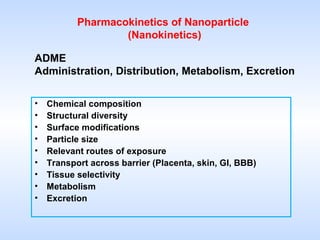
1. Pharmacokinetics of Nanoparticle
(Nanokinetics)
ADME
Administration, Distribution, Metabolism, Excretion
• Chemical composition
• Structural diversity
• Surface modifications
• Particle size
• Relevant routes of exposure
• Transport across barrier (Placenta, skin, GI, BBB)
• Tissue selectivity
• Metabolism
• Excretion
2. Hurdles
• Interaction of NP with plasma proteins,
coagulation factors, platelets, red and white
blood cells.
• Cellular uptake by diffusion, channels or
adhesive interactions and transmembrane active
processes.
• Binding to plasma components relevant for
distribution and excretion of NP.
4. Chemical composition
Nanoscale materials may possess unexpected
physical, chemical, optical, electrical and
mechanical properties, different from their
macrosized counterparts.
6. Surface modifications
PEGylated NP in “Brush ”
configuration attract less
Opsonins from plasma
Monuclear phagocyte system (MPS) is the major contributor for the clearance of
nanoparticles. Reducing the rate of MPS uptake by minimizing the opsonization
is the best strategy for prolonging the circulation of nanoparticles..
7. • opsonization
• NP is marked for ingestion and
destruction by phagocytes.
Opsonization involves the binding of
an opsonin. After opsonin binds to the
membrane, phagocytes are attracted.
12. Particle size
Arruebo M. et al. Nanotoday 2, 2007
NPs endowed with specific characteristics: size, way of conjugating the drug
(attached, adsorbed, encapsulated), surface chemistry, hydrophilicity/hydrophobicity,
surface functionalization, biodegradability, and physical response properties
(temperature, pH, electric charge, light, sound, magnetism).
13. Renal
elimination
Elimination by RES
(Reticuloendothelial system)
Spleen opsonization
100 cut-off
<5.5 200-250 nm
Optimal NP size
15. Routes of exposure
• Inhalation
• Absorption via the olfactory nervous system
• Oral administration
• Dermal absorption
• Systemic administration
16.
17. Inhalation exposure
• Distribution of inhalated NP was observed in
animal models, but not confirmed in human.
18. Inhalation exposure
• Particle deposition depends on particle size,
breathing force and the structure of the lungs.
• Brownian diffusion is also involved resulting in
the deep penetration of NP in the lungs and
diffusion in the alveolar region.
• NP >100 nm may be localized in the upper
airways before the transportation in the deep
lung.
20. Absorption via the olfactory nervous system
• This is an alternative port of entry of NP via
olfactory nerve into the brain which circunventes
the BBB.
• Neuronal absorption depends on chemical
composition, size and charge of NP.
21.
22. Absorption via the olfactory nervous
system
Surface enginnering of nanoparticles with lectins opened a
novel pathway to improve the brain uptake of agents
loaded by biodegradable PEG-PLA nanoparticles following
intranasal administration. Ulex europeus agglutinin I (UEA
I), specifically binding to L-fucose, which is largely located
in the olfactory epithelium was selected as ligand and
conjugated onto PEG-PLA nanoparticles surface.
23. Absorption via the olfactory nervous system
OLFACTORY BULB OLFACTORY TRACT
CEREBRUM CEREBELLUM
BLOOD
24. Oral absorption
• Gastrointestinal tract represents an important port of
entry of NP. The size and shape and the charge of NP
are critical for the passage into lymphatic and blood
circulation.
• 50 nm – 20 μm NP are generally absorbed through
Peyer’s patches of the small intestine
• NP must be stable to acidic pH and resistant to protease
action. Polymeric NP (e.g. PLGA ,polylactic-co-glycolic,
and SLN
• Small NP < 100 nm are more efficiently absorbed
• Positively charged NP are more effectively absorbed
than neutral or negatively charged ones.
25.
26. 26
Oral route
• Nature’s intended mode of
uptake of foreign material
• most convenient
• preferred route of
administration
• No pain (compared to
injections)
• Sterility not required
• Fewer regulatory issues
Nano-Systems
Direct uptake through the
intestine
Protection of encapsulated
drug
Slow and controlled release
Can aid delivery of drugs
with various
pharmacological and
physicochemical properties
27. 27
Lymphatic uptake of nanoparticles
Liver
NP
Blood vessel
Systemic circulation
PPs
Intestinal lumen
(II) (l)
(lll)
Mechanism of uptake of orally administered nanoparticles. NP: Nanoparticles
PPs: Peyers patches, (l) M-cells of the Peyer’s patches, (ll) Enterocytes, (lll)
Gut associated lymphoid tissue (GALT)
Bhardwaj et, al. Pharmaceutical Aspects of Polymeric Nanoparticles for Oral Delivery, Journal of Biomedical Nanotechnology (2005), 1, 1-23
29. Distribution following oral exposure
•Solid lipid nanoparticles (SLN).
•Wheat germ agglutinin-N-glutaryl-phosphatylethanolamine
(WGA-modified
SLN).
•WGA binds selectively to
intestinal cells lines.
30. Dermal absorption
• Dermal absorption is an important route for
vaccines and drug delivery.
• Size, shape, charge and material are critical
determinants for skin penetration.
• Negatively charged and small NP (<100nm)
cross more actively the epidermis than neutral or
positively charged ones.
35. Distribution following intravenous exposure
• NP kinetics depends on size charge and
functional coating.
• Delivery to RES tissues: liver, spleen, lungs and
bone marrow.
39. Metabolism
Inert NP are not metabolized (gold and silver,
fullerenes, carbon nanotubes).
Functionalized or “biocompatible” NP can be
metabolized effectively by enzymes in the body,
especially present in liver and kidney.
The intracellularly released drug is metabolized
according to the usual pathways.
40. POLYMERIC NANOPARTICLES
•Hydrolysis of ester bond; degradation products
alkylalcohol and poly(cyanoacrylic acid) are
eliminated by kidney filtration
41. GOLD NP
studies from the literature show that very little
gold is excreted from the body following
intravenous (i.v.) administration of gold
nanoparticles with a hydrodynamic (HD)
diameter exceeding 8 nm. This is in part a
consequence of the gold nanoparticles not
being composed of subunits that can be easily
broken down.
42. LIPOSOMES
are completely degraded
Phospholipids and cholesterol
follow lipid catabolic pathways
Fatty acids are oxidised
Cholesterol is degraded into bile
acids
43. Excretion
Data are not available regarding the accumulation
of NP in vivo.
The elimination route of absorbed NP remained
largely unknown and it is possible that not all
particles will be eliminated from the body.
Accumulation can take place at several sites in
the body. At low concentrations or single
exposure the accumulation may not be
significant, however high or long-term exposure
may play a relevant role in the therapeutical
effects of the active ingredient.
44. Mechanisms of Removal from Circulation
• Fast removal from circulation
-binding to cells, membranes, or plasma proteins
-uptake by phagocytes (macrophages)
-trapping in capillary bed (lungs)
• Renal clearance
-size restriction for kidney glomerulus is ~30-35 kDa for polymers
(~20-30 nm)
• Extravasation
-depends on the permeability of blood vessels
the primary route of excretion for nanoparticles greater than 8 nm is
through the hepatobiliary system in which the particles may be excreted
into bile by hepatocytes and eliminated in feces
. Additionally, nanoparticles may be phagocytosed by Kupffer cells of the
reticuloendothelial system (RES), and if not broken down by intercellular
processes, will remain in this body location long-term.2, 3 and 9
49. Defining dose for NP in vitro
• Particles are assumed to be spherical, or can be represented as spheres,
• d is the particle diameter in cm,
• surface area concentration is in cm2/ml media,
• mass concentration is in g/ml media,
• # indicates particle number, and particle density is in g/cm3.