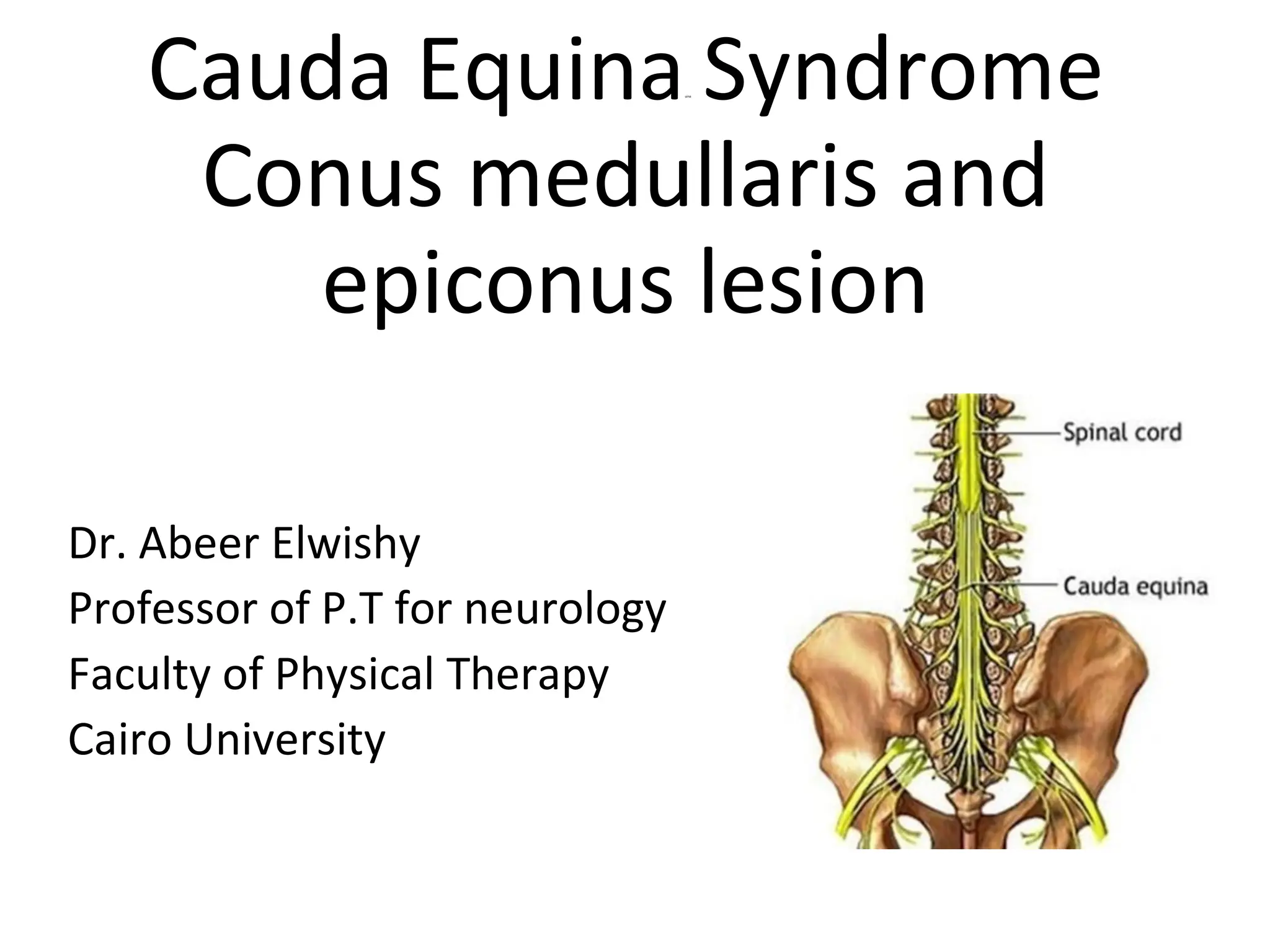
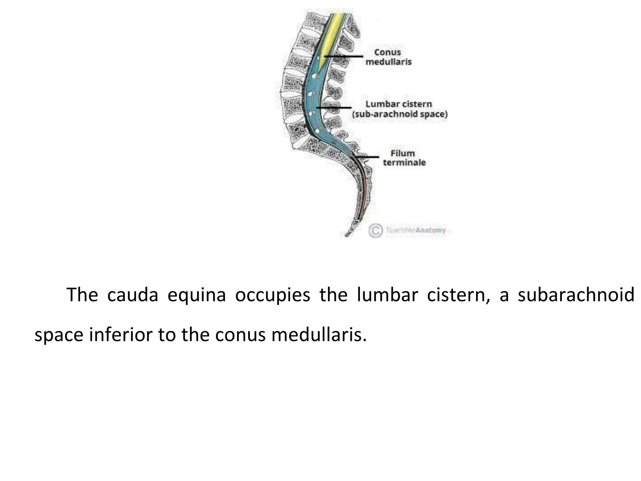
Cauda Equinauina
Syndrome
Conus medullaris and
epiconus lesion
Dr. Abeer Elwishy
Professor of P.T for neurology
Faculty of Physical Therapy
Cairo University
Nervous supply of the bladder
• The lower most three segments o the spinal cord (S3,4,5) are known
anatomically as the conus medullaris.
• The conus and epiconus form part of the spinal cord.
Cauda equina
It is collection of lumbo-sacral roots in the lower part of the spinal
canal . The cauda equina consists of nerve roots.
The collection of nerves at the end of
the spinal cord is known as the cauda
equina, due to its resemblance to a horse's
tail.
The individual nerve roots at the end of
the spinal cord that provide motor and
sensory function to the legs and the bladder
continue along in the spinal canal.
The cauda equina is the continuation of
these nerve roots in the lumbar and sacral
region. These nerves send and receive
messages to and from the lower limbs and
pelvic organs.
Cauda equina syndrome (CES) occurs
when there is dysfunction of multiple
lumbar and sacral nerve roots of the cauda
equina.
The cauda equina occupies the lumbar cistern, a subarachnoid
space inferior to the conus medullaris.
The nerves that compose
the cauda equina innervate the
pelvic organs and lower limbs
to include motor innervation of
the hips, knees, ankles, feet,
internal anal sphincter and
external
sphincter. In addition, the
cauda equina extends to
sensory innervation of the
perineum and,
anal
partially,
the
parasympathetic innervation of
bladder.
Cauda Equina Syndrome (CES)
Cauda equina syndrome is a rare disorder affecting the bundle of
nerve roots (cauda equina) at the lower (lumbar) end of the spinal cord,
is a surgical emergency.
Cauda equina syndrome occurs when the nerve roots in the lumbar
spine are compressed, disrupting sensation and movement.
Nerve roots that control the function of the bladder and bowel are
especially vulnerable to damage.
It can lead to permanent paralysis, impaired bladder and/or bowel
control, loss of sexual sensation, and other problems if left untreated. Even
with immediate treatment, some patients may not recover complete
function.
Symptoms of CES
CES is accompanied by a range of symptoms, the severity of which
depend on the degree of compression and the precise nerve roots
that are being compressed.
Patients with CES may experience some or all of these “red flag”
symptoms.
Urinary retention: the most common symptom. The patient’s
bladder fills with urine, but the patient does not experience the
normal sensation or urge to urinate.
Urinary and/or fecal incontinence. The overfull bladder can result in
incontinence of urine. Incontinence of stool can occur due to
dysfunction of the anal sphincter.
Patients with CES may experience some or all of these “red flag”
symptoms.
“Saddle anethesia” sensory disturbance, which can involve the anus,
genitals and buttock region.
.
Patients with CES may experience some or all of these “red flag”
symptoms.
Weakness or paralysis of usually more than one nerve root. The
weakness can affect lower extremities.
Pain in the back and/or legs (also known as sciatica).
Sexual dysfunction.
Causes of cauda equine lesions:
• Congenital: Spina bifida • Traumatic:
-Fracture or fracture dislocation of the lumbar vertebrae -Post
traumatic disc prolapsed.
• Inflammatory: Pott's disease o the lumbar vertebrae.
• Neoplastic:
a) Vertebral:
-Primary: Osteoma, haemangioma
-Secondary: Metastatic
b) Meningeal: Meningioma
c)Radicular: Neurofibroma
• Degenerative: Lumbar spondylosis.
Clinical picture of cauda equina lesions:
It is usually unilateral, if it is bilateral, it is asymmetrical.
Cauda equina lesions may present by one or more of the
following manifestations:
1- Motor manifestations
• There is motor weakness or paralysis in one or both lower limbs
• The weakness or paralysis is of a LMN nature (it is associated with
wasting , hypotonia and hyporeflexia.
• The motor weakness or paralysis will affect the muscles which are
supplied by affected root.
2-Sensory manifestations:
Cauda equine lesions usually have a painful onset. The pain
.
is radicular and is referred to the lower limbs, either along the
femoral distribution when the lesion affects the upper lumbar
roots or along the sciatic distribution when the lesion affects
the lower lumbar and sacral roots. Later on, there is
hyposthesia or anaesthesia in the dermatome supplied by the
affected root. The sensory impairment affects both superficial
and deep sensations.
.
3-Autonomic manifestations:
a) Sphincteric manifestations: are usually late unless the lesion is
bilateral and affects mainly S2,3,4 roots (roots of innervations of the
bladder).
The sphincter disturbances are in the form of
• Sensory atonic bladder.
• Motor atonic bladder.
• Autonomic bladder.
b) Vasomotor changes and trophic ulcers may occur in the lower
limb.
Neurogenic bladder:
Lesions affecting bladder function:
1-Lesions at the level of the reflex arc (LMNL):
A-Lesions in the afferent fibers-Sensory a tonic bladder
characterized by
-Absence of the sense of fullness of the bladder.
-Retention of urine associated with a huge size of the bladder.
-Dribbling of urine every now and then because of overflow.
-Neurogenic detrusor over activity.
Neurogenic bladder:
Lesions affecting bladder function:
1-Lesions at the level of the reflex arc (LMNL):
B- Lesions in the efferent fibers-Motor a tonic
bladder characterized by
-Preservation of the sense of fullness of the bladder. -
Retention of urine associated with a moderate size of the
bladder.
-Inability to evacuate bladder voluntarily.
-Dribbling of urine every now and then because of overflow.
-catheterization is usually quickly done.
Neurogenic bladder:
Lesions affecting bladder function:
1-Lesions at the level of the reflex arc (LMNL):
C-Lesion in both afferent and efferent fibers or in the
spinal center.
-Autonomic or Autonomous bladder characterized by
incomplete ,Irregular and Involuntary evacuation of the
bladder as the evacuation of the bladder depend on its
myogenic contraction.
Neurogenic bladder:
Lesions affecting bladder function:
2-Lesions above the level of the reflex arc (UMNL):
A-Acute: Retention with overflow.
B-Gradual:
1-Partial lesion: Precipitancy of micturition.
2-Complete lesion: Automatic bladder: this is characterized by
complete and regular evacuation of the bladder which works by
spinal reflex arc.
N.B: For any disturbance in bladder function to occur, the
lesion should be bilateral.
Clinical picture of conusmedullaris lesion(S3,4,5
segments)
1- Early urinary incontinence (autonomic bladder) and faecal
incontinence.
2- Impotence.
3- Impairment of sensation in the saddle-shaped area (usually of
a dissociated nature).
4- No motor or sensory disability in the lower limbs.
Clinical picture of epiconus lesion (L4,5,S1,S2 segments):
1-Weakness or paralysis in the lower limbs, in the muscles supplied
by L4,5 and S1,2 (dorsiflexors and plantar flexors of the ankle and
toes, the flexors of the knee and the extensors of the hip).
2-The ankle reflex is absent while the knee reflex is intact.
3-Sensory loss from L4 to S2 segment (usually of a dissociated
nature).
4-Bladder disturbances may occur in the form of precipitancy.
Physical therapy for cauda equina lesion
According to the patient's evaluation, the patient will need
some or all of the following:
• Passive movements
• Stretching ex's for shortened soft tissues.
• Strengthening ex's for weak muscles.
• Electrical stimulation for paralyzed muscles.
• Bracing to help walking.
• Bladder training if needed.
Bladder training
Bladder training or voiding
Started by regulation of the amount of fluids intake and counting the
quantity of fluids coming out.
-Advice the patient to stop fluid intake three hours before sleep. -
Catheterisation if needed.
- Help the patient to evacuate the bladder by the following
modalities to stimulate the bladder:
- Pressure over the lower
abdominal wall in an
upward downward
direction.
- Squeezing the upper
medial part of the thigh.
- Pulling the pubic hair.
- Let the patient listen to
the sound of a water tape.
1- Reflex (upper motor
neuron) bladder contract
and reflexely empty in
response to a certain level of
filling pressure. The reflex arc is intact. This reflex emptying
may be triggered by manual stimulation techniques such as
stroking, kneading or tapping the suprapubic region or thigh
and lower abdominal stroking, pinching or hair pulling.
.
2- Autonomous or non reflex
(lower motor neuron)
bladder are essentially flaccid as
there is no reflex action of the
detrusor muscle. This type of
bladder can be emptied by
increasing intra-abdominal
pressure using a valsalva
maneuver or by manually
compressing the lower abdomen
using the crede maneuver.
Crede maneuver
Try to strengthen the muscles controlling the bladder •
through:
•Giving lower abdominal exercises, pelvic floor ex’s or gluteal
region ex’s.
•Faradic stimulation.
•Interferential current to the bladder. Fours electrodes used (two
electrodes on upper medial aspects of thigh and other two
electrodes on lower abdominal wall). For 10-20 minutes.
Physical therapy for conus medullaris and epiconus lesion
- For conus medullaris (S3,4,5):
- The patient needs mainly bladder training and fecal training.
-For epiconus lesion (L4,5,S1,2):
The patient needs mainly bladder training + electrical stimulation
+ putting on short leg brace, according to the severity of the
lesion.
.
Patient Education
Patient education needs will vary with the type and severity of persistent
deficits, and may include the following:
-Training in self-catheterization and finger fecal dis-impaction, if required.
-Use of measures to prevent pressure ulcers, such as skin inspection/care,
positioning, turning and transferring tactics, use of skin protectors, or
pressure-reducing support surfaces.
-Maintenance of endurance and strength-training exercises.
-Regular follow-up by the consulting teams who treated the patient in the
hospital.
-Instructions on how and when medications should be taken and when
follow-up laboratory tests should be performed.
Prognosis of cauda equine
Morbidity and especially mortality rates are determined by the
underlying etiology.
Prognosis of cauda equine
The prognosis improves if a definitive cause is identified and
appropriate treatment occurs early in the course. Surgical
decompression may be performed emergently, or, in some
patients, delayed, depending on the etiology. Residual
weakness, incontinence, impotence, and/or sensory
abnormalities are potential problems if therapy is delayed.
Prognosis of cauda equina
• Patients with bilateral sciatica have
been reported to have a less
favorable prognosis than patients
with unilateral pain.
• Patients with complete perineal
anesthesia are more likely to have
worse outcomes
permanent paralysis of the bladder.
• Patients with unilateral deficits have a better prognosis
than patients with bilateral deficits.
• Females and patients with bowel dysfunction have
been reported to have postoperatively.
Prognosis can be predicted with the American Spinal
Injury Association (ASIA) impairment scale as follows:
ASIA A: 90% of patients remain incapable of functional ambulation
(reciprocal gait of 200 feet or more).
ASIA B: 72% of patients are unable to attain functional ambulation .
ASIA C/D: 13% are unable to attain functional ambulation 1 year
after injury .
Ambulatory motor index
is used to predict ambulatory capability
It is calculated by scoring hip flexion, hip abduction, hip extension, knee
extension, and knee flexion on both sides, using a 4-point scale (0=absent,
1=trace/poor, 2=fair, 3=good or normal); the score is expressed as a
percentage of the maximum score of 30.
Prognostic significance is as follows:
A patient with a score of 60% or more has a good chance for community
ambulation with no more than one knee-ankle-foot orthosis (KAFO).
A patient with a score of 79% or higher may not need an orthosis.
A patient with a score of 40% or less may require 2 KAFOs for community
ambulation.
cauda equina syndrome 2021 (lumbar) .pdf

cauda equina syndrome 2021 (lumbar) .pdf

  • 1.
    Cauda Equinauina Syndrome Conus medullarisand epiconus lesion Dr. Abeer Elwishy Professor of P.T for neurology Faculty of Physical Therapy Cairo University
  • 2.
    Nervous supply ofthe bladder
  • 3.
    • The lowermost three segments o the spinal cord (S3,4,5) are known anatomically as the conus medullaris.
  • 4.
    • The conusand epiconus form part of the spinal cord.
  • 5.
    Cauda equina It iscollection of lumbo-sacral roots in the lower part of the spinal canal . The cauda equina consists of nerve roots.
  • 6.
    The collection ofnerves at the end of the spinal cord is known as the cauda equina, due to its resemblance to a horse's tail. The individual nerve roots at the end of the spinal cord that provide motor and sensory function to the legs and the bladder continue along in the spinal canal.
  • 7.
    The cauda equinais the continuation of these nerve roots in the lumbar and sacral region. These nerves send and receive messages to and from the lower limbs and pelvic organs. Cauda equina syndrome (CES) occurs when there is dysfunction of multiple lumbar and sacral nerve roots of the cauda equina.
  • 8.
    The cauda equinaoccupies the lumbar cistern, a subarachnoid space inferior to the conus medullaris.
  • 9.
    The nerves thatcompose the cauda equina innervate the pelvic organs and lower limbs to include motor innervation of the hips, knees, ankles, feet, internal anal sphincter and external sphincter. In addition, the cauda equina extends to sensory innervation of the perineum and, anal partially, the
  • 10.
    parasympathetic innervation of bladder. CaudaEquina Syndrome (CES) Cauda equina syndrome is a rare disorder affecting the bundle of nerve roots (cauda equina) at the lower (lumbar) end of the spinal cord, is a surgical emergency. Cauda equina syndrome occurs when the nerve roots in the lumbar spine are compressed, disrupting sensation and movement. Nerve roots that control the function of the bladder and bowel are especially vulnerable to damage. It can lead to permanent paralysis, impaired bladder and/or bowel control, loss of sexual sensation, and other problems if left untreated. Even with immediate treatment, some patients may not recover complete function.
  • 11.
    Symptoms of CES CESis accompanied by a range of symptoms, the severity of which depend on the degree of compression and the precise nerve roots that are being compressed.
  • 12.
    Patients with CESmay experience some or all of these “red flag” symptoms. Urinary retention: the most common symptom. The patient’s bladder fills with urine, but the patient does not experience the normal sensation or urge to urinate. Urinary and/or fecal incontinence. The overfull bladder can result in incontinence of urine. Incontinence of stool can occur due to dysfunction of the anal sphincter. Patients with CES may experience some or all of these “red flag” symptoms. “Saddle anethesia” sensory disturbance, which can involve the anus, genitals and buttock region.
  • 13.
    . Patients with CESmay experience some or all of these “red flag” symptoms. Weakness or paralysis of usually more than one nerve root. The weakness can affect lower extremities. Pain in the back and/or legs (also known as sciatica). Sexual dysfunction.
  • 14.
    Causes of caudaequine lesions: • Congenital: Spina bifida • Traumatic: -Fracture or fracture dislocation of the lumbar vertebrae -Post traumatic disc prolapsed. • Inflammatory: Pott's disease o the lumbar vertebrae. • Neoplastic: a) Vertebral: -Primary: Osteoma, haemangioma -Secondary: Metastatic b) Meningeal: Meningioma
  • 15.
    c)Radicular: Neurofibroma • Degenerative:Lumbar spondylosis. Clinical picture of cauda equina lesions: It is usually unilateral, if it is bilateral, it is asymmetrical. Cauda equina lesions may present by one or more of the following manifestations: 1- Motor manifestations • There is motor weakness or paralysis in one or both lower limbs • The weakness or paralysis is of a LMN nature (it is associated with wasting , hypotonia and hyporeflexia. • The motor weakness or paralysis will affect the muscles which are supplied by affected root. 2-Sensory manifestations:
  • 16.
    Cauda equine lesionsusually have a painful onset. The pain . is radicular and is referred to the lower limbs, either along the femoral distribution when the lesion affects the upper lumbar roots or along the sciatic distribution when the lesion affects the lower lumbar and sacral roots. Later on, there is hyposthesia or anaesthesia in the dermatome supplied by the affected root. The sensory impairment affects both superficial and deep sensations. . 3-Autonomic manifestations:
  • 17.
    a) Sphincteric manifestations:are usually late unless the lesion is bilateral and affects mainly S2,3,4 roots (roots of innervations of the bladder). The sphincter disturbances are in the form of • Sensory atonic bladder. • Motor atonic bladder. • Autonomic bladder. b) Vasomotor changes and trophic ulcers may occur in the lower limb. Neurogenic bladder: Lesions affecting bladder function: 1-Lesions at the level of the reflex arc (LMNL):
  • 18.
    A-Lesions in theafferent fibers-Sensory a tonic bladder characterized by -Absence of the sense of fullness of the bladder. -Retention of urine associated with a huge size of the bladder. -Dribbling of urine every now and then because of overflow. -Neurogenic detrusor over activity. Neurogenic bladder: Lesions affecting bladder function: 1-Lesions at the level of the reflex arc (LMNL): B- Lesions in the efferent fibers-Motor a tonic bladder characterized by -Preservation of the sense of fullness of the bladder. - Retention of urine associated with a moderate size of the bladder. -Inability to evacuate bladder voluntarily.
  • 19.
    -Dribbling of urineevery now and then because of overflow. -catheterization is usually quickly done. Neurogenic bladder: Lesions affecting bladder function: 1-Lesions at the level of the reflex arc (LMNL): C-Lesion in both afferent and efferent fibers or in the spinal center. -Autonomic or Autonomous bladder characterized by incomplete ,Irregular and Involuntary evacuation of the bladder as the evacuation of the bladder depend on its myogenic contraction. Neurogenic bladder: Lesions affecting bladder function: 2-Lesions above the level of the reflex arc (UMNL):
  • 20.
    A-Acute: Retention withoverflow. B-Gradual: 1-Partial lesion: Precipitancy of micturition. 2-Complete lesion: Automatic bladder: this is characterized by complete and regular evacuation of the bladder which works by spinal reflex arc. N.B: For any disturbance in bladder function to occur, the lesion should be bilateral. Clinical picture of conusmedullaris lesion(S3,4,5 segments) 1- Early urinary incontinence (autonomic bladder) and faecal incontinence. 2- Impotence.
  • 21.
    3- Impairment ofsensation in the saddle-shaped area (usually of a dissociated nature). 4- No motor or sensory disability in the lower limbs. Clinical picture of epiconus lesion (L4,5,S1,S2 segments): 1-Weakness or paralysis in the lower limbs, in the muscles supplied by L4,5 and S1,2 (dorsiflexors and plantar flexors of the ankle and toes, the flexors of the knee and the extensors of the hip). 2-The ankle reflex is absent while the knee reflex is intact. 3-Sensory loss from L4 to S2 segment (usually of a dissociated nature). 4-Bladder disturbances may occur in the form of precipitancy.
  • 22.
    Physical therapy forcauda equina lesion According to the patient's evaluation, the patient will need some or all of the following: • Passive movements • Stretching ex's for shortened soft tissues. • Strengthening ex's for weak muscles. • Electrical stimulation for paralyzed muscles. • Bracing to help walking. • Bladder training if needed. Bladder training Bladder training or voiding
  • 23.
    Started by regulationof the amount of fluids intake and counting the quantity of fluids coming out. -Advice the patient to stop fluid intake three hours before sleep. - Catheterisation if needed. - Help the patient to evacuate the bladder by the following modalities to stimulate the bladder: - Pressure over the lower abdominal wall in an upward downward direction. - Squeezing the upper medial part of the thigh. - Pulling the pubic hair. - Let the patient listen to the sound of a water tape. 1- Reflex (upper motor neuron) bladder contract and reflexely empty in response to a certain level of
  • 24.
    filling pressure. Thereflex arc is intact. This reflex emptying may be triggered by manual stimulation techniques such as stroking, kneading or tapping the suprapubic region or thigh and lower abdominal stroking, pinching or hair pulling. .
  • 25.
    2- Autonomous ornon reflex (lower motor neuron) bladder are essentially flaccid as there is no reflex action of the detrusor muscle. This type of bladder can be emptied by increasing intra-abdominal pressure using a valsalva maneuver or by manually compressing the lower abdomen using the crede maneuver.
  • 26.
    Crede maneuver Try tostrengthen the muscles controlling the bladder • through:
  • 27.
    •Giving lower abdominalexercises, pelvic floor ex’s or gluteal region ex’s. •Faradic stimulation. •Interferential current to the bladder. Fours electrodes used (two electrodes on upper medial aspects of thigh and other two electrodes on lower abdominal wall). For 10-20 minutes. Physical therapy for conus medullaris and epiconus lesion - For conus medullaris (S3,4,5): - The patient needs mainly bladder training and fecal training.
  • 28.
    -For epiconus lesion(L4,5,S1,2): The patient needs mainly bladder training + electrical stimulation + putting on short leg brace, according to the severity of the lesion. . Patient Education Patient education needs will vary with the type and severity of persistent deficits, and may include the following: -Training in self-catheterization and finger fecal dis-impaction, if required. -Use of measures to prevent pressure ulcers, such as skin inspection/care, positioning, turning and transferring tactics, use of skin protectors, or pressure-reducing support surfaces. -Maintenance of endurance and strength-training exercises. -Regular follow-up by the consulting teams who treated the patient in the hospital.
  • 29.
    -Instructions on howand when medications should be taken and when follow-up laboratory tests should be performed. Prognosis of cauda equine Morbidity and especially mortality rates are determined by the underlying etiology.
  • 30.
    Prognosis of caudaequine The prognosis improves if a definitive cause is identified and appropriate treatment occurs early in the course. Surgical decompression may be performed emergently, or, in some patients, delayed, depending on the etiology. Residual
  • 31.
    weakness, incontinence, impotence,and/or sensory abnormalities are potential problems if therapy is delayed. Prognosis of cauda equina • Patients with bilateral sciatica have been reported to have a less favorable prognosis than patients with unilateral pain. • Patients with complete perineal anesthesia are more likely to have worse outcomes
  • 32.
    permanent paralysis ofthe bladder. • Patients with unilateral deficits have a better prognosis than patients with bilateral deficits. • Females and patients with bowel dysfunction have been reported to have postoperatively. Prognosis can be predicted with the American Spinal Injury Association (ASIA) impairment scale as follows: ASIA A: 90% of patients remain incapable of functional ambulation (reciprocal gait of 200 feet or more). ASIA B: 72% of patients are unable to attain functional ambulation . ASIA C/D: 13% are unable to attain functional ambulation 1 year after injury . Ambulatory motor index is used to predict ambulatory capability It is calculated by scoring hip flexion, hip abduction, hip extension, knee extension, and knee flexion on both sides, using a 4-point scale (0=absent,
  • 33.
    1=trace/poor, 2=fair, 3=goodor normal); the score is expressed as a percentage of the maximum score of 30. Prognostic significance is as follows: A patient with a score of 60% or more has a good chance for community ambulation with no more than one knee-ankle-foot orthosis (KAFO). A patient with a score of 79% or higher may not need an orthosis. A patient with a score of 40% or less may require 2 KAFOs for community ambulation.