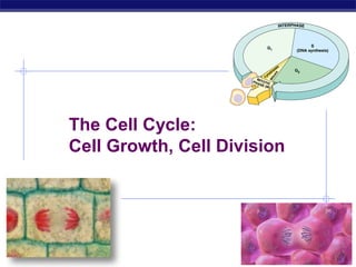
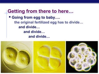
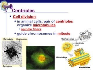
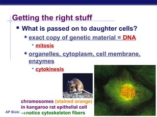
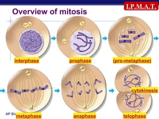
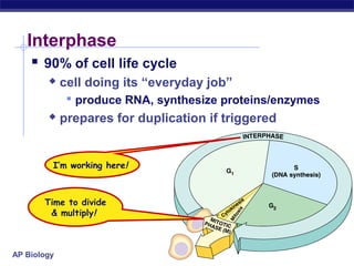
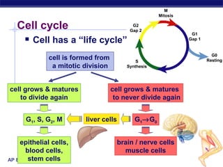
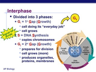
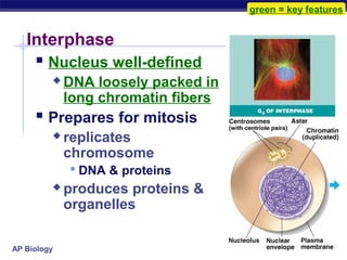
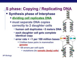
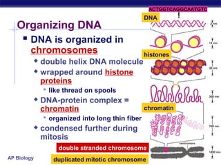
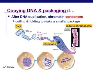
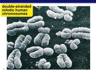
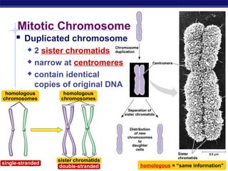
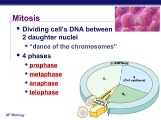
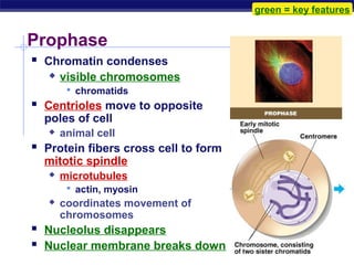
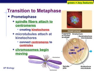
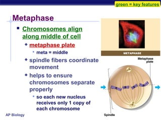
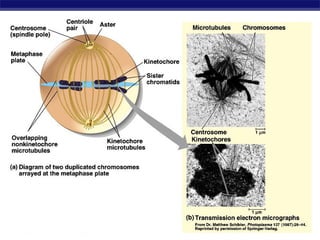
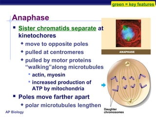
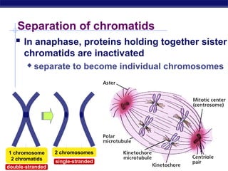
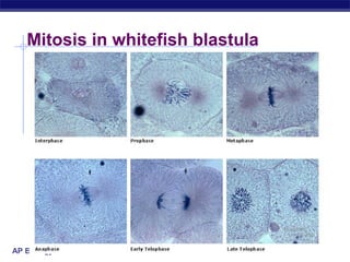
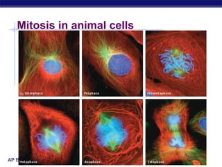
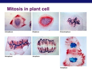
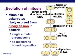
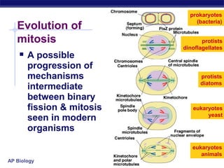
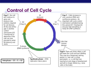
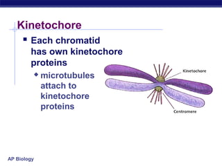
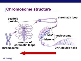
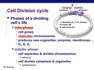
The document describes the process of cell division through mitosis. It begins with cells in interphase, where the cell grows and duplicates its DNA in S phase. The cell then enters prophase of mitosis, where the chromosomes condense and spindle fibers form. In metaphase, the chromosomes align along the center of the cell. In anaphase, the sister chromatids are separated and moved to opposite poles by spindle fibers.
The presentation includes about the basic knowledge of Deoxyribonucleic Acid or DNA. It involves the definition, structure, occurence, quantity, chemical composition, stability, variety, types, molecular weight, complementary of base pairs, absorbance, viscosity, ionic interactions, alternative forms and functions of DNA.
bacteria reproduce by binary fission , which is a single cell, divides into two identical daughter cells. it occurs through formation of the Z ring that recruits additional proteins to form the septa ring.
1. Biology is the only subject
in which multiplication is
the same thing as division…
AP Biology 2007-2008
2. The Cell Cycle:
Cell Growth, Cell Division
AP Biology 2007-2008
3. Where it all began…
You started as a cell smaller than
a period at the end of a sentence…
AP Biology
4. And now look at you…
How did you
get from there
AP Biology to here?
5. Getting from there to here…
Going from egg to baby….
the original fertilized egg has to divide…
and divide…
and divide…
and divide…
AP Biology
6. Why do cells divide?
For reproduction
asexual reproduction
one-celled organisms
For growth
from fertilized egg to
multi-celled organism
amoeba
For repair & renewal
replace cells that die
from normal wear & QuickTime™ and a
TIFF (Uncompressed) decompressor
are needed to see this picture.
tear or from injury
AP Biology
7. Making new cells
Nucleus
chromosomes
DNA
Cytoskeleton
centrioles
in animals
microtubule
spindle fibers
AP Biology
8. DNA
Nucleus
Function chromosome
protects DNA
histone protein
Structure
nuclear envelope
double membrane
membrane fused in spots to create pores
allows large macromolecules to pass through
nuclear
pores
nuclear
pore
What kind of
molecules need to
pass through? nucleolus
AP Biology nuclear envelope
14. Getting the right stuff
What is passed on to daughter cells?
exact copy of genetic material = DNA
mitosis
organelles, cytoplasm, cell membrane,
enzymes
cytokinesis
chromosomes (stained orange)
in kangaroo rat epithelial cell
→notice cytoskeleton fibers
AP Biology
15. I.P.M.A.T.
Overview of mitosis
interphase prophase (pro-metaphase)
cytokinesis
AP Biology
metaphase anaphase telophase
16. Interphase
90% of cell life cycle
cell doing its “everyday job”
produce RNA, synthesize proteins/enzymes
prepares for duplication if triggered
I’m working here!
Time to divide
& multiply!
AP Biology
17. M
Mitosis
Cell cycle G2
Gap 2
G1
Cell has a “life cycle” Gap 1
G0
cell is formed from S Resting
a mitotic division Synthesis
cell grows & matures cell grows & matures
to divide again to never divide again
G1, S, G2, M liver cells G1→G0
epithelial cells, brain / nerve cells
blood cells, muscle cells
stem cells
AP Biology
18. Interphase
Divided into 3 phases: G0
G1 = 1st Gap (Growth)
cell doing its “everyday job”
o cell grows
n al te
sig ivid S = DNA Synthesis
d
copies chromosomes
G2 = 2nd Gap (Growth)
prepares for division
cell grows (more)
produces organelles,
proteins, membranes
AP Biology
19. green = key features
Interphase
Nucleus well-defined
DNA loosely packed in
long chromatin fibers
Prepares for mitosis
replicates
chromosome
DNA & proteins
produces proteins &
organelles
AP Biology
20. S phase: Copying / Replicating DNA
Synthesis phase of Interphase
dividing cell replicates DNA
must separate DNA copies
correctly to 2 daughter cells
human cell duplicates ~3 meters DNA
each daughter cell gets complete
identical copy
error rate = ~1 per 100 million bases
3 billion base pairs in mammalian
genome
~30 errors per cell cycle
mutations (to somatic (body) cells)
AP Biology
21. ACTGGTCAGGCAATGTC
DNA
Organizing DNA
DNA is organized in
chromosomes histones
double helix DNA molecule
wrapped around histone
proteins
like thread on spools
DNA-protein complex =
chromatin chromatin
organized into long thin fiber
condensed further during
mitosis
double stranded chromosome
AP Biology duplicated mitotic chromosome
22. Copying DNA & packaging it…
After DNA duplication, chromatin condenses
coiling & folding to make a smaller package
DNA mitotic chromosome
chromatin
AP Biology
24. Mitotic Chromosome
Duplicated chromosome
2 sister chromatids
narrow at centromeres
contain identical
copies of original DNA
homologous homologous
chromosomes chromosomes
sister chromatids
single-stranded
AP Biology double-stranded homologous = “same information”
25. Mitosis
Dividing cell’s DNA between
2 daughter nuclei
“dance of the chromosomes”
4 phases
prophase
metaphase
anaphase
telophase
AP Biology
26. green = key features
Prophase
Chromatin condenses
visible chromosomes
chromatids
Centrioles move to opposite
poles of cell
animal cell
Protein fibers cross cell to form
mitotic spindle
microtubules
actin, myosin
coordinates movement of
chromosomes
Nucleolus disappears
Nuclear membrane breaks down
AP Biology
27. green = key features
Transition to Metaphase
Prometaphase
spindle fibers attach to
centromeres
creating kinetochores
microtubules attach at
kinetochores
connect centromeres to
centrioles
chromosomes begin
moving
AP Biology
28. green = key features
Metaphase
Chromosomes align
along middle of cell
metaphase plate
meta = middle
spindle fibers coordinate
movement
helps to ensure
chromosomes separate
properly
so each new nucleus
receives only 1 copy of
each chromosome
AP Biology
30. green = key features
Anaphase
Sister chromatids separate at
kinetochores
move to opposite poles
pulled at centromeres
pulled by motor proteins
“walking”along microtubules
actin, myosin
increased production of
ATP by mitochondria
Poles move farther apart
polar microtubules lengthen
AP Biology
31. Separation of chromatids
In anaphase, proteins holding together sister
chromatids are inactivated
separate to become individual chromosomes
1 chromosome 2 chromosomes
2 chromatids single-stranded
AP Biology
double-stranded
32. Chromosome movement
Kinetochores use
motor proteins that
“walk” chromosome
along attached
microtubule
microtubule
shortens by
dismantling at
kinetochore
(chromosome) end
AP Biology
33. green = key features
Telophase
Chromosomes arrive at
opposite poles
daughter nuclei form
nucleoli form
chromosomes disperse
no longer visible under
light microscope
Spindle fibers disperse
Cytokinesis begins
cell division
AP Biology
34. Cytokinesis
Animals
constriction belt of
actin microfilaments
around equator of cell
cleavage furrow forms
splits cell in two
like tightening a draw
string
AP Biology
38. Cytokinesis in Plants
Plants
cell plate forms
vesicles line up at
equator
derived from Golgi
vesicles fuse to form
2 cell membranes
new cell wall laid
down between
membranes
new cell wall fuses
with existing cell wall
AP Biology
42. Origin of
replication
Evolution of mitosis
chromosome:
Mitosis in double-stranded replication
DNA of DNA
eukaryotes
likely evolved from
binary fission in elongation of cell
bacteria
single circular ring of
proteins
chromosome
no membrane-
bound organelles
cell pinches
in two
AP Biology
43. prokaryotes
(bacteria)
Evolution of
mitosis protists
dinoflagellates
A possible
progression of
mechanisms protists
diatoms
intermediate
between binary
fission & mitosis eukaryotes
yeast
seen in modern
organisms
eukaryotes
animals
AP Biology
49. Kinetochore
Each chromatid
has own kinetochore
proteins
microtubules
attach to
kinetochore
proteins
AP Biology
50. Chromosome structure
scaffold chromatin loop
protein
n m
30
DNA nucleosome
histone
rosettes of
chromatin loops
chromosome DNA double helix
AP Biology
51. M
metaphase anaphase
Cell Division cycle prophase telophase
C
G2
Phases of a dividing
cell’s life interphase (G1, S, G2 phases)
mitosis (M)
interphase S
cytokinesis (C) G1
cell grows
replicates chromosomes
produces new organelles, enzymes, membranes…
G1, S, G2
mitotic phase
cell separates & divides chromosomes
mitosis
cell divides cytoplasm & organelles
cytokinesis
AP Biology
52. Substitute Slides
for Student Print version
(for student note-taking)
AP Biology 2007-2008
53. And now look at you…
How did you
get from there
AP Biology to here?
Editor's Notes
Unicellular organisms Cell division = reproduction Reproduces entire organism& increase population Multicellular organisms Cell division provides for growth & development in a multicellular organism that begins as a fertilized egg Also use cell division to repair & renew cells that die from normal wear & tear or accidents
Centromeres are segments of DNA which have long series of tandem repeats = 100,000s of bases long. The sequence of the repeated bases is quite variable. It has proven difficult to sequence.
Microtubules are NOT reeled in to centrioles like line on a fishing rod. The motor proteins walk along the microtubule like little hanging robots on a clothes line. In dividing animal cells, non-kinetochore microtubules are responsible for elongating the whole cell during anaphase, readying fro cytokinesis
Division of cytoplasm happens quickly.
Prokaryotes (bacteria) No nucleus; single circular chromosome. After DNA is replicated, it is partitioned in the cell. After cell elongation, FtsZ protein assembles into a ring and facilitates septation and cell division. Protists (dinoflagellates) Nucleus present and nuclear envelope remains intact during cell division. Chromosomes linear. Fibers called microtubules, composed of the protein tubulin, pass through tunnels in the nuclear membrane and set up an axis for separation of replicated chromosomes, and cell division. Protists (diatoms) A spindle of microtubules forms between two pairs of centrioles at opposite ends of the cell. The spindle passes through one tunnel in the intact nuclear envelope. Kinetochore microtubules form between kinetochores on the chromosomes and the spindle poles and pull the chromosomes to each pole. Eukaryotes (yeast) Nuclear envelope remains intact; spindle microtubules form inside the nucleus between spindle pole bodies. A single kinetochore microtubule attaches to each chromosome and pulls each to a pole. Eukaryotes (animals) Spindle microtubules begin to form between centrioles outside of nucleus. As these centrioles move to the poles, the nuclear envelope breaks down, and kinetochore microtubules attach kinetochores of chromosomes to spindle poles. Polar microtubules extend toward the center of the cell and overlap.