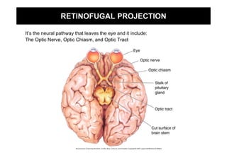
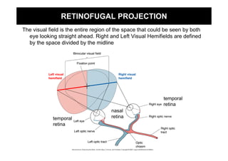
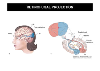
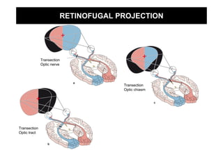
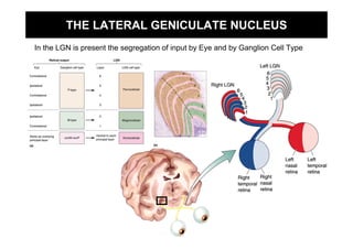
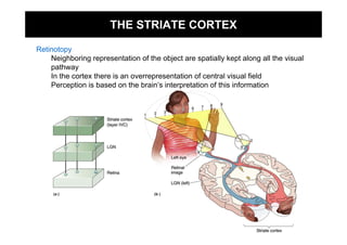
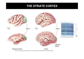
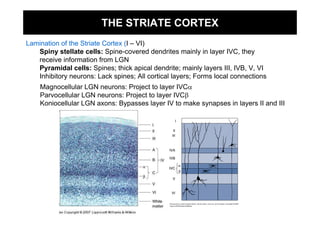
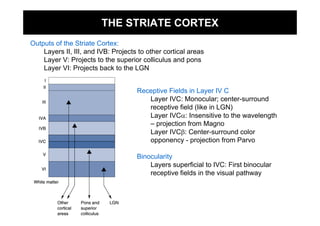
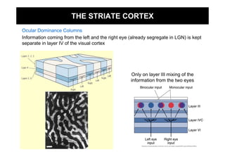
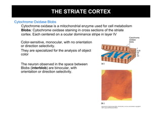
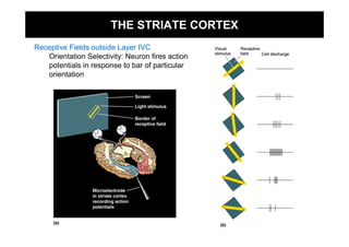
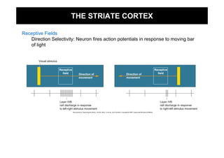
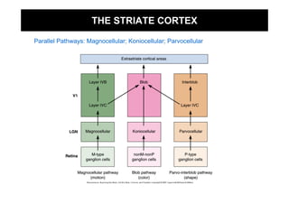
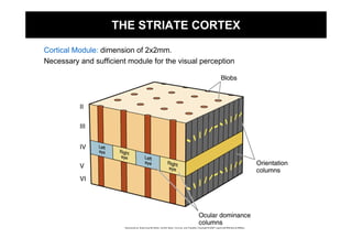
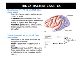
THE CENTRAL VISUAL SYSTEM
RETINOFUGAL PROJECTION

It’s the neural pathway that leaves the eye and it include:
The Optic Nerve, Optic Chiasm, and Optic Tract
RETINOFUGAL PROJECTION
The visual field is the entire region of the space that could be seen by both
  eye looking straight ahead. Right and Left Visual Hemifields are defined
  by the space divided by the midline


                                                            temporal
                                                            retina
                                      nasal
             temporal                 retina
             retina
RETINOFUGAL PROJECTION


           LGN
             Optic radiation

                                  R optic tract
retina              V1
                                          R LGN

                                                R optic
                                                radiation


                                           V1
RETINOFUGAL PROJECTION


              Transection
              Optic nerve


                                     Transection
                                     Optic chiasm


Transection
Optic tract
THE LATERAL GENICULATE NUCLEUS
In the LGN is present the segregation of input by Eye and by Ganglion Cell Type
THE STRIATE CORTEX

Retinotopy
    Neighboring representation of the object are spatially kept along all the visual
    pathway
    In the cortex there is an overrepresentation of central visual field
    Perception is based on the brain’s interpretation of this information
THE STRIATE CORTEX
THE STRIATE CORTEX
Lamination of the Striate Cortex (I – VI)
   Spiny stellate cells: Spine-covered dendrites mainly in layer IVC, they
   receive information from LGN
   Pyramidal cells: Spines; thick apical dendrite; mainly layers III, IVB, V, VI
   Inhibitory neurons: Lack spines; All cortical layers; Forms local connections
    Magnocellular LGN neurons: Project to layer IVCα
    Parvocellular LGN neurons: Project to layer IVCβ
    Koniocellular LGN axons: Bypasses layer IV to make synapses in layers II and III
THE STRIATE CORTEX
Outputs of the Striate Cortex:
    Layers II, III, and IVB: Projects to other cortical areas
    Layer V: Projects to the superior colliculus and pons
    Layer VI: Projects back to the LGN


                                     Receptive Fields in Layer IV C
                                        Layer IVC: Monocular; center-surround
                                        receptive field (like in LGN)
                                        Layer IVCα: Insensitive to the wavelength
                                        – projection from Magno
                                        Layer IVCβ: Center-surround color
                                        opponency - projection from Parvo

                                     Binocularity
                                         Layers superficial to IVC: First binocular
                                         receptive fields in the visual pathway
THE STRIATE CORTEX
Ocular Dominance Columns
Information coming from the left and the right eye (already segregate in LGN) is kept
separate in layer IV of the visual cortex


                                                   Only on layer III mixing of the
                                                   information from the two eyes
THE STRIATE CORTEX
Cytochrome Oxidase Blobs
    Cytochrome oxidase is a mitochondrial enzyme used for cell metabolism
    Blobs: Cytochrome oxidase staining in cross sections of the striate
    cortex. Each centered on a ocular dominance stripe in layer IV

    Color-sensitive, monocular, with no orientation
    or direction selectivity.
    They are specialized for the analysis of object
    color

    The neuron observed in the space between
    Blobs (interblob) are binocular, with
    orientation or direction selectivity.
THE STRIATE CORTEX
Receptive Fields outside Layer IVC
   Orientation Selectivity: Neuron fires action
   potentials in response to bar of particular
   orientation
THE STRIATE CORTEX

Receptive Fields
   Direction Selectivity: Neuron fires action potentials in response to moving bar
   of light
THE STRIATE CORTEX

Parallel Pathways: Magnocellular; Koniocellular; Parvocellular
THE STRIATE CORTEX

Cortical Module: dimension of 2x2mm.
Necessary and sufficient module for the visual perception
THE EXTRASTRIATE CORTEX
Dorsal stream (V1, V2, V3, MT, MST, Other
dorsal areas)
    Analysis of visual motion and the visual
    control of action
    In Area MT (temporal lobe) most cells:
    Direction-selective; Respond more to the
    motion of objects than their shape
    Area MST (parietal lobe) for navigation,
    directing eye movements, motion
    perception


Ventral stream (V1, V2, V3, V4, IT, Other
ventral areas)
    Perception of the visual world and the
    recognition of objects,
    Area V4 orientation and perception of
    color
    Area IT is major output of V4. Receptive
    fields respond to a wide variety of colors
    and abstract shapes. Important also for
    memory

Ch10

  • 1.
  • 2.
    RETINOFUGAL PROJECTION It’s theneural pathway that leaves the eye and it include: The Optic Nerve, Optic Chiasm, and Optic Tract
  • 3.
    RETINOFUGAL PROJECTION The visualfield is the entire region of the space that could be seen by both eye looking straight ahead. Right and Left Visual Hemifields are defined by the space divided by the midline temporal retina nasal temporal retina retina
  • 4.
    RETINOFUGAL PROJECTION LGN Optic radiation R optic tract retina V1 R LGN R optic radiation V1
  • 5.
    RETINOFUGAL PROJECTION Transection Optic nerve Transection Optic chiasm Transection Optic tract
  • 6.
    THE LATERAL GENICULATENUCLEUS In the LGN is present the segregation of input by Eye and by Ganglion Cell Type
  • 7.
    THE STRIATE CORTEX Retinotopy Neighboring representation of the object are spatially kept along all the visual pathway In the cortex there is an overrepresentation of central visual field Perception is based on the brain’s interpretation of this information
  • 8.
  • 9.
    THE STRIATE CORTEX Laminationof the Striate Cortex (I – VI) Spiny stellate cells: Spine-covered dendrites mainly in layer IVC, they receive information from LGN Pyramidal cells: Spines; thick apical dendrite; mainly layers III, IVB, V, VI Inhibitory neurons: Lack spines; All cortical layers; Forms local connections Magnocellular LGN neurons: Project to layer IVCα Parvocellular LGN neurons: Project to layer IVCβ Koniocellular LGN axons: Bypasses layer IV to make synapses in layers II and III
  • 10.
    THE STRIATE CORTEX Outputsof the Striate Cortex: Layers II, III, and IVB: Projects to other cortical areas Layer V: Projects to the superior colliculus and pons Layer VI: Projects back to the LGN Receptive Fields in Layer IV C Layer IVC: Monocular; center-surround receptive field (like in LGN) Layer IVCα: Insensitive to the wavelength – projection from Magno Layer IVCβ: Center-surround color opponency - projection from Parvo Binocularity Layers superficial to IVC: First binocular receptive fields in the visual pathway
  • 11.
    THE STRIATE CORTEX OcularDominance Columns Information coming from the left and the right eye (already segregate in LGN) is kept separate in layer IV of the visual cortex Only on layer III mixing of the information from the two eyes
  • 12.
    THE STRIATE CORTEX CytochromeOxidase Blobs Cytochrome oxidase is a mitochondrial enzyme used for cell metabolism Blobs: Cytochrome oxidase staining in cross sections of the striate cortex. Each centered on a ocular dominance stripe in layer IV Color-sensitive, monocular, with no orientation or direction selectivity. They are specialized for the analysis of object color The neuron observed in the space between Blobs (interblob) are binocular, with orientation or direction selectivity.
  • 13.
    THE STRIATE CORTEX ReceptiveFields outside Layer IVC Orientation Selectivity: Neuron fires action potentials in response to bar of particular orientation
  • 14.
    THE STRIATE CORTEX ReceptiveFields Direction Selectivity: Neuron fires action potentials in response to moving bar of light
  • 15.
    THE STRIATE CORTEX ParallelPathways: Magnocellular; Koniocellular; Parvocellular
  • 16.
    THE STRIATE CORTEX CorticalModule: dimension of 2x2mm. Necessary and sufficient module for the visual perception
  • 17.
    THE EXTRASTRIATE CORTEX Dorsalstream (V1, V2, V3, MT, MST, Other dorsal areas) Analysis of visual motion and the visual control of action In Area MT (temporal lobe) most cells: Direction-selective; Respond more to the motion of objects than their shape Area MST (parietal lobe) for navigation, directing eye movements, motion perception Ventral stream (V1, V2, V3, V4, IT, Other ventral areas) Perception of the visual world and the recognition of objects, Area V4 orientation and perception of color Area IT is major output of V4. Receptive fields respond to a wide variety of colors and abstract shapes. Important also for memory