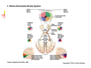
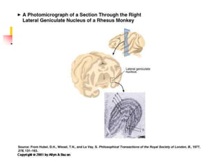
THE VISUAL SYSTEM: VISUAL
COGNITION AND BRAIN
THE PERCEPTION OF
CONTRAST AND COLOR
 Outline
 1. Contrast: The Perception of Edges
 a. Lateral Inhibition: The Physiological Basis of Contrast Enhancement
 2. Brightness-Contrast Detectors in the Mammalian Visual System
 a. Mapping Receptive Fields
 b. Receptive Fields of Neurons in the Retina-Geniculate-Striate Pathway
 c. Simple Cortical Cells
 d. Complex Cortical Cells
 e. Hubel and Wiesel's Model of Striate-Cortex Organization
 3. Seeing Color
 a. Component Theory
 b. Opponent Process Theory
Receptive Fields of Neurons in the
Retina-Geniculate-Striate Pathway
 Most retinal ganglion cells, lateral geniculate
nucleus neurons, and the neurons in lower layer
IV of the striate cortex have similar receptive fields
 They are circular
 They are smaller in the fovea area (larger in the periphery)
 Indicative of greater acuity in the fovea
 They are monocular
 They have both an excitatory and an inhibitory area
separated by a circular boundary.
 Neurons in these regions have 2 patterns of
responding:
 (1) on firing
 (2) inhibition followed by off firing
Simple Cortical Cells
 Respond best to bars or edges of light
in a particular location in the receptive
field and in a particular orientation
(e.g., 45 degrees)
 All simple cells are monocular
 They also have on and off areas
Complex Cortical Cells
 Most of the cells in the striate cortex are
complex cells
 more numerous
 like simple cells in that they respond best to
straight-line stimuli in a particular orientation
 unlike simple cells in that the position of the
stimulus within the receptive field does not matter
 the cell responds to the appropriate stimulus no matter
where it is in its large receptive field
 Over half of the complex cells are binocular,
and about half of those that are binocular
display ocular dominance
Hubel and Wiesel's Model of
Striate-Cortex Organization
 When you record from visual cortex using vertical
electrode passes, you find
 (1) As the electrode moves up or down through the layers of
the striate cortex it finds neurons that respond to stimulation
from about the same location on the retina
 (2) simple and complex, cells that all prefer the same
orientation – the cells respond to line orientations that are at
the same degree
 Info. flows from on and off cells in lower layer IV to simple
cells and then to complex cells
 (3) binocular complex cells that are all dominated by the
same eye (if they display ocular dominance)
Hubel and Wiesel's Model of
Striate-Cortex Organization
 When you record from visual cortex
using horizontal electrode passes
you find
 (1) receptive field location shifts slightly
with each electrode advance
 (2) orientation preference shifts slightly
with each electrode advance
 (3) ocular dominance periodically shifts to
the other eye with electrode advances
CORTICAL MECHANISMS
OF VISION
 Outline
 1. The Parallel, Functionally Segregated
Hierarchical Model of Perception
 2. Cortical Mechanisms of Vision
 a. Scotomas and Blindsight
 b. Completion
 c. Secondary Visual Cortex and Association Cortex
 d. Visual Agnosia
 3. Selective Attention
The Current Parallel, Functionally Segregated
Hierarchical Model of Sensory-
System Organization
 Parallel: sensory systems are organized so that information
flows between different structures simultaneously along multiple
pathways
 Explicit –– a sensory system for consciously seeing things
 Implicit –– a sensory system that helps us interact with objects in
space.
 Functionally Segregated: There are divisions of labor at the
different hierarchical levels
 Some neurons respond to motion, some to color, and so on.
 Hierarchical: information flows through brain structures in
order of their increasing neuroanatomical and functional
complexity.
 Primary
 Secondary
 Association
Vertemnus (1591) by
Guiseppe Arcimboldo
The Vegetable Gardener
(1590)
Neural Correlates?
 There is evidence that selective attention occurs by strengthening the
neural responses to attended-to aspects and by weakening the
response to others.
 Anticipation of a stimulus increases neural activity in the same circuits
affected by the stimulus itself.
 When attempting to recognize faces - ventral stream is activated
 Does this face belong to the same person?
 face recognition activates the ventral visual pathway – is this face the same
person you saw before?
 fMRI
 When attending to location - dorsal stream is activated.
 Is this face in the same position as the last face you saw?
 face position activates the dorsal visual pathway – is this face in the same
location as the face you saw before?
 fMRI
 Primates
 Recorded from neurons in the prestriate area
that is part of the ventral stream that are
particular responsive to color.
 Some cells responded to red; some to green
 Monkeys were taught to perform a task that required
attention to a red cue.
 The activity of red neurons was increased, whereas the
activity of green neurons was reduced

visualsystem.ppt

  • 1.
    THE VISUAL SYSTEM:VISUAL COGNITION AND BRAIN
  • 18.
    THE PERCEPTION OF CONTRASTAND COLOR  Outline  1. Contrast: The Perception of Edges  a. Lateral Inhibition: The Physiological Basis of Contrast Enhancement  2. Brightness-Contrast Detectors in the Mammalian Visual System  a. Mapping Receptive Fields  b. Receptive Fields of Neurons in the Retina-Geniculate-Striate Pathway  c. Simple Cortical Cells  d. Complex Cortical Cells  e. Hubel and Wiesel's Model of Striate-Cortex Organization  3. Seeing Color  a. Component Theory  b. Opponent Process Theory
  • 22.
    Receptive Fields ofNeurons in the Retina-Geniculate-Striate Pathway  Most retinal ganglion cells, lateral geniculate nucleus neurons, and the neurons in lower layer IV of the striate cortex have similar receptive fields  They are circular  They are smaller in the fovea area (larger in the periphery)  Indicative of greater acuity in the fovea  They are monocular  They have both an excitatory and an inhibitory area separated by a circular boundary.  Neurons in these regions have 2 patterns of responding:  (1) on firing  (2) inhibition followed by off firing
  • 25.
    Simple Cortical Cells Respond best to bars or edges of light in a particular location in the receptive field and in a particular orientation (e.g., 45 degrees)  All simple cells are monocular  They also have on and off areas
  • 27.
    Complex Cortical Cells Most of the cells in the striate cortex are complex cells  more numerous  like simple cells in that they respond best to straight-line stimuli in a particular orientation  unlike simple cells in that the position of the stimulus within the receptive field does not matter  the cell responds to the appropriate stimulus no matter where it is in its large receptive field  Over half of the complex cells are binocular, and about half of those that are binocular display ocular dominance
  • 29.
    Hubel and Wiesel'sModel of Striate-Cortex Organization  When you record from visual cortex using vertical electrode passes, you find  (1) As the electrode moves up or down through the layers of the striate cortex it finds neurons that respond to stimulation from about the same location on the retina  (2) simple and complex, cells that all prefer the same orientation – the cells respond to line orientations that are at the same degree  Info. flows from on and off cells in lower layer IV to simple cells and then to complex cells  (3) binocular complex cells that are all dominated by the same eye (if they display ocular dominance)
  • 31.
    Hubel and Wiesel'sModel of Striate-Cortex Organization  When you record from visual cortex using horizontal electrode passes you find  (1) receptive field location shifts slightly with each electrode advance  (2) orientation preference shifts slightly with each electrode advance  (3) ocular dominance periodically shifts to the other eye with electrode advances
  • 37.
    CORTICAL MECHANISMS OF VISION Outline  1. The Parallel, Functionally Segregated Hierarchical Model of Perception  2. Cortical Mechanisms of Vision  a. Scotomas and Blindsight  b. Completion  c. Secondary Visual Cortex and Association Cortex  d. Visual Agnosia  3. Selective Attention
  • 38.
    The Current Parallel,Functionally Segregated Hierarchical Model of Sensory- System Organization  Parallel: sensory systems are organized so that information flows between different structures simultaneously along multiple pathways  Explicit –– a sensory system for consciously seeing things  Implicit –– a sensory system that helps us interact with objects in space.  Functionally Segregated: There are divisions of labor at the different hierarchical levels  Some neurons respond to motion, some to color, and so on.  Hierarchical: information flows through brain structures in order of their increasing neuroanatomical and functional complexity.  Primary  Secondary  Association
  • 43.
  • 45.
  • 53.
    Neural Correlates?  Thereis evidence that selective attention occurs by strengthening the neural responses to attended-to aspects and by weakening the response to others.  Anticipation of a stimulus increases neural activity in the same circuits affected by the stimulus itself.  When attempting to recognize faces - ventral stream is activated  Does this face belong to the same person?  face recognition activates the ventral visual pathway – is this face the same person you saw before?  fMRI  When attending to location - dorsal stream is activated.  Is this face in the same position as the last face you saw?  face position activates the dorsal visual pathway – is this face in the same location as the face you saw before?  fMRI
  • 54.
     Primates  Recordedfrom neurons in the prestriate area that is part of the ventral stream that are particular responsive to color.  Some cells responded to red; some to green  Monkeys were taught to perform a task that required attention to a red cue.  The activity of red neurons was increased, whereas the activity of green neurons was reduced