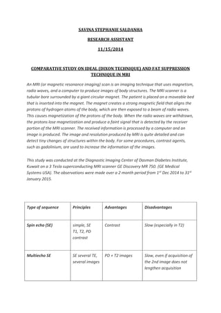
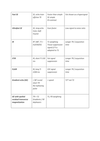
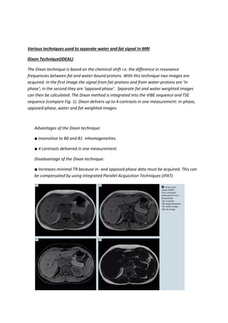
SlideShare a Scribd company logo
1 of 7
Download to read offline
SAVINA STEPHANIE SALDANHA
RESEARCH ASSISTANT
11/15/2014
COMPARATIVE STUDY ON IDEAL (DIXON TECHNIQUE) AND FAT SUPPRESSION
TECHNIQUE IN MRI
An MRI (or magnetic resonance imaging) scan is an imaging technique that uses magnetism,
radio waves, and a computer to produce images of body structures. The MRI scanner is a
tubular bore surrounded by a giant circular magnet. The patient is placed on a moveable bed
that is inserted into the magnet. The magnet creates a strong magnetic field that aligns the
protons of hydrogen atoms of the body, which are then exposed to a beam of radio waves.
This causes magnetization of the protons of the body. When the radio waves are withdrawn,
the protons lose magnetization and produce a faint signal that is detected by the receiver
portion of the MRI scanner. The received information is processed by a computer and an
image is produced. The image and resolution produced by MRI is quite detailed and can
detect tiny changes of structures within the body. For some procedures, contrast agents,
such as gadolinium, are used to increase the information of the images.
This study was conducted at the Diagnostic Imaging Center of Dasman Diabetes Institute,
Kuwait on a 3 Tesla superconducting MRI scanner GE Discovery MR 750. (GE Medical
Systems USA). The observations were made over a 2 month period from 1st Dec 2014 to 31st
January 2015.
Type of sequence Principles Advantages Disadvantages
Spin echo (SE) simple, SE
T1, T2, PD
contrast
Contrast Slow (especially in T2)
Multiecho SE SE several TE,
several images
PD + T2 images Slow, even if acquisition of
the 2nd image does not
lengthen acquisition
Fast SE SE, echo train
effctive TE
Faster than simple
SE simple
ES contrast
Fat shown as a hypersignal
Ultrafast SE SE, long echo
train, half-
Fourier
Even faster Low signal to noise ratio
IR RF 180°, TI +
ES/ESR/EG
T1 weighting
Tissue suppression
signal if TI is
adapted to T1
Longer TR / acquisition
time
STIR IR, short TI 150
ms
Fat signal
suppression
Longer TR / acquisition
time
FLAIR IR, long TI
2200 ms
CSF signal
suppression
Longer TR / acquisition
time
Gradient echo (GE) < 90° α and
short TR
No rephasing
pulse
+ speed T2* not T2
GE with spoiled
residual transverse
magnetization
TR < T2
Gradients / RF
dephasers
T1, PD weighting
Various techniques used to separate water and fat signal in MRI
Dixon Technique(IDEAL):
The Dixon technique is based on the chemical shift i.e. the difference in resonance
frequencies between fat and water-bound protons. With this technique two images are
acquired. In the first image the signal from fat-protons and from water-protons are ‘in
phase’; in the second they are ‘opposed phase’. Separate fat and water weighted images
can then be calculated. The Dixon method is integrated into the VIBE sequence and TSE
sequence (compare Fig. 1). Dixon delivers up to 4 contrasts in one measurement: in-phase,
opposed-phase, water and fat weighted images.
Advantages of the Dixon technique:
■ Insensitive to B0 and B1 inhomogeneities.
■ 4 contrasts delivered in one measurement.
Disadvantage of the Dixon technique:
■ Increases minimal TR because in- and opposed phase data must be acquired. This can
be compensated by using integrated Parallel Acquisition Techniques (iPAT)
Spectral Fat Saturation:
This technique is based on the chemical shift (3.4 ppm) i.e. the difference in resonance
frequencies between fat- and water-bound protons. The application of a narrow band
frequency selective radiofrequency (RF) pulse excites mainly fatbound protons. This
transversal magnetization is destroyed afterwards by spoiler gradients, thus no fat
magnetization is left for imaging (compare figure 2). For spectral fat saturation, a ‘Quick-Fat
Sat’ setting is available. If this feature is selected, not every slice excitation is preceded by a
preparation pulse. This means:
■ Shorter possible TR;
■ Shorter breath-hold examinations (e.g. VIBE, recommended 40 lines/shot).
Two Fat Sat modes (strong/weak) are also available. Basically, the user can select how much
of the fat signal is contributing to the MR image. In the ‘strong’ mode a nearly full
suppression is achieved, whereas in the ‘weak’ modus, anatomical information of fatty tissue
is partially preserved.
Advantages of spectral fat saturation:
■ Tissue contrast is not affected;
■ Quick-Fat Sat can be applied for increased performance.
Disadvantages of spectral fat saturation:
■ Sensitive to B0 and B1 inhomogeneities;
■ Additional preparation pulse increases minimal TR and total measurement time or reduces
maximum number of slices (partially compensated by Quick Fat Sat).
Water excitation:
This technique is based on the chemical shift i.e. the difference in resonance frequencies
between fat and water-bound protons. No additional preparation pulse is necessary.
Instead, a special excitation pulse (binomial pulse) is used with the spectral excitation profile
(minimum excitation of fat bound protons, maximum excitation of water-bound protons)
Advantages of water excitation:
■ Reduced sensitivity to B1 inhomogeneities.
Disadvantages of water excitation:
■ Increased min TE, TR and total measurement time or reduced maximum number of slices.
Fat suppression with Inversion Recovery In clinical routine, two types of inversion recovery
techniques are applied: SPAIR (Spectrally Adiabatic Inversion Recovery) method, and Short TI
Inversion Recovery (STIR; in principal identical to TIRM (Turbo Inversion Recovery
Magnetization)
STIR (Short TI Inversion Recovery):
This relaxation-dependant technique is based on the different relaxation behaviour of water
and fat tissue. Fat has a much shorter T1 relaxation time than other tissues. Prior to the
excitation pulse of the sequence an inversion pulse ( = 180°) is applied which inverts the spins
of all tissue and fat protons. This is followed by T1 relaxation. When choosing TI such that
the longitudinal magnetization of fat at the time when the excitation pulse is applied is zero,
the fat spins will not contribute to the MR signal. STIR images have an inverted T1 contrast:
Tissue with long T1 appears brighter than tissue with short T1 (compare Fig. 5).
Advantages of STIR:
■ Insensitive to B0 Inhomogeneities.
Disadvantages of STIR:
■ Additional inversion pulse increases minimal TR and total measurement time or reduces
maximum number of slices;
■ Tissue contrast is affected, SNR is reduced.
SPIN ECHO WITH FAT SUPPRESSION
The original 2-point Dixon method was developed for fat and water image separation by
utilizing differences in chemical shifts but was sensitive to field inhomogeneity. Modified
Dixon techniques include a third acquisition that results in a robust sequence that is able to
reliably provide for fat and water separation, even in the presence of magnetic field
inhomogeneity. There has been increasing interest in using 3PD methods in clinical practice
because uniform and reliable fat suppression can be of substantial clinical value. The 3PD
method may be implemented in a variety of pulse sequences. Most of our head and neck
protocols include fast spin-echo (FSE) T1- or T2-weighted images. Thus, in this work, we
utilize the FSE-IDEAL technique (Iterative Decomposition of water and fat with Echo
Asymmetry and Least squares estimation), a prerelease General Electric 3PD-FSE pulse
sequence that can be used for both T1 and T2-weighted imaging.
In general, 3PD imaging consists of a repetitive, phase-shifted acquisition (pulse sequence)
and specialized reconstruction software. For each echo train acquired by a conventional FSE
sequence, the modified 3PD sequence repeats the acquisition 3 times, with differing echo
shifts for each repetition. For FSE-IDEAL, echo shifts of −π/6, π/2, and 7π/6 are chosen, an
asymmetric sampling scheme that optimizes the signal intensity–to-noise ratio (SNR). The
echo shift is obtained by increasing the interecho spacing of the readout train.
Specialized reconstruction software is required to analyze the phase information from the 3
acquisitions to calculate separate fat and water images. Postprocessing of the acquired data
is performed at the scanner console. The reconstruction software uses an iterative phase
correction algorithm.In this way, the phase offset due to inhomogeneity can be calculated
and eliminated, allowing the additional information from phase shifting to be used to
generate separate fat and water images. For multichannel coils, the data from each coil
element are analyzed separately and then combined to form the final image.

More Related Content

What's hot

L10 Patient Dose
L10 Patient DoseL10 Patient Dose
L10 Patient Dose
lidgor
 
Implementation of an audit and dose reduction program for ct matyagin
Implementation of an audit and dose reduction program for ct matyaginImplementation of an audit and dose reduction program for ct matyagin
Implementation of an audit and dose reduction program for ct matyagin
Leishman Associates
 
Adaptive Radiation Therapy With Tomo Therapy
Adaptive Radiation Therapy With Tomo TherapyAdaptive Radiation Therapy With Tomo Therapy
Adaptive Radiation Therapy With Tomo Therapy
fondas vakalis
 

What's hot (20)

Ultrasound assignment 3 mark question and answer
Ultrasound assignment 3 mark question and answer Ultrasound assignment 3 mark question and answer
Ultrasound assignment 3 mark question and answer
 
CT radiation dose concepts and radiation dose optimization- Avinesh Shrestha
CT radiation dose concepts and radiation dose optimization- Avinesh Shrestha CT radiation dose concepts and radiation dose optimization- Avinesh Shrestha
CT radiation dose concepts and radiation dose optimization- Avinesh Shrestha
 
Dr. masciotra speed of sound role in us studies
Dr. masciotra   speed of sound role in us studiesDr. masciotra   speed of sound role in us studies
Dr. masciotra speed of sound role in us studies
 
Basic principles of CT scanning
Basic principles of CT scanningBasic principles of CT scanning
Basic principles of CT scanning
 
Artefacts mr & ct
Artefacts   mr & ctArtefacts   mr & ct
Artefacts mr & ct
 
Mri in ent
Mri in entMri in ent
Mri in ent
 
TomoTherapy
TomoTherapyTomoTherapy
TomoTherapy
 
Automatic Extraction of Diaphragm Motion and Respiratory Pattern from Time-se...
Automatic Extraction of Diaphragm Motion and Respiratory Pattern from Time-se...Automatic Extraction of Diaphragm Motion and Respiratory Pattern from Time-se...
Automatic Extraction of Diaphragm Motion and Respiratory Pattern from Time-se...
 
Basic principle of ct and ct generations
Basic principle of ct and ct generationsBasic principle of ct and ct generations
Basic principle of ct and ct generations
 
Dose reduction in MDCT . Daniel J.P , Khorfakhan hospital . UAE
Dose reduction in MDCT . Daniel J.P , Khorfakhan hospital . UAEDose reduction in MDCT . Daniel J.P , Khorfakhan hospital . UAE
Dose reduction in MDCT . Daniel J.P , Khorfakhan hospital . UAE
 
Dose reduction
Dose reductionDose reduction
Dose reduction
 
Ultrasound & MRI method of examination
Ultrasound & MRI method of examinationUltrasound & MRI method of examination
Ultrasound & MRI method of examination
 
L10 Patient Dose
L10 Patient DoseL10 Patient Dose
L10 Patient Dose
 
Implementation of an audit and dose reduction program for ct matyagin
Implementation of an audit and dose reduction program for ct matyaginImplementation of an audit and dose reduction program for ct matyagin
Implementation of an audit and dose reduction program for ct matyagin
 
Adaptive Radiation Therapy With Tomo Therapy
Adaptive Radiation Therapy With Tomo TherapyAdaptive Radiation Therapy With Tomo Therapy
Adaptive Radiation Therapy With Tomo Therapy
 
CT SCAN - 3 Marks - QUESTION AND ANSWERS
CT SCAN - 3 Marks - QUESTION AND ANSWERSCT SCAN - 3 Marks - QUESTION AND ANSWERS
CT SCAN - 3 Marks - QUESTION AND ANSWERS
 
CT scan
CT scanCT scan
CT scan
 
MRI artifacts
MRI artifactsMRI artifacts
MRI artifacts
 
Mri 2
Mri 2Mri 2
Mri 2
 
Design of ultrasound transducer
Design of ultrasound transducer Design of ultrasound transducer
Design of ultrasound transducer
 

Similar to Project Kuwait

STIR Short TI OR tau Inversion Recovery.pptx
STIR Short TI OR tau Inversion Recovery.pptxSTIR Short TI OR tau Inversion Recovery.pptx
STIR Short TI OR tau Inversion Recovery.pptx
aliahmadi9
 

Similar to Project Kuwait (20)

STIR Short TI OR tau Inversion Recovery.pptx
STIR Short TI OR tau Inversion Recovery.pptxSTIR Short TI OR tau Inversion Recovery.pptx
STIR Short TI OR tau Inversion Recovery.pptx
 
MRI PULSE SEQUENCE.pptx
MRI PULSE SEQUENCE.pptxMRI PULSE SEQUENCE.pptx
MRI PULSE SEQUENCE.pptx
 
Resonancia Magnetica
Resonancia Magnetica Resonancia Magnetica
Resonancia Magnetica
 
Presentation1, mr pulse sequences.
Presentation1, mr pulse sequences.Presentation1, mr pulse sequences.
Presentation1, mr pulse sequences.
 
Basic Pulse Sequences In MRI
Basic Pulse Sequences In MRIBasic Pulse Sequences In MRI
Basic Pulse Sequences In MRI
 
1basicpulsesequencesinmri-181215050852.pdf
1basicpulsesequencesinmri-181215050852.pdf1basicpulsesequencesinmri-181215050852.pdf
1basicpulsesequencesinmri-181215050852.pdf
 
Ir sequence
Ir sequenceIr sequence
Ir sequence
 
SPIN ECHO SEQUENCES MRI PPT
SPIN ECHO SEQUENCES MRI PPTSPIN ECHO SEQUENCES MRI PPT
SPIN ECHO SEQUENCES MRI PPT
 
Introduction to mri
Introduction to mriIntroduction to mri
Introduction to mri
 
Advantages At 3 T Jb Aedits
Advantages At 3 T   Jb AeditsAdvantages At 3 T   Jb Aedits
Advantages At 3 T Jb Aedits
 
BASIC MRI SEQUENCES
BASIC MRI SEQUENCESBASIC MRI SEQUENCES
BASIC MRI SEQUENCES
 
Gradient echo pulse sequences hjn.pptx
Gradient echo pulse sequences hjn.pptxGradient echo pulse sequences hjn.pptx
Gradient echo pulse sequences hjn.pptx
 
MRI
MRIMRI
MRI
 
Emmanuel w. fiagbedzi presentation on MRI ARTIFACTS(WATER FAT SHIFT)
Emmanuel w. fiagbedzi presentation on MRI ARTIFACTS(WATER FAT SHIFT)Emmanuel w. fiagbedzi presentation on MRI ARTIFACTS(WATER FAT SHIFT)
Emmanuel w. fiagbedzi presentation on MRI ARTIFACTS(WATER FAT SHIFT)
 
MRI Centauri 3000
MRI  Centauri  3000MRI  Centauri  3000
MRI Centauri 3000
 
Mra , mrv
Mra , mrvMra , mrv
Mra , mrv
 
FLAIR,STIR,FLASH,PDW Sequences presented by waseem javid
FLAIR,STIR,FLASH,PDW Sequences presented by waseem javidFLAIR,STIR,FLASH,PDW Sequences presented by waseem javid
FLAIR,STIR,FLASH,PDW Sequences presented by waseem javid
 
Ultrasonography ppt[1]
Ultrasonography ppt[1]Ultrasonography ppt[1]
Ultrasonography ppt[1]
 
ANALYTICAL STUDY OF BRAIN MRI PROTOCOLS, SEQUENCES AND PARAMETERS FOR DETECTI...
ANALYTICAL STUDY OF BRAIN MRI PROTOCOLS, SEQUENCES AND PARAMETERS FOR DETECTI...ANALYTICAL STUDY OF BRAIN MRI PROTOCOLS, SEQUENCES AND PARAMETERS FOR DETECTI...
ANALYTICAL STUDY OF BRAIN MRI PROTOCOLS, SEQUENCES AND PARAMETERS FOR DETECTI...
 
BASIC MRI SEQUENCES
BASIC MRI SEQUENCESBASIC MRI SEQUENCES
BASIC MRI SEQUENCES
 

Project Kuwait

  • 1. SAVINA STEPHANIE SALDANHA RESEARCH ASSISTANT 11/15/2014 COMPARATIVE STUDY ON IDEAL (DIXON TECHNIQUE) AND FAT SUPPRESSION TECHNIQUE IN MRI An MRI (or magnetic resonance imaging) scan is an imaging technique that uses magnetism, radio waves, and a computer to produce images of body structures. The MRI scanner is a tubular bore surrounded by a giant circular magnet. The patient is placed on a moveable bed that is inserted into the magnet. The magnet creates a strong magnetic field that aligns the protons of hydrogen atoms of the body, which are then exposed to a beam of radio waves. This causes magnetization of the protons of the body. When the radio waves are withdrawn, the protons lose magnetization and produce a faint signal that is detected by the receiver portion of the MRI scanner. The received information is processed by a computer and an image is produced. The image and resolution produced by MRI is quite detailed and can detect tiny changes of structures within the body. For some procedures, contrast agents, such as gadolinium, are used to increase the information of the images. This study was conducted at the Diagnostic Imaging Center of Dasman Diabetes Institute, Kuwait on a 3 Tesla superconducting MRI scanner GE Discovery MR 750. (GE Medical Systems USA). The observations were made over a 2 month period from 1st Dec 2014 to 31st January 2015. Type of sequence Principles Advantages Disadvantages Spin echo (SE) simple, SE T1, T2, PD contrast Contrast Slow (especially in T2) Multiecho SE SE several TE, several images PD + T2 images Slow, even if acquisition of the 2nd image does not lengthen acquisition
  • 2. Fast SE SE, echo train effctive TE Faster than simple SE simple ES contrast Fat shown as a hypersignal Ultrafast SE SE, long echo train, half- Fourier Even faster Low signal to noise ratio IR RF 180°, TI + ES/ESR/EG T1 weighting Tissue suppression signal if TI is adapted to T1 Longer TR / acquisition time STIR IR, short TI 150 ms Fat signal suppression Longer TR / acquisition time FLAIR IR, long TI 2200 ms CSF signal suppression Longer TR / acquisition time Gradient echo (GE) < 90° α and short TR No rephasing pulse + speed T2* not T2 GE with spoiled residual transverse magnetization TR < T2 Gradients / RF dephasers T1, PD weighting
  • 3. Various techniques used to separate water and fat signal in MRI Dixon Technique(IDEAL): The Dixon technique is based on the chemical shift i.e. the difference in resonance frequencies between fat and water-bound protons. With this technique two images are acquired. In the first image the signal from fat-protons and from water-protons are ‘in phase’; in the second they are ‘opposed phase’. Separate fat and water weighted images can then be calculated. The Dixon method is integrated into the VIBE sequence and TSE sequence (compare Fig. 1). Dixon delivers up to 4 contrasts in one measurement: in-phase, opposed-phase, water and fat weighted images. Advantages of the Dixon technique: ■ Insensitive to B0 and B1 inhomogeneities. ■ 4 contrasts delivered in one measurement. Disadvantage of the Dixon technique: ■ Increases minimal TR because in- and opposed phase data must be acquired. This can be compensated by using integrated Parallel Acquisition Techniques (iPAT)
  • 4. Spectral Fat Saturation: This technique is based on the chemical shift (3.4 ppm) i.e. the difference in resonance frequencies between fat- and water-bound protons. The application of a narrow band frequency selective radiofrequency (RF) pulse excites mainly fatbound protons. This transversal magnetization is destroyed afterwards by spoiler gradients, thus no fat magnetization is left for imaging (compare figure 2). For spectral fat saturation, a ‘Quick-Fat Sat’ setting is available. If this feature is selected, not every slice excitation is preceded by a preparation pulse. This means: ■ Shorter possible TR; ■ Shorter breath-hold examinations (e.g. VIBE, recommended 40 lines/shot). Two Fat Sat modes (strong/weak) are also available. Basically, the user can select how much of the fat signal is contributing to the MR image. In the ‘strong’ mode a nearly full suppression is achieved, whereas in the ‘weak’ modus, anatomical information of fatty tissue is partially preserved. Advantages of spectral fat saturation: ■ Tissue contrast is not affected; ■ Quick-Fat Sat can be applied for increased performance. Disadvantages of spectral fat saturation: ■ Sensitive to B0 and B1 inhomogeneities; ■ Additional preparation pulse increases minimal TR and total measurement time or reduces maximum number of slices (partially compensated by Quick Fat Sat). Water excitation: This technique is based on the chemical shift i.e. the difference in resonance frequencies between fat and water-bound protons. No additional preparation pulse is necessary.
  • 5. Instead, a special excitation pulse (binomial pulse) is used with the spectral excitation profile (minimum excitation of fat bound protons, maximum excitation of water-bound protons) Advantages of water excitation: ■ Reduced sensitivity to B1 inhomogeneities. Disadvantages of water excitation: ■ Increased min TE, TR and total measurement time or reduced maximum number of slices. Fat suppression with Inversion Recovery In clinical routine, two types of inversion recovery techniques are applied: SPAIR (Spectrally Adiabatic Inversion Recovery) method, and Short TI Inversion Recovery (STIR; in principal identical to TIRM (Turbo Inversion Recovery Magnetization) STIR (Short TI Inversion Recovery): This relaxation-dependant technique is based on the different relaxation behaviour of water and fat tissue. Fat has a much shorter T1 relaxation time than other tissues. Prior to the excitation pulse of the sequence an inversion pulse ( = 180°) is applied which inverts the spins of all tissue and fat protons. This is followed by T1 relaxation. When choosing TI such that the longitudinal magnetization of fat at the time when the excitation pulse is applied is zero, the fat spins will not contribute to the MR signal. STIR images have an inverted T1 contrast: Tissue with long T1 appears brighter than tissue with short T1 (compare Fig. 5). Advantages of STIR: ■ Insensitive to B0 Inhomogeneities. Disadvantages of STIR: ■ Additional inversion pulse increases minimal TR and total measurement time or reduces maximum number of slices; ■ Tissue contrast is affected, SNR is reduced.
  • 6. SPIN ECHO WITH FAT SUPPRESSION The original 2-point Dixon method was developed for fat and water image separation by utilizing differences in chemical shifts but was sensitive to field inhomogeneity. Modified Dixon techniques include a third acquisition that results in a robust sequence that is able to reliably provide for fat and water separation, even in the presence of magnetic field inhomogeneity. There has been increasing interest in using 3PD methods in clinical practice because uniform and reliable fat suppression can be of substantial clinical value. The 3PD method may be implemented in a variety of pulse sequences. Most of our head and neck protocols include fast spin-echo (FSE) T1- or T2-weighted images. Thus, in this work, we utilize the FSE-IDEAL technique (Iterative Decomposition of water and fat with Echo Asymmetry and Least squares estimation), a prerelease General Electric 3PD-FSE pulse sequence that can be used for both T1 and T2-weighted imaging. In general, 3PD imaging consists of a repetitive, phase-shifted acquisition (pulse sequence) and specialized reconstruction software. For each echo train acquired by a conventional FSE sequence, the modified 3PD sequence repeats the acquisition 3 times, with differing echo shifts for each repetition. For FSE-IDEAL, echo shifts of −π/6, π/2, and 7π/6 are chosen, an asymmetric sampling scheme that optimizes the signal intensity–to-noise ratio (SNR). The echo shift is obtained by increasing the interecho spacing of the readout train. Specialized reconstruction software is required to analyze the phase information from the 3 acquisitions to calculate separate fat and water images. Postprocessing of the acquired data is performed at the scanner console. The reconstruction software uses an iterative phase correction algorithm.In this way, the phase offset due to inhomogeneity can be calculated and eliminated, allowing the additional information from phase shifting to be used to
  • 7. generate separate fat and water images. For multichannel coils, the data from each coil element are analyzed separately and then combined to form the final image.