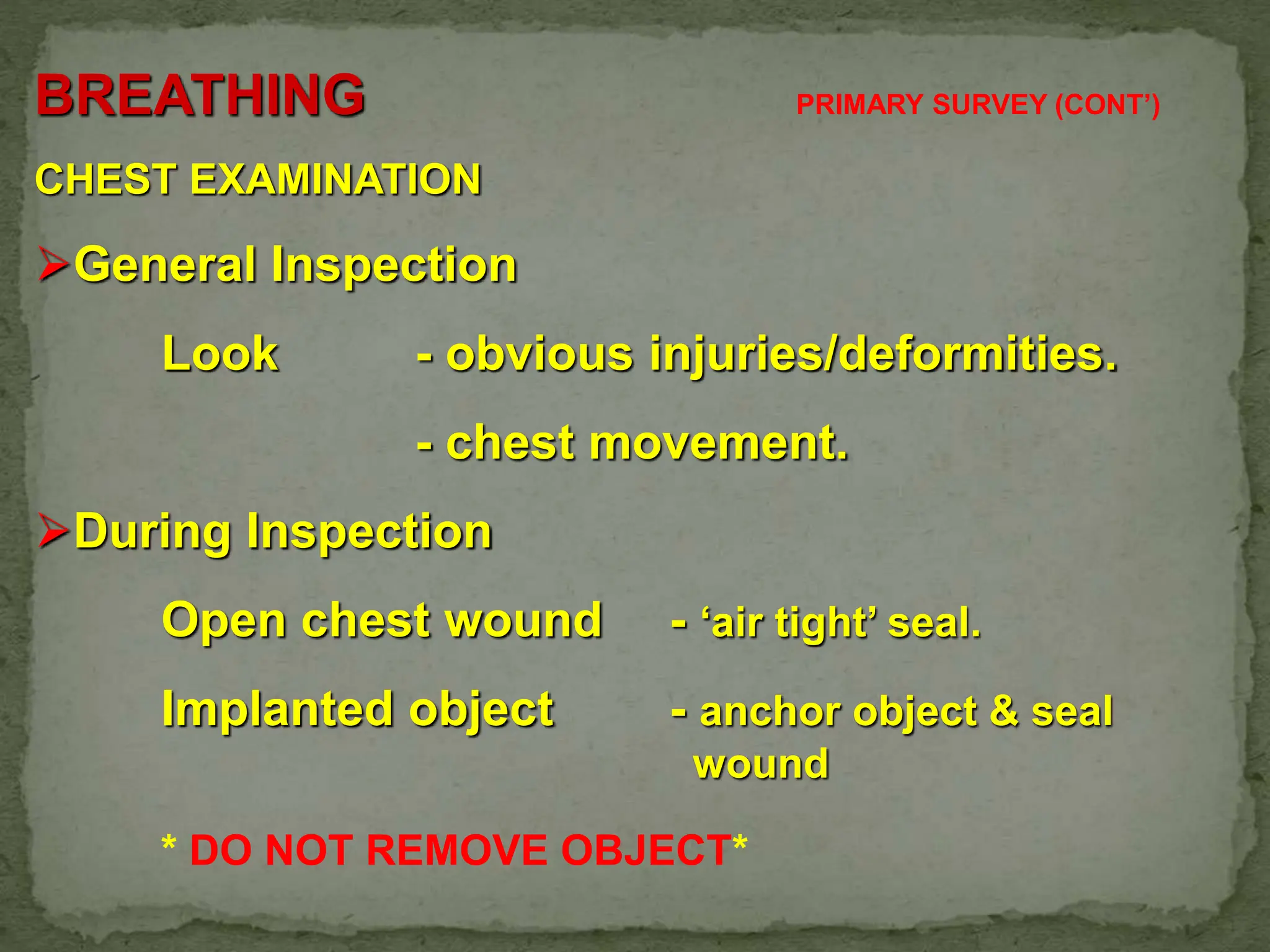
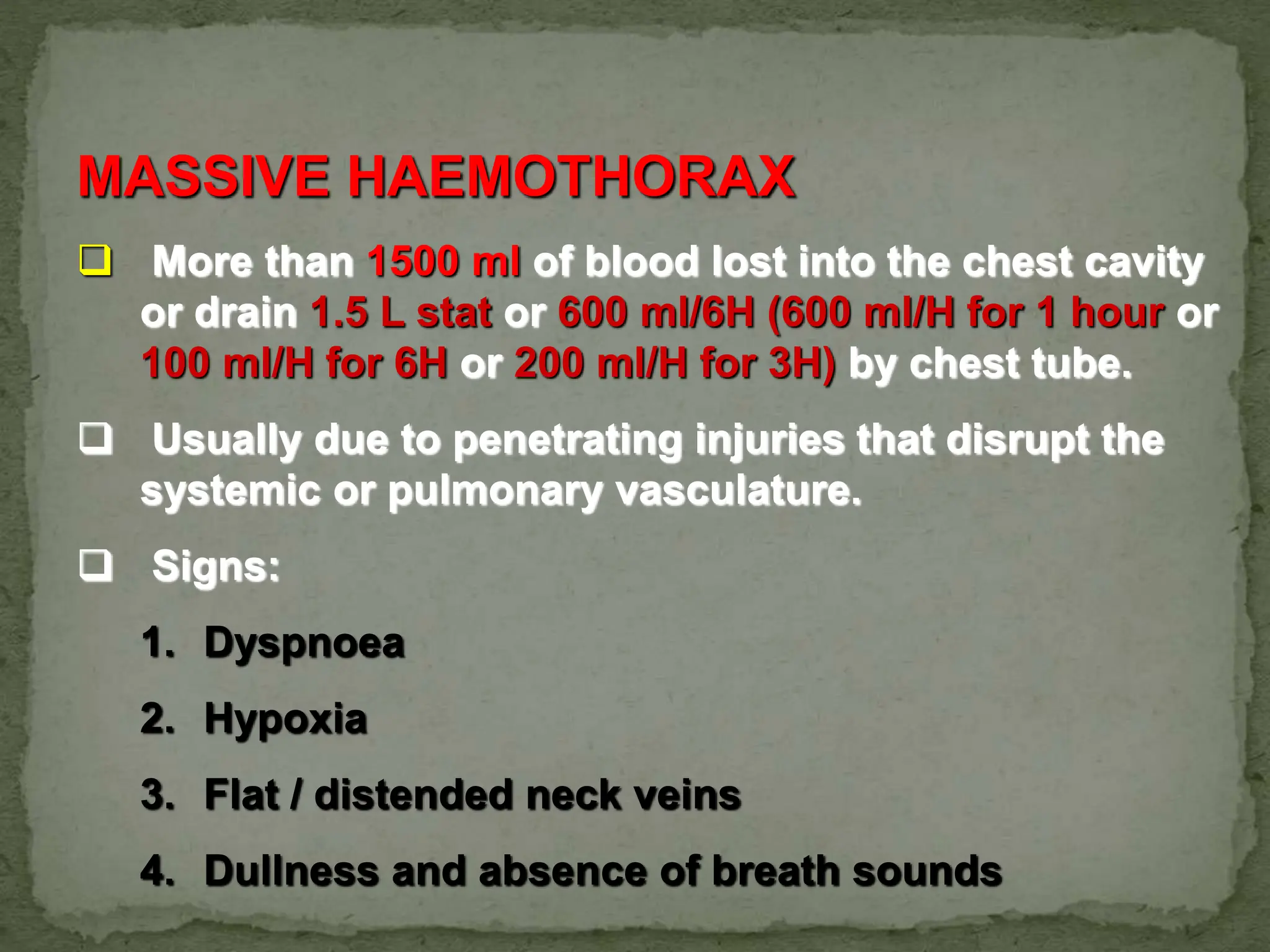
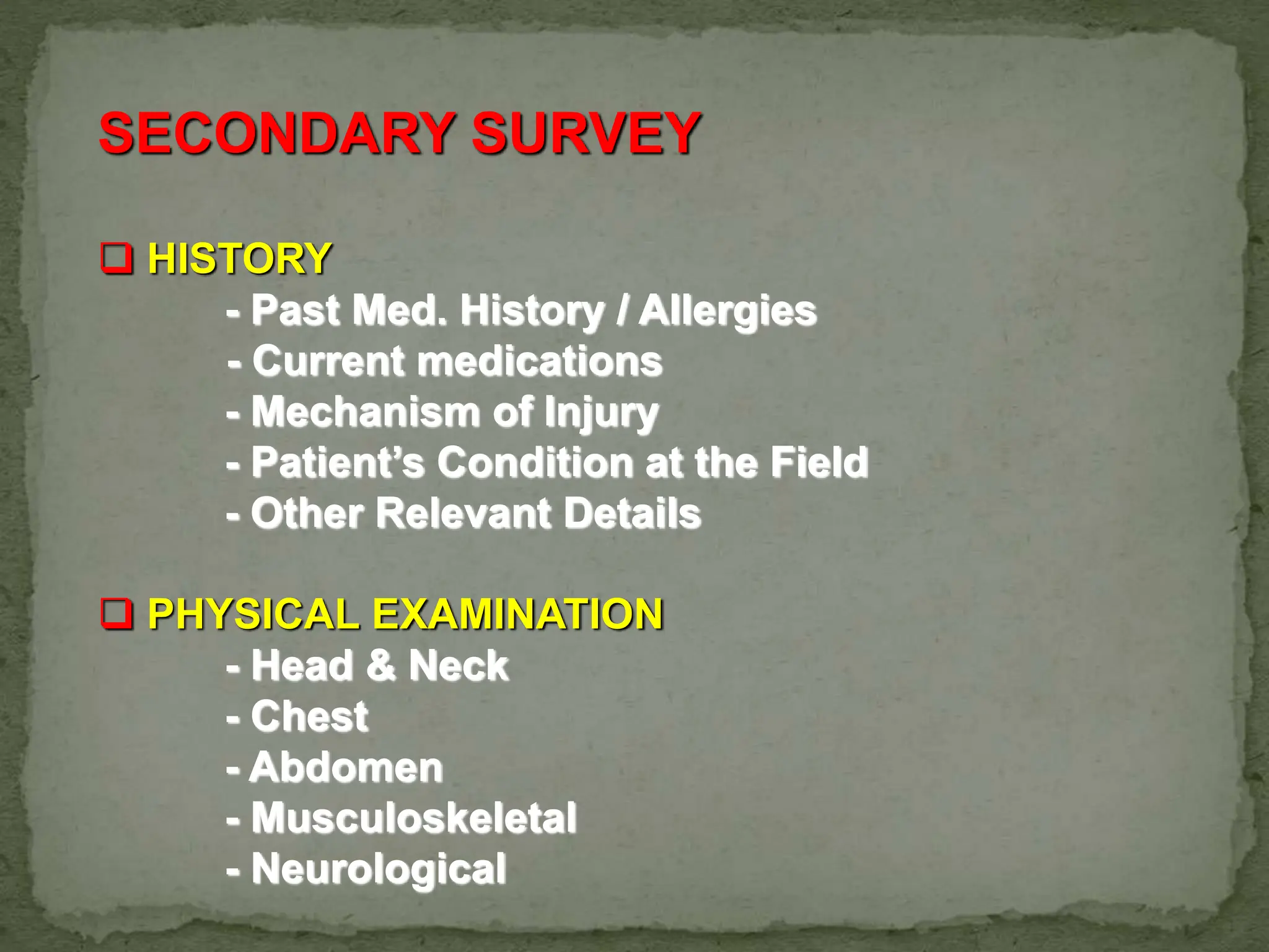
POLYTRAUMA
PRIMARY AND SECONDARY
SURVEY
3
0
10
20
30
40
50
60
70
seconds 30 min 1 hours 4hours 8 hours day 5 week
Line 1
4
TRAUMA DEATH
The First Peak of Death is within seconds to minutes
of injury.
Causes:
1. Laceration of the Brain.
2. Brain Stem injury.
3. High Spinal Cord Injury.
4. Heart, Aorta and Large vessels lacerations.
* Only prevention can reduce this peak trauma related death
* Usually non-salvageable
Undergraduates CME 5
Undergraduates CME 6
The Second Death Peak occurs within minutes to
several hours after injury.
Main focus of Trauma Life Support is in this peak.
This period is referred to as the “Golden Hour”.
Conceptionally, “Golden Hour - First Hour”
Characterized by;
1. Rapid Transportation
2. Rapid assessment and stabilization
3. Rapid definitive care
TRAUMA DEATH
Undergraduates CME 7
Second Peak of Trauma Deaths are
preventable and managable.
Conditions include:
A. Subdural/Extradural Haematoma.
B. Haemo-pneumothorax.
C. Ruptured Spleen/Liver lacerations.
D. Pelvic Fractures.
E. Multiple injuries associated with significant
blood loss.
TRAUMA DEATH
8
The Third Peak of Death occurs several
days or weeks after initial injury.
Causes:
1. Sepsis.
2. Organ Failure.
TRAUMA DEATH
9
WHAT IS POLYTRAUMA ?
10
POLYTRAUMA
Definition :
A Clinical syndrome where a patient
sustained serious injuries involving
two or more major organ and
physiological systems.
Injury
Reevaluation
Resuscitation
Adjuncts
Primary Survey
Adjuncts
Secondary Survey
Reevaluation
Optimize patient
status
Transfer
12
PRIMARY SURVEY :-
Definition :-
The Preliminary Assessment of a patient which
is conducted in a systematic manner with the
objective of identifying life threatening
conditions and managing them as soon as they
are found.
13
THE FIRST PERSON TO
ASSESS THE PATIENT CAN
AFFECT THE FINAL
OUTCOME
14
Primary survey and
resuscitation of vital
functions are done
simultaneously.
15
PRIMARY SURVEY
Should not take more than 2 minutes
should not be interrupted unless there
is airway obstruction or cardiac arrest
16
PRIMARY SURVEY
IMMEDIATE ASSESSMENT
A. AIRWAY & CERVICAL SPINE CONTROL
B. BREATHING & VENTILATION
C. CIRCULATORY FUNCTION & HEMORRHAGE
CONTROL
D. DISABILITY & NEUROLOGICAL STATUS
E. EXPOSURE & UNDRESS COMPLETELY
17
PRIMARY SURVEY - AIRWAY
1. GENERAL INSPECTION
•ANY LIFE THREATENING CONDITION DETECTED MUST BE
CORRECTED SIMULTANEOUSLY *
-ATOMFC-
18
Airway Obstruction
Causes:
Presence of secretions and foreign matter
in the mouth.
Tongue falling back in the unconscious
patient.
Deformity and injury to the airway
including maxillofacial injuries.
Swelling and inflammation of the airway as
in burns injury or ingestion of toxic
substances.
laryngospasm
PRIMARY SURVEY
1. Blood/Secretions - Suction/removal of debris.
2. Floppy Tongue - Oropharyngeal airway
Airway and bag
3. Maxillo-facial injury - Attempt reduction,
intubation or
cricothyrodotomy.
4. Mechanical blockade - Finger sweep and removal of
object.
5. Partially Airway Obst. - jaw thrust / chin-lift
AIRWAY MANAGEMENT
20
Protection of the C-spine
Assume that the C-spine is damaged in any
injury above the clavicle.
Examination of the neck is to be done
together with manual immobilization.
Note any injury to the neck
e.g tracheal deviation
surgical emphysema
the neck collar must be rigid and of the
correct size.
December 12, 2011
Undergraduates CME 21
22
PROTECTION OF THE C-SPINE
23
PROTECTION OF THE C-SPINE
BREATHING PRIMARY SURVEY (CONT’)
CHEST EXAMINATION
General Inspection
Look - obvious injuries/deformities.
- chest movement.
During Inspection
Open chest wound - ‘air tight’ seal.
Implanted object - anchor object & seal
wound
* DO NOT REMOVE OBJECT*
Examination of Chest
Apex beat, chest expansion & percussion note.
‘Spring Test’
Positive - conscious - tenderness at
fracture site.
- unconscious - laxity of rib-cage
Auscultation
- Apex site
- Quality of heart sound.
- Air entry and Abnormal sounds.
PRIMARY SURVEY (CONT’D)
‘Life threatening conditions must be
diagnosed and treated immediately’.
1. Airway Obstruction.
2. Tension Pneumothorax.
3. Open Pneumothorax.
4. Massive Haemothorax.
5. Flail Chest.
6. Cardiac Tamponade.
27
TENSION
PNEUMOTHORAX
* One-way valve
mechanism
* Air trapped in
pleural space
* Increase press. in
pleural space
* Lung collapse
with mediastinal
shift
Undergraduates CME 28
TENSION PNEUMOTHORAX
 SIGNS
1. Tracheal Deviation
2. Respiratory Distress
• Absence of breath sounds - Unilateral
• Distended Neck Veins
• Cyanosis – Late
 DIAGNOSIS - Clinically, NOT Radiological
 MANAGEMENT
• Needle Thoracocentesis
• Chest Tube Insertion
MASSIVE HAEMOTHORAX
 More than 1500 ml of blood lost into the chest cavity
or drain 1.5 L stat or 600 ml/6H (600 ml/H for 1 hour or
100 ml/H for 6H or 200 ml/H for 3H) by chest tube.
 Usually due to penetrating injuries that disrupt the
systemic or pulmonary vasculature.
 Signs:
1. Dyspnoea
2. Hypoxia
3. Flat / distended neck veins
4. Dullness and absence of breath sounds
MASSIVE HAEMOTHORAX
MANAGEMENT
1. Rapid Volume Restoration
2. Chest Tube Insertion
3. Thoracotomy - consider if blood lost
is more than 600ml/6H
OPEN PNEUMOTHORAX
Large defects / open
wounds (diameter of
wound > than trachea)
causing ‘sucking’ chest
wounds.
Equilibration between
intrathoracic and
atmospheric pressure
resulting in impairment of
effective ventilation.
OPEN PNEUMOTHORAX
MANAGEMENT:
1. Cover defect with sterile occlusive
dressing.
2. Chest tube insertion.
3. Definitive surgical closure.
OPEN PNEUMOTHORAX ( SUCKING CHEST WOUND )
37
December 12, 2011 38
Flail Chest
When a segment of chest
wall does not have bony
continuity with the rest of
the thoracic cage
Usually defined as at least
two fractures per rib (
producing a free segment)
in at least two ribs
FLAIL CHEST
MANAGEMENT
 Adequate ventilation & Oxygen
 Volume restoration
 Analgesia
CARDIAC TAMPONADE
COMMON CAUSES
- Penetrating injuries
- Blunt injury
 CHARACTERISTIC
• BECK’S TRIAD
- Elevated JVP
- Muffled Heart Sounds
- Distended Neck Veins
• Narrowed Pulse Pressure
CARDIAC TAMPONADE
In trauma, as little of 150 ml – 200 ml of blood in
pericardium can caused sign of cardiac
tamponade
 MANAGEMENT
- PERICARDIOCENTESIS
- OPEN THORACOTOMY
CARDIAC TAMPONADE
PRIMARY SURVEY (CONT’D)
CIRCULATION & HEMORRHAGE CONTROL
 GENERAL ASSESSMENT
• skin color & temperature
• pulse rate, blood pressure
• capillary refill
• identify source of bleeding
•Inspect, palpate and auscultate abdomen
•Pelvic spring
* DON’T WAIT UNTIL THE BLOOD PRESSURE FALLS TO
SUSPECT SHOCK AND BEGIN TREATMENT *
MANAGEMENT OF THE CIRCULATING SYSTEM
• Control bleeding site by pressure bandage
• Insert 2 large caliber IV cannula
• Rapid infusion of volume replacement
• Send blood for blood GXM
• Insert urinary catheter & naso-gastric tube
(unless contraindicated)
45
DISABILITY & NEUROLOGICAL
STATUS
AVPU/GCS
Pupillary signs
Spine tenderness and per rectal
examination
46
EXPOSURE/ENVIRONMENT
Undress patient completely
 - Thorough examination so that not miss any injury
- Pelvis
- Groin
- Genitalia
- Back
Keep patient warm
47
Reassessment
Reevaluate ABCDE
Reevaluate vital signs
Undergraduates CME 48
Primary Survey
Primary survey adjuncts
– Blood Ix , glucose
– ECG , ABG
– Plain xray
Cervical spine
Chest
Pelvis
– FAST
Focused assessment with sonography in trauma
For detection of fluid in peritoneal and pericardial
space.
SECONDARY SURVEY
 HISTORY
- Past Med. History / Allergies
- Current medications
- Mechanism of Injury
- Patient’s Condition at the Field
- Other Relevant Details
 PHYSICAL EXAMINATION
- Head & Neck
- Chest
- Abdomen
- Musculoskeletal
- Neurological
 CT scan
 Ultrasound
 Bronchoscopy
RE-EVALUATION
Because of the dynamic state of the
physiological systems, the condition may
change within a short period of time. Hence,
after each primary & secondary survey a
complete RE-EVALUATION of all the vital
systems must be carried out.
Always Work in Team
THANK YOU

365577706-Primary-and-Secondary-Survey-in-Trauma.pptx

  • 1.
  • 3.
    3 0 10 20 30 40 50 60 70 seconds 30 min1 hours 4hours 8 hours day 5 week Line 1
  • 4.
    4 TRAUMA DEATH The FirstPeak of Death is within seconds to minutes of injury. Causes: 1. Laceration of the Brain. 2. Brain Stem injury. 3. High Spinal Cord Injury. 4. Heart, Aorta and Large vessels lacerations. * Only prevention can reduce this peak trauma related death * Usually non-salvageable
  • 5.
  • 6.
    Undergraduates CME 6 TheSecond Death Peak occurs within minutes to several hours after injury. Main focus of Trauma Life Support is in this peak. This period is referred to as the “Golden Hour”. Conceptionally, “Golden Hour - First Hour” Characterized by; 1. Rapid Transportation 2. Rapid assessment and stabilization 3. Rapid definitive care TRAUMA DEATH
  • 7.
    Undergraduates CME 7 SecondPeak of Trauma Deaths are preventable and managable. Conditions include: A. Subdural/Extradural Haematoma. B. Haemo-pneumothorax. C. Ruptured Spleen/Liver lacerations. D. Pelvic Fractures. E. Multiple injuries associated with significant blood loss. TRAUMA DEATH
  • 8.
    8 The Third Peakof Death occurs several days or weeks after initial injury. Causes: 1. Sepsis. 2. Organ Failure. TRAUMA DEATH
  • 9.
  • 10.
    10 POLYTRAUMA Definition : A Clinicalsyndrome where a patient sustained serious injuries involving two or more major organ and physiological systems.
  • 11.
  • 12.
    12 PRIMARY SURVEY :- Definition:- The Preliminary Assessment of a patient which is conducted in a systematic manner with the objective of identifying life threatening conditions and managing them as soon as they are found.
  • 13.
    13 THE FIRST PERSONTO ASSESS THE PATIENT CAN AFFECT THE FINAL OUTCOME
  • 14.
    14 Primary survey and resuscitationof vital functions are done simultaneously.
  • 15.
    15 PRIMARY SURVEY Should nottake more than 2 minutes should not be interrupted unless there is airway obstruction or cardiac arrest
  • 16.
    16 PRIMARY SURVEY IMMEDIATE ASSESSMENT A.AIRWAY & CERVICAL SPINE CONTROL B. BREATHING & VENTILATION C. CIRCULATORY FUNCTION & HEMORRHAGE CONTROL D. DISABILITY & NEUROLOGICAL STATUS E. EXPOSURE & UNDRESS COMPLETELY
  • 17.
    17 PRIMARY SURVEY -AIRWAY 1. GENERAL INSPECTION •ANY LIFE THREATENING CONDITION DETECTED MUST BE CORRECTED SIMULTANEOUSLY * -ATOMFC-
  • 18.
    18 Airway Obstruction Causes: Presence ofsecretions and foreign matter in the mouth. Tongue falling back in the unconscious patient. Deformity and injury to the airway including maxillofacial injuries. Swelling and inflammation of the airway as in burns injury or ingestion of toxic substances. laryngospasm
  • 19.
    PRIMARY SURVEY 1. Blood/Secretions- Suction/removal of debris. 2. Floppy Tongue - Oropharyngeal airway Airway and bag 3. Maxillo-facial injury - Attempt reduction, intubation or cricothyrodotomy. 4. Mechanical blockade - Finger sweep and removal of object. 5. Partially Airway Obst. - jaw thrust / chin-lift AIRWAY MANAGEMENT
  • 20.
    20 Protection of theC-spine Assume that the C-spine is damaged in any injury above the clavicle. Examination of the neck is to be done together with manual immobilization. Note any injury to the neck e.g tracheal deviation surgical emphysema the neck collar must be rigid and of the correct size.
  • 21.
  • 22.
  • 23.
  • 24.
    BREATHING PRIMARY SURVEY(CONT’) CHEST EXAMINATION General Inspection Look - obvious injuries/deformities. - chest movement. During Inspection Open chest wound - ‘air tight’ seal. Implanted object - anchor object & seal wound * DO NOT REMOVE OBJECT*
  • 25.
    Examination of Chest Apexbeat, chest expansion & percussion note. ‘Spring Test’ Positive - conscious - tenderness at fracture site. - unconscious - laxity of rib-cage Auscultation - Apex site - Quality of heart sound. - Air entry and Abnormal sounds.
  • 26.
    PRIMARY SURVEY (CONT’D) ‘Lifethreatening conditions must be diagnosed and treated immediately’. 1. Airway Obstruction. 2. Tension Pneumothorax. 3. Open Pneumothorax. 4. Massive Haemothorax. 5. Flail Chest. 6. Cardiac Tamponade.
  • 27.
    27 TENSION PNEUMOTHORAX * One-way valve mechanism *Air trapped in pleural space * Increase press. in pleural space * Lung collapse with mediastinal shift
  • 28.
  • 29.
    TENSION PNEUMOTHORAX  SIGNS 1.Tracheal Deviation 2. Respiratory Distress • Absence of breath sounds - Unilateral • Distended Neck Veins • Cyanosis – Late  DIAGNOSIS - Clinically, NOT Radiological  MANAGEMENT • Needle Thoracocentesis • Chest Tube Insertion
  • 30.
    MASSIVE HAEMOTHORAX  Morethan 1500 ml of blood lost into the chest cavity or drain 1.5 L stat or 600 ml/6H (600 ml/H for 1 hour or 100 ml/H for 6H or 200 ml/H for 3H) by chest tube.  Usually due to penetrating injuries that disrupt the systemic or pulmonary vasculature.  Signs: 1. Dyspnoea 2. Hypoxia 3. Flat / distended neck veins 4. Dullness and absence of breath sounds
  • 32.
    MASSIVE HAEMOTHORAX MANAGEMENT 1. RapidVolume Restoration 2. Chest Tube Insertion 3. Thoracotomy - consider if blood lost is more than 600ml/6H
  • 33.
    OPEN PNEUMOTHORAX Large defects/ open wounds (diameter of wound > than trachea) causing ‘sucking’ chest wounds. Equilibration between intrathoracic and atmospheric pressure resulting in impairment of effective ventilation.
  • 34.
    OPEN PNEUMOTHORAX MANAGEMENT: 1. Coverdefect with sterile occlusive dressing. 2. Chest tube insertion. 3. Definitive surgical closure.
  • 36.
    OPEN PNEUMOTHORAX (SUCKING CHEST WOUND )
  • 37.
  • 38.
    December 12, 201138 Flail Chest When a segment of chest wall does not have bony continuity with the rest of the thoracic cage Usually defined as at least two fractures per rib ( producing a free segment) in at least two ribs
  • 39.
    FLAIL CHEST MANAGEMENT  Adequateventilation & Oxygen  Volume restoration  Analgesia
  • 40.
    CARDIAC TAMPONADE COMMON CAUSES -Penetrating injuries - Blunt injury  CHARACTERISTIC • BECK’S TRIAD - Elevated JVP - Muffled Heart Sounds - Distended Neck Veins • Narrowed Pulse Pressure
  • 41.
    CARDIAC TAMPONADE In trauma,as little of 150 ml – 200 ml of blood in pericardium can caused sign of cardiac tamponade  MANAGEMENT - PERICARDIOCENTESIS - OPEN THORACOTOMY
  • 42.
  • 43.
    PRIMARY SURVEY (CONT’D) CIRCULATION& HEMORRHAGE CONTROL  GENERAL ASSESSMENT • skin color & temperature • pulse rate, blood pressure • capillary refill • identify source of bleeding •Inspect, palpate and auscultate abdomen •Pelvic spring * DON’T WAIT UNTIL THE BLOOD PRESSURE FALLS TO SUSPECT SHOCK AND BEGIN TREATMENT *
  • 44.
    MANAGEMENT OF THECIRCULATING SYSTEM • Control bleeding site by pressure bandage • Insert 2 large caliber IV cannula • Rapid infusion of volume replacement • Send blood for blood GXM • Insert urinary catheter & naso-gastric tube (unless contraindicated)
  • 45.
    45 DISABILITY & NEUROLOGICAL STATUS AVPU/GCS Pupillarysigns Spine tenderness and per rectal examination
  • 46.
    46 EXPOSURE/ENVIRONMENT Undress patient completely - Thorough examination so that not miss any injury - Pelvis - Groin - Genitalia - Back Keep patient warm
  • 47.
  • 48.
    Undergraduates CME 48 PrimarySurvey Primary survey adjuncts – Blood Ix , glucose – ECG , ABG – Plain xray Cervical spine Chest Pelvis – FAST Focused assessment with sonography in trauma For detection of fluid in peritoneal and pericardial space.
  • 49.
    SECONDARY SURVEY  HISTORY -Past Med. History / Allergies - Current medications - Mechanism of Injury - Patient’s Condition at the Field - Other Relevant Details  PHYSICAL EXAMINATION - Head & Neck - Chest - Abdomen - Musculoskeletal - Neurological
  • 50.
     CT scan Ultrasound  Bronchoscopy
  • 51.
    RE-EVALUATION Because of thedynamic state of the physiological systems, the condition may change within a short period of time. Hence, after each primary & secondary survey a complete RE-EVALUATION of all the vital systems must be carried out.
  • 52.
  • 53.