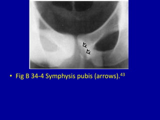
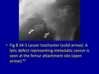
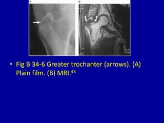
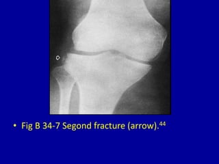
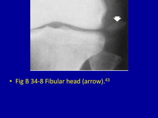
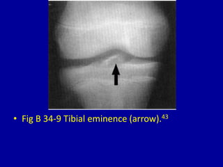
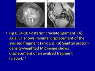
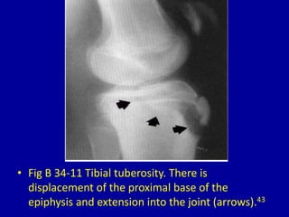
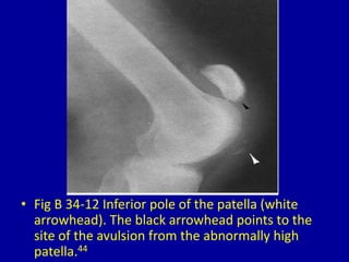
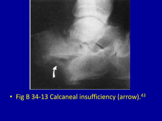
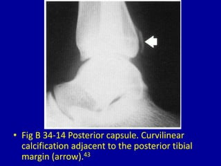
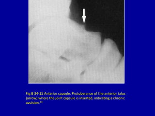
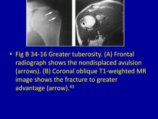
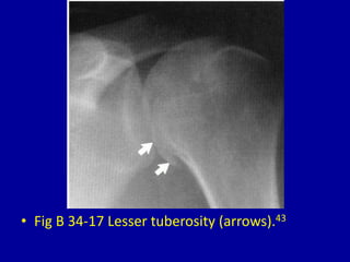
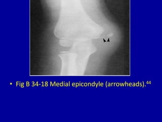
34Avulsion Injuries*
CLINICAL IMAGAGING
AN ATLAS OF DIFFERENTIAL DAIGNOSIS
EISENBERG
DR. Muhammad Bin Zulfiqar
PGR-FCPS III SIMS/SHL
• Fig B 34-1 Ischial tuberosity. Bilateral chronic
avulsions. Note the protuberant bone (closed
arrows) and a large, smooth fragment (open
arrows).43
• Fig B 34-2 Anterior superior iliac spine
(arrowhead).44
• Fig B 34-3 Anterior inferior iliac spine
(arrowhead).44
• Fig B 34-4 Symphysis pubis (arrows).43
• Fig B 34-5 Lesser trochanter (solid arrow). A
lytic defect representing metastatic cancer is
seen at the femur attachment site (open
arrow).43
• Fig B 34-6 Greater trochanter (arrows). (A)
Plain film. (B) MRI.43
• Fig B 34-7 Segond fracture (arrow).44
• Fig B 34-8 Fibular head (arrow).43
• Fig B 34-9 Tibial eminence (arrow).43
• Fig B 34-10 Posterior cruciate ligament. (A)
Axial CT shows minimal displacement of the
avulsed fragment (arrows). (B) Sagittal proton-
density-weighted MR image shows
displacement of an avulsed fragment
(arrow).43
• Fig B 34-11 Tibial tuberosity. There is
displacement of the proximal base of the
epiphysis and extension into the joint (arrows).43
• Fig B 34-12 Inferior pole of the patella (white
arrowhead). The black arrowhead points to the
site of the avulsion from the abnormally high
patella.44
• Fig B 34-13 Calcaneal insufficiency (arrow).43
• Fig B 34-14 Posterior capsule. Curvilinear
calcification adjacent to the posterior tibial
margin (arrow).43
Fig B 34-15 Anterior capsule. Protuberance of the anterior talus
(arrow) where the joint capsule is inserted, indicating a chronic
avulsion.43
• Fig B 34-16 Greater tuberosity. (A) Frontal
radiograph shows the nondisplaced avulsion
(arrows). (B) Coronal oblique T1-weighted MR
image shows the fracture to greater
advantage (arrow).43
• Fig B 34-17 Lesser tuberosity (arrows).43
• Fig B 34-18 Medial epicondyle (arrowheads).44

34 avulsion injuries

  • 1.
  • 2.
    CLINICAL IMAGAGING AN ATLASOF DIFFERENTIAL DAIGNOSIS EISENBERG DR. Muhammad Bin Zulfiqar PGR-FCPS III SIMS/SHL
  • 3.
    • Fig B34-1 Ischial tuberosity. Bilateral chronic avulsions. Note the protuberant bone (closed arrows) and a large, smooth fragment (open arrows).43
  • 4.
    • Fig B34-2 Anterior superior iliac spine (arrowhead).44
  • 5.
    • Fig B34-3 Anterior inferior iliac spine (arrowhead).44
  • 6.
    • Fig B34-4 Symphysis pubis (arrows).43
  • 7.
    • Fig B34-5 Lesser trochanter (solid arrow). A lytic defect representing metastatic cancer is seen at the femur attachment site (open arrow).43
  • 8.
    • Fig B34-6 Greater trochanter (arrows). (A) Plain film. (B) MRI.43
  • 9.
    • Fig B34-7 Segond fracture (arrow).44
  • 10.
    • Fig B34-8 Fibular head (arrow).43
  • 11.
    • Fig B34-9 Tibial eminence (arrow).43
  • 12.
    • Fig B34-10 Posterior cruciate ligament. (A) Axial CT shows minimal displacement of the avulsed fragment (arrows). (B) Sagittal proton- density-weighted MR image shows displacement of an avulsed fragment (arrow).43
  • 13.
    • Fig B34-11 Tibial tuberosity. There is displacement of the proximal base of the epiphysis and extension into the joint (arrows).43
  • 14.
    • Fig B34-12 Inferior pole of the patella (white arrowhead). The black arrowhead points to the site of the avulsion from the abnormally high patella.44
  • 15.
    • Fig B34-13 Calcaneal insufficiency (arrow).43
  • 16.
    • Fig B34-14 Posterior capsule. Curvilinear calcification adjacent to the posterior tibial margin (arrow).43
  • 17.
    Fig B 34-15Anterior capsule. Protuberance of the anterior talus (arrow) where the joint capsule is inserted, indicating a chronic avulsion.43
  • 18.
    • Fig B34-16 Greater tuberosity. (A) Frontal radiograph shows the nondisplaced avulsion (arrows). (B) Coronal oblique T1-weighted MR image shows the fracture to greater advantage (arrow).43
  • 19.
    • Fig B34-17 Lesser tuberosity (arrows).43
  • 20.
    • Fig B34-18 Medial epicondyle (arrowheads).44