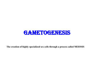
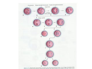
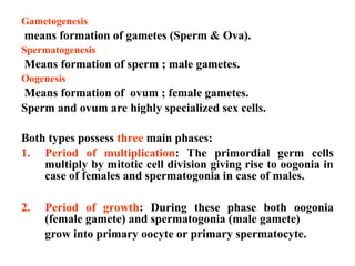
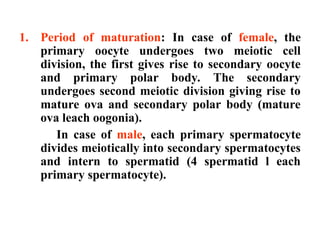
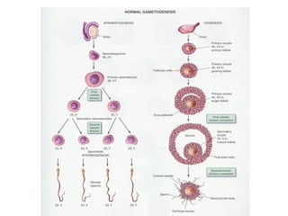
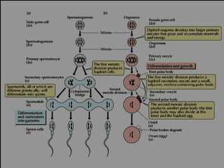
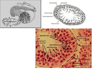
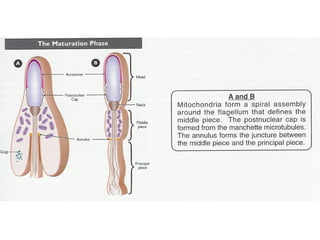
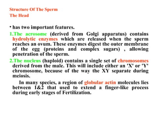
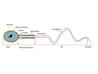
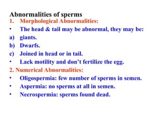
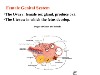
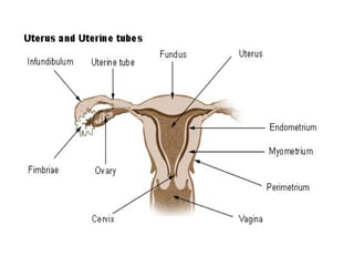
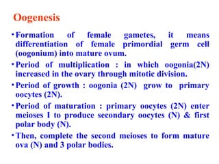
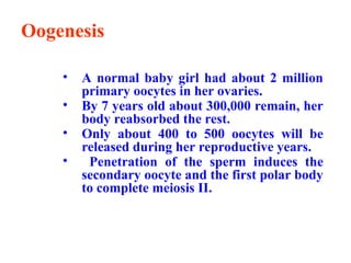
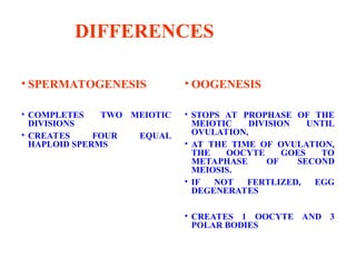
The document discusses gametogenesis, focusing on the processes of spermatogenesis and oogenesis, which create male and female gametes, respectively. It details the phases involved in each process, including multiplication, growth, and maturation, as well as providing an overview of the male and female reproductive systems. Key differences between spermatogenesis and oogenesis are highlighted, emphasizing the outcomes and stages of each process.

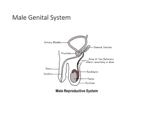
![• Prostate Gland (Ejaculatory Duct)
•Contributes Milky Alkaline Fluid that assists
Sperm Activation
• Cowpers Gland [Bilbourethral Gland]
•Contributes Mucus to Semen
• Urethra (Penis)
•Organ of Copulation](https://image.slidesharecdn.com/3-250130194338-5590a4c6/85/3-Embryology-Gametogenesis-Gametogenesis-ppt-12-320.jpg)

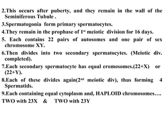


![CTEV [ clubfoot] DR ARUN LAL ,DR MOHAMED ASHRAF travancore medical college k...](https://cdn.slidesharecdn.com/ss_thumbnails/ctevclubfootdrarunlaldrmohamedashraftravancoremedicalcollegekollamkeralaindia-260208063247-18fc466c-thumbnail.jpg?width=640&height=640&fit=bounds)
