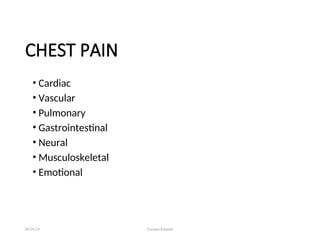
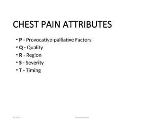
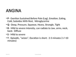
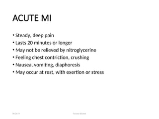
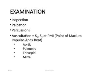
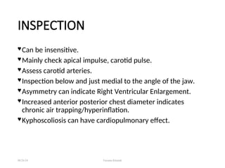
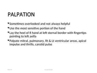
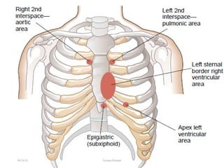
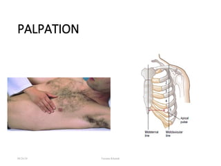
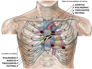
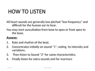
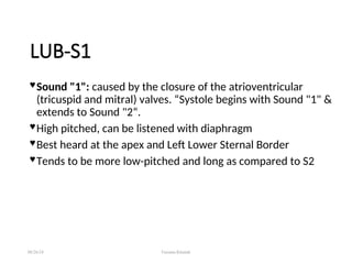
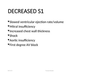
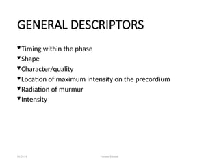
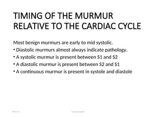
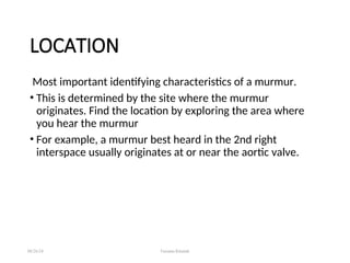
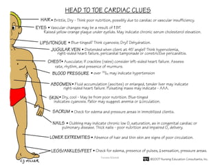
UNIT-III: ASSESSMENT
OF CARDIOVASCULAR
SYSTEM
By: Farzana Kausar Khattak
Lecturer
INS-KMU
• Present illness, chief complaint
• Pain: Onset, course duration, quality precipitating &
alleviating factors
• Fatigue
• Palpitation
• Pain
• Dyspnea
• Cough
• Exercise
08/26/24 Farzana Khattak
HISTORY
• Syncope
• Dependent Edema.
• Weight gain
• Nocturia
• Hemoptysis
• Cyanosis
08/26/24 Farzana Khattak
HISTORY
PAST MEDICAL HISTORY
• Co-morbids / known case
• Previous illness
• Hospitalization
• Surgeries
• Use of drugs, recreational drug use, herbs
• Allergies
08/26/24 Farzana Khattak
FAMILY HISTORY
• HTN
• Diabetes
• Stroke
• Kidney disease
• Siblings & parents health
08/26/24 Farzana Khattak
PSYCOSOCIAL HISTORY
• Occupation
• Education
• Stress tolerance
• Coping
• Marital status
• Health habits, drugs, smoking etc.
08/26/24 Farzana Khattak
HEART PHYSICAL ASSESSMENT
• General
• BP
• Arterial Pulse
• JVD
• Inspection, Palpation, Percussion & Auscultation
• Edema
08/26/24 Farzana Khattak
GENERAL APPEARANCE
• Patient Position
• Facial Expression
• Restless
• Quiet
• Pallor
• Cyanosis
• Level Of Consciousness
08/26/24 Farzana Khattak
CHEST PAIN
• Cardiac
• Vascular
• Pulmonary
• Gastrointestinal
• Neural
• Musculoskeletal
• Emotional
08/26/24 Farzana Khattak
CHEST PAIN ATTRIBUTES
• P - Provocative-palliative Factors
• Q - Quality
• R - Region
• S - Severity
• T - Timing
08/26/24 Farzana Khattak
ANGINA
• P - Exertion Sustained Before Pain (Lag), Emotion, Eating,
Cold, Subsides With Rest, Nitroglycerine
• Q - Deep, Pressure, Squeeze, Heavy, Strangle, Tight
• R - Mild to severe intensity, can radiate to Jaw, arms, neck,
back: Diffuse
• S - Mild to severe
• T - Episodic, “seizes”, Duration is short: 2-3 minutes (>/<10
minutes)
08/26/24 Farzana Khattak
ACUTE MI
• Steady, deep pain
• Lasts 20 minutes or longer
• May not be relieved by nitroglycerine
• Feeling chest contriction, crushing
• Nausea, vomiting, diaphoresis
• May occur at rest, with exertion or stress
08/26/24 Farzana Khattak
PERICARDITIS
• Deep constant or pleuritic pain
• Pericardial friction, may be related to resp.
• Increases with cough
• Sharp, stabbing
• Fever or recent infection
• Shallow breathing, sitting up, leaning forward relieves
08/26/24 Farzana Khattak
• Palpitation is an abnormality of heartbeat that ranges from
often unnoticed skipped beats or accelerated heart rate to
very noticeable changes.
• May not indicate serious disease.
• Cardiac
• Thyrotoxicosis (hyperthyroidism)
• Hypoglycemia
• Fever
• Anemia
• Anxiety
• Other Factors: Caffeine, Tobacco, Drugs
08/26/24 Farzana Khattak
PALPITATIONS: ARRHYTHMIAS
SYNCOPE
• Fainting, Dizziness, Blackout
• Cardiac
• Metabolic
• Psychiatric
• Neurologic
• Orthostatic Hypertension
08/26/24 Farzana Khattak
FATIGUE (MOST COMMON)
• Decreased cardiac output
• CHF
• Mitral valvular disease
• Anxiety & depression
• Anemia or chronic diseases
08/26/24 Farzana Khattak
DEPENDENT EDEMA
• CHF
• Worse as day progresses
• SOB
EDEMA GRADING
• +1 = 2mm
• +2 = 4mm
• +3 = 6 mm
• +4 = 8 mm
08/26/24 Farzana Khattak
GENERAL EXAMINATION
GUIDELINES
08/26/24 Farzana Khattak
THE PATIENT
Should have their shirt(s) off, or wear an
examination gown
Females nine years old and older should wear a
gown with the opening in the front
Should be calm and quiet
POSITION OF PATIENT
• Supine, with the head elevated 30°
•Left lateral decubitus
•Sitting, leaning forward, after full exhalation
08/26/24 Farzana Khattak
THE STETHOSCOPE
• Should have a separate bell and diaphragm
• Bell allows in all sounds
• Diaphragm lets in middle and high frequency sounds
• Bell should be used relatively lightly (avoid diaphragm
effect)
08/26/24 Farzana Khattak
THE ENVIRONMENT
 Should be quiet (patient, family, clinic attendants, exam
room, surrounding areas)
 Should be well lighted.
08/26/24 Farzana Khattak
EXAMINATION
•Inspection
•Palpation
•Percussion?
•Auscultation = S1, S2 at PMI (Point of Maxium
Impulse-Apex Beat)
• Aortic
• Pulmonic
• Tricuspid
• Mitral
08/26/24 Farzana Khattak
INSPECTION
 Can be insensitive.
 Mainly check apical impulse, carotid pulse.
 Assess carotid arteries.
 Inspection below and just medial to the angle of the jaw.
 Asymmetry can indicate Right Ventricular Enlargement.
 Increased anterior posterior chest diameter indicates
chronic air trapping/hyperinflation.
 Kyphoscoliosis can have cardiopulmonary effect.
08/26/24 Farzana Khattak
PALPATION
 Sometimes overlooked and not always helpful
 Use the most sensitive portion of the hand
 Lay the heel of R hand at left sternal border with fingertips
pointing to left axilla
 Palpate mitral, pulmonary, Rt & Lt ventricular areas, apical
impulse and thrills, carotid pulse
08/26/24 Farzana Khattak
PALPATION
08/26/24 Farzana Khattak
PERCUSSION
• Percussion of the heart is not commonly done since chest X
ray study is a more accurate measure of heart enlargement.
• Usually not performed for cardiac borders, but for lung
fields.
• The sound will change from resonance (over the lungs) to
dullness (over the heart).
• Should be done in the upright position (even infants can be
held upright).
08/26/24 Farzana Khattak
AUSCULTATION
WHERE TO LISTEN:
Apex/5LICS (mitral area)
Left lower sternal border/4LICS (tricuspid and secondary
aortic area)
Right middle sternal border/2RICS (aortic area)
Left middle sternal border/2LICS (pulmonary area)
08/26/24 Farzana Khattak
• Aortic: 2nd right intercostal space.
• Pulmonary: 2nd left intercostal space.
• Tricuspid: 4th intercostal space, at lower left
sternal border.
• Mitral: 5th left intercostal space, 1 cm medial
to midclavicular line.
08/26/24 Farzana Khattak
HOW TO LISTEN
All heart sounds are generally low pitched “low frequency” and
difficult for the human ear to hear.
You may start auscultation from base to apex or from apex to
the base.
Assess:
1. Rate and rhythm of the beat.
2. Concentrates initially on sound "1", noting its intensity and
variations.
3. Then listen to Sound "2" for same characteristics.
4. Finally listen for extra sounds and for murmurs
08/26/24 Farzana Khattak
LUB-S1
 Sound "1": caused by the closure of the atrioventricular
(tricuspid and mitral) valves. “Systole begins with Sound "1" &
extends to Sound "2“.
 High pitched, can be listened with diaphragm
 Best heard at the apex and Left Lower Sternal Border
 Tends to be more low-pitched and long as compared to S2
08/26/24 Farzana Khattak
DECREASED S1
 Slowed ventricular ejection rate/volume
 Mitral insufficiency
 Increased chest wall thickness
 Shock
 Aortic insufficiency
 First degree AV block
08/26/24 Farzana Khattak
INCREASED S1
• Increased cardiac output
• Increased A-V valve flow velocity (acquired mitral
stenosis, but not congenital MS)
08/26/24 Farzana Khattak
DUB-S2
• Results from closing of the aortic & pulmonary valves.
• Sound 2 louder than Sound 1 at the base of heart, and is
quieter than Sound 1 at the apex.
• Divided into A2 and P2 (aortic and pulmonary closure
sounds) Normally split due to different impedance of
systemic and pulmonary vascular bed.
• Best heard at 2LICS
 Higher pitched than S1-better heard with diaphragm
08/26/24 Farzana Khattak
S2 SPLITTING (NORMAL)
 Normally split due to different impedance of systemic and
pulmonary vascular bed.
08/26/24 Farzana Khattak
EXTRA HEART SOUNDS
S3 (GALLOP)
Usually physiologic
Low pitched sound, occurs with rapid filling of ventricles in
early diastole causes vibrations of ventricular walls , and this
known as sound "3" .
•Sound "3" best heard at the apex with bell of stethoscope.
Best heard with patient supine or in left lateral decubitus
Increased by exercise, abdominal pressure, or lifting legs
LV S3 heard at apex and RV S3 heard at LLSB
08/26/24 Farzana Khattak
S4 (GALLOP)
 Nearly always pathologic
 Can be normal in elderly or athletes
 Low pitched sound in late diastole
 Sound "4": occur after Sound "3" (late diastolic filling), occur
from vibrations of ventricular wall or vibrations of the
valves.
 Better heard at the apex in the supine or left lateral
decubitus position
 Occurs separate from S3
08/26/24 Farzana Khattak
S4 ASSOCIATIONS
 Congestive cardiac failure(CCF)
 Severe systemic HTN
 Pulmonary HTN
 Myocarditis
08/26/24 Farzana Khattak
EJECTION / SYSTOLIC CLICK
 Usually pathologic
 Snappy, high pitched sound usually in early systole
 Due to vibrations in the artery distal to a stenotic valve
08/26/24 Farzana Khattak
WHOOP (SOMETIMES CALLED A
HONK)
 Loud, variable intensity, musical sound heard at the apex in
late systole
 Classically associated w/ and MR(Mitral Valve Regargitation)
 Seen with VSD’s closing, w/ an aneurysm,
 Some whoops evolve to become systolic murmurs
08/26/24 Farzana Khattak
FRICTION RUB
 Creaking sound heard with pericardial inflammation.
 The sound of a pericardial rub resembles the sound of
squeaky leather and is often described as grating, scratching,
or rasping.
 Changes with position, louder with inspiration.
08/26/24 Farzana Khattak
MURMUR
 Sounds made by turbulence in the heart or blood stream
 Can be benign (innocent, flow, functional) or pathologic
 Murmurs are the leading cause for referral for further
evaluation
 Don’t let murmurs distract you from the rest of the exam!!
08/26/24 Farzana Khattak
CARDIAC EXAM AND MURMUR
GENERAL DESCRIPTORS
 Various combinations used for all normal and abnormal
heart sounds
08/26/24 Farzana Khattak
GENERAL DESCRIPTORS
 Timing within the phase
 Shape
 Character/quality
 Location of maximum intensity on the precordium
 Radiation of murmur
 Intensity
08/26/24 Farzana Khattak
TIMING OF THE MURMUR
RELATIVE TO THE CARDIAC CYCLE
Most benign murmurs are early to mid systolic.
• Diastolic murmurs almost always indicate pathology.
• A systolic murmur is present between S1 and S2
• A diastolic murmur is present between S2 and S1
• A continuous murmur is present in systole and diastole
08/26/24 Farzana Khattak
LOCATION
Most important identifying characteristics of a murmur.
• This is determined by the site where the murmur
originates. Find the location by exploring the area where
you hear the murmur
• For example, a murmur best heard in the 2nd right
interspace usually originates at or near the aortic valve.
08/26/24 Farzana Khattak
RADIATION
• This reflects not only the site of origin but also the intensity
of the murmur and the direction of blood flow.
• For example A loud murmur of aortic stenosis often radiates
into the neck (in the direction of arterial flow).
08/26/24 Farzana Khattak
INTENSITY
• Intensity is synonymous with the loudness or amplitude of a
sound wave.
• Grade 1: very soft and heard with difficulty
• Grade 2: soft but readily heard with stethoscope
• Grade 3: moderately loud, no thrill.
• Grade 4: Loud with thrill (palpable vibration of the chest wall).
Louder than the first and second heart sounds.
• Grade 5: Thrill, very loud, but not audible without a
stethoscope
• Grade 6: Thrill, audible without a stethoscope
08/26/24 Farzana Khattak
QUALITY
This is described in terms such as blowing, harsh, rumbling, and
musical.
PITCH
•The frequency of a murmur depends on the pressure gradient
across a valve or narrowing.
•Low-pitched murmurs are heard best with a bell, and high-
pitched murmurs are heard best with a diaphragm.
08/26/24 Farzana Khattak
OTHER ASSESSMENTS
JUGULAR VEIN PRESSURE
Assess JVD which reflects increased filling volume and
pressure on (R) side of heart
 JVD associated with (R) HF,
•(Normal is 4cm)
PULSE DEFICIT
•The difference between apical HR and peripheral pulse
associated with heart blocks
PULSE PRESSURE
The difference between systolic & diastolic pressure
08/26/24 Farzana Khattak
AGE RELATED CHANGES
• Decreased myocardial contractility
• Thickening of endocardium & valves
• Coronary arteries rigid & thickened
• Decreased elasticity of vessel walls
• Decreased internal diameter of vessels
08/26/24 Farzana Khattak
DOCUMENTATION
• No visible pulsation on anterior chest.
• PMI palpable at left 5th
ICS.
• Heart Auscultation: rate 68 beats/ min, regular rhythm,
S1and S2 identified.
• No extra heat sounds, murmur or rubs.
August 26, 2024 Unit-III (Cardiac Assessment) || By: Noor 54
08/26/24 Farzana Khattak

3-CARDIOVASCULAR ASSESSMENt presentation. ppt

  • 1.
    UNIT-III: ASSESSMENT OF CARDIOVASCULAR SYSTEM By:Farzana Kausar Khattak Lecturer INS-KMU
  • 2.
    • Present illness,chief complaint • Pain: Onset, course duration, quality precipitating & alleviating factors • Fatigue • Palpitation • Pain • Dyspnea • Cough • Exercise 08/26/24 Farzana Khattak HISTORY
  • 3.
    • Syncope • DependentEdema. • Weight gain • Nocturia • Hemoptysis • Cyanosis 08/26/24 Farzana Khattak HISTORY
  • 4.
    PAST MEDICAL HISTORY •Co-morbids / known case • Previous illness • Hospitalization • Surgeries • Use of drugs, recreational drug use, herbs • Allergies 08/26/24 Farzana Khattak
  • 5.
    FAMILY HISTORY • HTN •Diabetes • Stroke • Kidney disease • Siblings & parents health 08/26/24 Farzana Khattak
  • 6.
    PSYCOSOCIAL HISTORY • Occupation •Education • Stress tolerance • Coping • Marital status • Health habits, drugs, smoking etc. 08/26/24 Farzana Khattak
  • 7.
    HEART PHYSICAL ASSESSMENT •General • BP • Arterial Pulse • JVD • Inspection, Palpation, Percussion & Auscultation • Edema 08/26/24 Farzana Khattak
  • 8.
    GENERAL APPEARANCE • PatientPosition • Facial Expression • Restless • Quiet • Pallor • Cyanosis • Level Of Consciousness 08/26/24 Farzana Khattak
  • 9.
    CHEST PAIN • Cardiac •Vascular • Pulmonary • Gastrointestinal • Neural • Musculoskeletal • Emotional 08/26/24 Farzana Khattak
  • 10.
    CHEST PAIN ATTRIBUTES •P - Provocative-palliative Factors • Q - Quality • R - Region • S - Severity • T - Timing 08/26/24 Farzana Khattak
  • 11.
    ANGINA • P -Exertion Sustained Before Pain (Lag), Emotion, Eating, Cold, Subsides With Rest, Nitroglycerine • Q - Deep, Pressure, Squeeze, Heavy, Strangle, Tight • R - Mild to severe intensity, can radiate to Jaw, arms, neck, back: Diffuse • S - Mild to severe • T - Episodic, “seizes”, Duration is short: 2-3 minutes (>/<10 minutes) 08/26/24 Farzana Khattak
  • 12.
    ACUTE MI • Steady,deep pain • Lasts 20 minutes or longer • May not be relieved by nitroglycerine • Feeling chest contriction, crushing • Nausea, vomiting, diaphoresis • May occur at rest, with exertion or stress 08/26/24 Farzana Khattak
  • 13.
    PERICARDITIS • Deep constantor pleuritic pain • Pericardial friction, may be related to resp. • Increases with cough • Sharp, stabbing • Fever or recent infection • Shallow breathing, sitting up, leaning forward relieves 08/26/24 Farzana Khattak
  • 14.
    • Palpitation isan abnormality of heartbeat that ranges from often unnoticed skipped beats or accelerated heart rate to very noticeable changes. • May not indicate serious disease. • Cardiac • Thyrotoxicosis (hyperthyroidism) • Hypoglycemia • Fever • Anemia • Anxiety • Other Factors: Caffeine, Tobacco, Drugs 08/26/24 Farzana Khattak PALPITATIONS: ARRHYTHMIAS
  • 15.
    SYNCOPE • Fainting, Dizziness,Blackout • Cardiac • Metabolic • Psychiatric • Neurologic • Orthostatic Hypertension 08/26/24 Farzana Khattak
  • 16.
    FATIGUE (MOST COMMON) •Decreased cardiac output • CHF • Mitral valvular disease • Anxiety & depression • Anemia or chronic diseases 08/26/24 Farzana Khattak
  • 17.
    DEPENDENT EDEMA • CHF •Worse as day progresses • SOB EDEMA GRADING • +1 = 2mm • +2 = 4mm • +3 = 6 mm • +4 = 8 mm 08/26/24 Farzana Khattak
  • 18.
  • 19.
    THE PATIENT Should havetheir shirt(s) off, or wear an examination gown Females nine years old and older should wear a gown with the opening in the front Should be calm and quiet POSITION OF PATIENT • Supine, with the head elevated 30° •Left lateral decubitus •Sitting, leaning forward, after full exhalation 08/26/24 Farzana Khattak
  • 20.
    THE STETHOSCOPE • Shouldhave a separate bell and diaphragm • Bell allows in all sounds • Diaphragm lets in middle and high frequency sounds • Bell should be used relatively lightly (avoid diaphragm effect) 08/26/24 Farzana Khattak
  • 21.
    THE ENVIRONMENT  Shouldbe quiet (patient, family, clinic attendants, exam room, surrounding areas)  Should be well lighted. 08/26/24 Farzana Khattak
  • 22.
    EXAMINATION •Inspection •Palpation •Percussion? •Auscultation = S1,S2 at PMI (Point of Maxium Impulse-Apex Beat) • Aortic • Pulmonic • Tricuspid • Mitral 08/26/24 Farzana Khattak
  • 23.
    INSPECTION  Can beinsensitive.  Mainly check apical impulse, carotid pulse.  Assess carotid arteries.  Inspection below and just medial to the angle of the jaw.  Asymmetry can indicate Right Ventricular Enlargement.  Increased anterior posterior chest diameter indicates chronic air trapping/hyperinflation.  Kyphoscoliosis can have cardiopulmonary effect. 08/26/24 Farzana Khattak
  • 24.
    PALPATION  Sometimes overlookedand not always helpful  Use the most sensitive portion of the hand  Lay the heel of R hand at left sternal border with fingertips pointing to left axilla  Palpate mitral, pulmonary, Rt & Lt ventricular areas, apical impulse and thrills, carotid pulse 08/26/24 Farzana Khattak
  • 25.
  • 26.
  • 27.
    PERCUSSION • Percussion ofthe heart is not commonly done since chest X ray study is a more accurate measure of heart enlargement. • Usually not performed for cardiac borders, but for lung fields. • The sound will change from resonance (over the lungs) to dullness (over the heart). • Should be done in the upright position (even infants can be held upright). 08/26/24 Farzana Khattak
  • 28.
    AUSCULTATION WHERE TO LISTEN: Apex/5LICS(mitral area) Left lower sternal border/4LICS (tricuspid and secondary aortic area) Right middle sternal border/2RICS (aortic area) Left middle sternal border/2LICS (pulmonary area) 08/26/24 Farzana Khattak
  • 29.
    • Aortic: 2ndright intercostal space. • Pulmonary: 2nd left intercostal space. • Tricuspid: 4th intercostal space, at lower left sternal border. • Mitral: 5th left intercostal space, 1 cm medial to midclavicular line. 08/26/24 Farzana Khattak
  • 30.
  • 31.
    HOW TO LISTEN Allheart sounds are generally low pitched “low frequency” and difficult for the human ear to hear. You may start auscultation from base to apex or from apex to the base. Assess: 1. Rate and rhythm of the beat. 2. Concentrates initially on sound "1", noting its intensity and variations. 3. Then listen to Sound "2" for same characteristics. 4. Finally listen for extra sounds and for murmurs 08/26/24 Farzana Khattak
  • 32.
    LUB-S1  Sound "1":caused by the closure of the atrioventricular (tricuspid and mitral) valves. “Systole begins with Sound "1" & extends to Sound "2“.  High pitched, can be listened with diaphragm  Best heard at the apex and Left Lower Sternal Border  Tends to be more low-pitched and long as compared to S2 08/26/24 Farzana Khattak
  • 33.
    DECREASED S1  Slowedventricular ejection rate/volume  Mitral insufficiency  Increased chest wall thickness  Shock  Aortic insufficiency  First degree AV block 08/26/24 Farzana Khattak
  • 34.
    INCREASED S1 • Increasedcardiac output • Increased A-V valve flow velocity (acquired mitral stenosis, but not congenital MS) 08/26/24 Farzana Khattak
  • 35.
    DUB-S2 • Results fromclosing of the aortic & pulmonary valves. • Sound 2 louder than Sound 1 at the base of heart, and is quieter than Sound 1 at the apex. • Divided into A2 and P2 (aortic and pulmonary closure sounds) Normally split due to different impedance of systemic and pulmonary vascular bed. • Best heard at 2LICS  Higher pitched than S1-better heard with diaphragm 08/26/24 Farzana Khattak
  • 36.
    S2 SPLITTING (NORMAL) Normally split due to different impedance of systemic and pulmonary vascular bed. 08/26/24 Farzana Khattak
  • 37.
    EXTRA HEART SOUNDS S3(GALLOP) Usually physiologic Low pitched sound, occurs with rapid filling of ventricles in early diastole causes vibrations of ventricular walls , and this known as sound "3" . •Sound "3" best heard at the apex with bell of stethoscope. Best heard with patient supine or in left lateral decubitus Increased by exercise, abdominal pressure, or lifting legs LV S3 heard at apex and RV S3 heard at LLSB 08/26/24 Farzana Khattak
  • 38.
    S4 (GALLOP)  Nearlyalways pathologic  Can be normal in elderly or athletes  Low pitched sound in late diastole  Sound "4": occur after Sound "3" (late diastolic filling), occur from vibrations of ventricular wall or vibrations of the valves.  Better heard at the apex in the supine or left lateral decubitus position  Occurs separate from S3 08/26/24 Farzana Khattak
  • 39.
    S4 ASSOCIATIONS  Congestivecardiac failure(CCF)  Severe systemic HTN  Pulmonary HTN  Myocarditis 08/26/24 Farzana Khattak
  • 40.
    EJECTION / SYSTOLICCLICK  Usually pathologic  Snappy, high pitched sound usually in early systole  Due to vibrations in the artery distal to a stenotic valve 08/26/24 Farzana Khattak
  • 41.
    WHOOP (SOMETIMES CALLEDA HONK)  Loud, variable intensity, musical sound heard at the apex in late systole  Classically associated w/ and MR(Mitral Valve Regargitation)  Seen with VSD’s closing, w/ an aneurysm,  Some whoops evolve to become systolic murmurs 08/26/24 Farzana Khattak
  • 42.
    FRICTION RUB  Creakingsound heard with pericardial inflammation.  The sound of a pericardial rub resembles the sound of squeaky leather and is often described as grating, scratching, or rasping.  Changes with position, louder with inspiration. 08/26/24 Farzana Khattak
  • 43.
    MURMUR  Sounds madeby turbulence in the heart or blood stream  Can be benign (innocent, flow, functional) or pathologic  Murmurs are the leading cause for referral for further evaluation  Don’t let murmurs distract you from the rest of the exam!! 08/26/24 Farzana Khattak
  • 44.
    CARDIAC EXAM ANDMURMUR GENERAL DESCRIPTORS  Various combinations used for all normal and abnormal heart sounds 08/26/24 Farzana Khattak
  • 45.
    GENERAL DESCRIPTORS  Timingwithin the phase  Shape  Character/quality  Location of maximum intensity on the precordium  Radiation of murmur  Intensity 08/26/24 Farzana Khattak
  • 46.
    TIMING OF THEMURMUR RELATIVE TO THE CARDIAC CYCLE Most benign murmurs are early to mid systolic. • Diastolic murmurs almost always indicate pathology. • A systolic murmur is present between S1 and S2 • A diastolic murmur is present between S2 and S1 • A continuous murmur is present in systole and diastole 08/26/24 Farzana Khattak
  • 47.
    LOCATION Most important identifyingcharacteristics of a murmur. • This is determined by the site where the murmur originates. Find the location by exploring the area where you hear the murmur • For example, a murmur best heard in the 2nd right interspace usually originates at or near the aortic valve. 08/26/24 Farzana Khattak
  • 48.
    RADIATION • This reflectsnot only the site of origin but also the intensity of the murmur and the direction of blood flow. • For example A loud murmur of aortic stenosis often radiates into the neck (in the direction of arterial flow). 08/26/24 Farzana Khattak
  • 49.
    INTENSITY • Intensity issynonymous with the loudness or amplitude of a sound wave. • Grade 1: very soft and heard with difficulty • Grade 2: soft but readily heard with stethoscope • Grade 3: moderately loud, no thrill. • Grade 4: Loud with thrill (palpable vibration of the chest wall). Louder than the first and second heart sounds. • Grade 5: Thrill, very loud, but not audible without a stethoscope • Grade 6: Thrill, audible without a stethoscope 08/26/24 Farzana Khattak
  • 50.
    QUALITY This is describedin terms such as blowing, harsh, rumbling, and musical. PITCH •The frequency of a murmur depends on the pressure gradient across a valve or narrowing. •Low-pitched murmurs are heard best with a bell, and high- pitched murmurs are heard best with a diaphragm. 08/26/24 Farzana Khattak
  • 51.
    OTHER ASSESSMENTS JUGULAR VEINPRESSURE Assess JVD which reflects increased filling volume and pressure on (R) side of heart  JVD associated with (R) HF, •(Normal is 4cm) PULSE DEFICIT •The difference between apical HR and peripheral pulse associated with heart blocks PULSE PRESSURE The difference between systolic & diastolic pressure 08/26/24 Farzana Khattak
  • 52.
  • 53.
    AGE RELATED CHANGES •Decreased myocardial contractility • Thickening of endocardium & valves • Coronary arteries rigid & thickened • Decreased elasticity of vessel walls • Decreased internal diameter of vessels 08/26/24 Farzana Khattak
  • 54.
    DOCUMENTATION • No visiblepulsation on anterior chest. • PMI palpable at left 5th ICS. • Heart Auscultation: rate 68 beats/ min, regular rhythm, S1and S2 identified. • No extra heat sounds, murmur or rubs. August 26, 2024 Unit-III (Cardiac Assessment) || By: Noor 54
  • 55.

Editor's Notes

  • #22 S1 lub 1 heart sound CLOSER OF Mitral & tricusid near to the apex of heart S2 dub 2heart sound closer of aortic & pulmonic