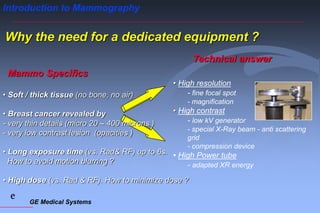
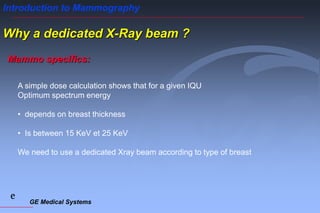
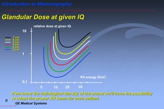
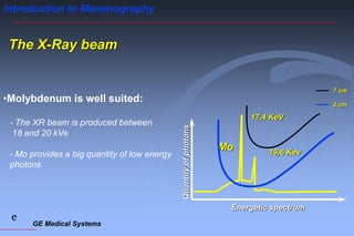
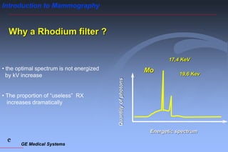
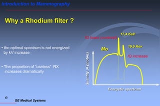
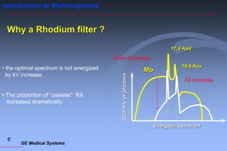
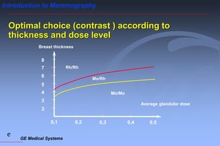
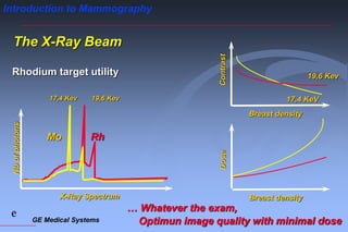
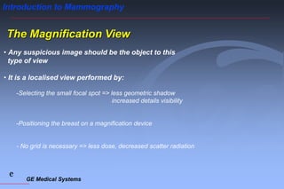
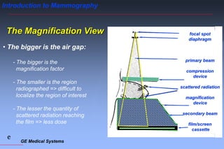
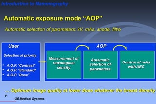
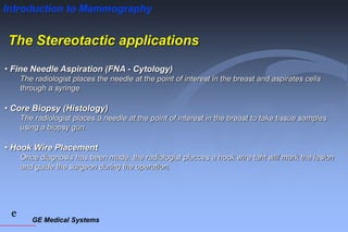
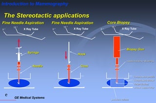
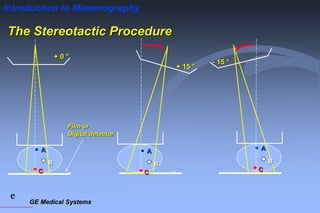
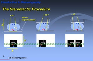
GE Medical Systems
e
Introduction to Mammography
The Basics of mammography
GE Medical Systems
e
Introduction to Mammography
Radiological caracteristics of lesions
Fibro glandular Tissues 1,035 0,8
Fat 0,93 0,45
Skin 1,09 0,80
Carcinomas 1,045 0,85
Micro calcifications 2,2 12,5
density absorption
GE Medical Systems
e
Introduction to Mammography
Physically speaking we can differentiate:
• 3 types of tissues : - skin
- fat
- fibro – glandular tissues
Radiologically we will see:
• 4 types of lesions : - “black » images
- micro-calcifications
- opacities
- fibrous signals
GE Medical Systems
e
Introduction to Mammography
Mammography: Different Types of Examinations
• Screening
- Health program organised by government
- 2 exposures: 1 incidence on each breast
• Diagnostic
- Personal decision process
- Usually 4 exposures: 2 incidences on each breast and
eventually magnified views.
• Interventional procedures
- Stereotactic biopsy procedures
GE Medical Systems
e
Introduction to Mammography
Why the need for a dedicated equipment ?
• Soft / thick tissue (no bone, no air)
• Breast cancer revealed by
- very thin details (micro 20 – 400 microns )
- very low contrast lesion (opacities )
• Long exposure time (vs. Rad& RF) up to 6s.
How to avoid motion blurring ?
• High dose (vs. Rad & RF). How to minimize dose ?
Mammo Specifics
GE Medical Systems
e
Introduction to Mammography
• High resolution
- fine focal spot
- magnification
• High contrast
- low kV generator
- special X-Ray beam - anti scattering
grid
- compression device
• High Power tube
- adapted XR energy
Technical answer
Why the need for a dedicated equipment ?
• Soft / thick tissue (no bone, no air)
• Breast cancer revealed by
- very thin details (micro 20 – 400 microns )
- very low contrast lesion (opacities )
• Long exposure time (vs. Rad& RF) up to 6s.
How to avoid motion blurring ?
• High dose (vs. Rad & RF). How to minimize dose ?
Mammo Specifics
GE Medical Systems
e
Introduction to Mammography
Why a dedicated X-Ray beam ?
Mammo specifics:
A simple dose calculation shows that for a given IQU
Optimum spectrum energy
• depends on breast thickness
• Is between 15 KeV et 25 KeV
We need to use a dedicated Xray beam according to type of breast
GE Medical Systems
e
Introduction to Mammography
Glandular Dose at given IQ
If we know the radiological density of the breast we’ll have the possibility
to adapt the proper XR beam for each patient.
RX energy (KeV)
relative dose at given IQ
0,1
1
10
15 25
5 35
7 cm
6 cm
5 cm
4 cm
3 cm
2 cm
GE Medical Systems
e
Introduction to Mammography
The X-Ray beam
•Molybdenum is well suited:
- The XR beam is produced between
18 and 20 kVe
- Mo provides a big quantity of low energy
photons
Mo
Quantity
of
photons
Energetic spectrum
17,4 KeV
19,6 Kev
7 cm
2 cm
GE Medical Systems
e
Introduction to Mammography
Why a Rhodium filter ?
• the optimal spectrum is not energized
by kV increase
• The proportion of “useless” RX
increases dramatically
Mo
17,4 KeV
19,6 Kev
Quantity
of
photons
Energetic spectrum
GE Medical Systems
e
Introduction to Mammography
Mo
17,4 KeV
19,6 Kev
IQ increase
IQ loses (contrast)
Why a Rhodium filter ?
• the optimal spectrum is not energized
by kV increase
• The proportion of “useless” RX
increases dramatically
Quantity
of
photons
Energetic spectrum
GE Medical Systems
e
Introduction to Mammography
Mo
17,4 KeV
19,6 Kev
IQ increase
Dose decrease
Why a Rhodium filter ?
• the optimal spectrum is not energized
by kV increase
• The proportion of “useless” RX
increases dramatically
Quantity
of
photons Energetic spectrum
GE Medical Systems
e
Introduction to Mammography
17,4 Kev
20,2 Kev
Mo Rh
Why a Rhodium target ?
• the useful spectrum is more energetic
• the energetic spectrum is close to
molybdenum
7 cm
2 cm
Quantity
of
photons
Energetic spectrum
GE Medical Systems
e
Introduction to Mammography
Optimal choice (contrast ) according to
thickness and dose level
0,2 0,3
0,1 0,4 0,5
2
3
4
5
6
7
8
Average glandular dose
Breast thickness
Mo/Mo
Rh/Rh
Mo/Rh
GE Medical Systems
e
Introduction to Mammography
19,6 Kev
17,4 KeV
Breast density
Contrast
Breast density
Dose
Nb
of
photons
X-Ray Spectrum
17,4 Kev 19,6 Kev
Mo Rh
… Whatever the exam,
Optimun image quality with minimal dose
The X-Ray Beam
Rhodium target utility
GE Medical Systems
e
Introduction to Mammography
• Geometric unsharpness increases with focal spot
• The small focal spot is used to magnify suspicious
regions - geometric shadow is extremely low.
The Tube - Focal spot size
Large Focal Spot Small Focal Spot
Object
Shadow Shadow
GE Medical Systems
e
Introduction to Mammography
The Filtration
• Energy peak represent
from 1/3 to 1/2 of total
radiation
• Mo filtration
contributes to :
- Suppression of low energies
(<10keV) which are absorbed
by the breast ( increase dose but
do not contribute to the image
formation)
- Suppression of high energies
(>20keV) which blacken the film.
Unfiltered
Filtered
Photon energy KeV
0 25 50 75 100
GE Medical Systems
e
Introduction to Mammography
The Grid
• Scattered radiation
reduces contrast
• Scattered radiation can
be absorbed by a grid to
maintain contrast:
- The lead strips stop the
scattered radiation
- The strips are angled to
point to the focal spot
- The grid is moved by a motor
to avoid strip projection on
the film
Focal Spot
Scattered
radiation
Grid movement
Grid
GE Medical Systems
e
Introduction to Mammography
The Compression
• Eliminates motion
blurring
• Reduces geometric
unsharpness
• Reduces the formation
of scattered radiation
• Reduces skin doses
• Enables imaging of the
entire breast including
deep layers Breast
Support
plate
compression
device
GE Medical Systems
e
Introduction to Mammography
The Magnification View
• Any suspicious image should be the object to this
type of view
• It is a localised view performed by:
-Selecting the small focal spot => less geometric shadow
increased details visibility
-Positioning the breast on a magnification device
- No grid is necessary => less dose, decreased scatter radiation
GE Medical Systems
e
Introduction to Mammography
The Magnification View
• The bigger is the air gap:
- The bigger is the
magnification factor
- The smaller is the region
radiographed => difficult to
localize the region of interest
- The lesser the quantity of
scattered radiation reaching
the film => less dose
focal spot
diaphragm
primary beam
compression
device
scattered radiation
magnification
device
secondary beam
film/screen
cassette
GE Medical Systems
e
Introduction to Mammography
Automatic exposure mode “AOP”
Automatic selection of parameters: kV, mAs, anode, filtre
Selection of priority
User
 A.O.P. "Contrast"
 A.O.P. "Standard"
 A.O.P. "Dose"
AOP
Measurement of
radiological
density
Automatic
selection of
parameters
Control of mAs
with AEC
… Optimum image quality at lower dose whatever the breast density
GE Medical Systems
e
Introduction to Mammography
The Stereotactic applications
• Fine Needle Aspiration (FNA - Cytology)
The radiologist places the needle at the point of interest in the breast and aspirates cells
through a syringe
• Core Biopsy (Histology)
The radiologist places a needle at the point of interest in the breast to take tissue samples
using a biopsy gun.
• Hook Wire Placement
Once diagnosis has been made, the radiologist placces a hook wire taht will mark the lesion
and guide the surgeon during the operation.
GE Medical Systems
e
Introduction to Mammography
The Stereotactic applications
0 °
15 °
X Ray Tube
Syringe
Needle
Fine Needle Aspiration
0 °
15 °
X Ray Tube
Coax
Hook
Fine Needle Aspiration
Breast Support tray
Compression paddle
Compressed Breast
Target (lesion)
needle holders + Bushings
0 °
15 °
puncture volume
X Ray Tube
Biopsy Gun
Core Biopsy
GE Medical Systems
e
Introduction to Mammography
The Stereotactic Procedure
Film or
Digital detector
+ 0 °
A
B
C
+ 15 °
A
B
C
- 15 °
A
B
C
GE Medical Systems
e
Introduction to Mammography
The Stereotactic Procedure
Film or
Digital detector
+ 0 °
A
B
C
X
Y
Z
+ 15 °
A
B
C
- 15 °
A
B
C
GE Medical Systems
e
Introduction to Mammography
28/09/2021 27
Thanks for listening

25632789 01-basics1-advanced-mammo-system

  • 1.
    GE Medical Systems e Introductionto Mammography The Basics of mammography
  • 2.
    GE Medical Systems e Introductionto Mammography Radiological caracteristics of lesions Fibro glandular Tissues 1,035 0,8 Fat 0,93 0,45 Skin 1,09 0,80 Carcinomas 1,045 0,85 Micro calcifications 2,2 12,5 density absorption
  • 3.
    GE Medical Systems e Introductionto Mammography Physically speaking we can differentiate: • 3 types of tissues : - skin - fat - fibro – glandular tissues Radiologically we will see: • 4 types of lesions : - “black » images - micro-calcifications - opacities - fibrous signals
  • 4.
    GE Medical Systems e Introductionto Mammography Mammography: Different Types of Examinations • Screening - Health program organised by government - 2 exposures: 1 incidence on each breast • Diagnostic - Personal decision process - Usually 4 exposures: 2 incidences on each breast and eventually magnified views. • Interventional procedures - Stereotactic biopsy procedures
  • 5.
    GE Medical Systems e Introductionto Mammography Why the need for a dedicated equipment ? • Soft / thick tissue (no bone, no air) • Breast cancer revealed by - very thin details (micro 20 – 400 microns ) - very low contrast lesion (opacities ) • Long exposure time (vs. Rad& RF) up to 6s. How to avoid motion blurring ? • High dose (vs. Rad & RF). How to minimize dose ? Mammo Specifics
  • 6.
    GE Medical Systems e Introductionto Mammography • High resolution - fine focal spot - magnification • High contrast - low kV generator - special X-Ray beam - anti scattering grid - compression device • High Power tube - adapted XR energy Technical answer Why the need for a dedicated equipment ? • Soft / thick tissue (no bone, no air) • Breast cancer revealed by - very thin details (micro 20 – 400 microns ) - very low contrast lesion (opacities ) • Long exposure time (vs. Rad& RF) up to 6s. How to avoid motion blurring ? • High dose (vs. Rad & RF). How to minimize dose ? Mammo Specifics
  • 7.
    GE Medical Systems e Introductionto Mammography Why a dedicated X-Ray beam ? Mammo specifics: A simple dose calculation shows that for a given IQU Optimum spectrum energy • depends on breast thickness • Is between 15 KeV et 25 KeV We need to use a dedicated Xray beam according to type of breast
  • 8.
    GE Medical Systems e Introductionto Mammography Glandular Dose at given IQ If we know the radiological density of the breast we’ll have the possibility to adapt the proper XR beam for each patient. RX energy (KeV) relative dose at given IQ 0,1 1 10 15 25 5 35 7 cm 6 cm 5 cm 4 cm 3 cm 2 cm
  • 9.
    GE Medical Systems e Introductionto Mammography The X-Ray beam •Molybdenum is well suited: - The XR beam is produced between 18 and 20 kVe - Mo provides a big quantity of low energy photons Mo Quantity of photons Energetic spectrum 17,4 KeV 19,6 Kev 7 cm 2 cm
  • 10.
    GE Medical Systems e Introductionto Mammography Why a Rhodium filter ? • the optimal spectrum is not energized by kV increase • The proportion of “useless” RX increases dramatically Mo 17,4 KeV 19,6 Kev Quantity of photons Energetic spectrum
  • 11.
    GE Medical Systems e Introductionto Mammography Mo 17,4 KeV 19,6 Kev IQ increase IQ loses (contrast) Why a Rhodium filter ? • the optimal spectrum is not energized by kV increase • The proportion of “useless” RX increases dramatically Quantity of photons Energetic spectrum
  • 12.
    GE Medical Systems e Introductionto Mammography Mo 17,4 KeV 19,6 Kev IQ increase Dose decrease Why a Rhodium filter ? • the optimal spectrum is not energized by kV increase • The proportion of “useless” RX increases dramatically Quantity of photons Energetic spectrum
  • 13.
    GE Medical Systems e Introductionto Mammography 17,4 Kev 20,2 Kev Mo Rh Why a Rhodium target ? • the useful spectrum is more energetic • the energetic spectrum is close to molybdenum 7 cm 2 cm Quantity of photons Energetic spectrum
  • 14.
    GE Medical Systems e Introductionto Mammography Optimal choice (contrast ) according to thickness and dose level 0,2 0,3 0,1 0,4 0,5 2 3 4 5 6 7 8 Average glandular dose Breast thickness Mo/Mo Rh/Rh Mo/Rh
  • 15.
    GE Medical Systems e Introductionto Mammography 19,6 Kev 17,4 KeV Breast density Contrast Breast density Dose Nb of photons X-Ray Spectrum 17,4 Kev 19,6 Kev Mo Rh … Whatever the exam, Optimun image quality with minimal dose The X-Ray Beam Rhodium target utility
  • 16.
    GE Medical Systems e Introductionto Mammography • Geometric unsharpness increases with focal spot • The small focal spot is used to magnify suspicious regions - geometric shadow is extremely low. The Tube - Focal spot size Large Focal Spot Small Focal Spot Object Shadow Shadow
  • 17.
    GE Medical Systems e Introductionto Mammography The Filtration • Energy peak represent from 1/3 to 1/2 of total radiation • Mo filtration contributes to : - Suppression of low energies (<10keV) which are absorbed by the breast ( increase dose but do not contribute to the image formation) - Suppression of high energies (>20keV) which blacken the film. Unfiltered Filtered Photon energy KeV 0 25 50 75 100
  • 18.
    GE Medical Systems e Introductionto Mammography The Grid • Scattered radiation reduces contrast • Scattered radiation can be absorbed by a grid to maintain contrast: - The lead strips stop the scattered radiation - The strips are angled to point to the focal spot - The grid is moved by a motor to avoid strip projection on the film Focal Spot Scattered radiation Grid movement Grid
  • 19.
    GE Medical Systems e Introductionto Mammography The Compression • Eliminates motion blurring • Reduces geometric unsharpness • Reduces the formation of scattered radiation • Reduces skin doses • Enables imaging of the entire breast including deep layers Breast Support plate compression device
  • 20.
    GE Medical Systems e Introductionto Mammography The Magnification View • Any suspicious image should be the object to this type of view • It is a localised view performed by: -Selecting the small focal spot => less geometric shadow increased details visibility -Positioning the breast on a magnification device - No grid is necessary => less dose, decreased scatter radiation
  • 21.
    GE Medical Systems e Introductionto Mammography The Magnification View • The bigger is the air gap: - The bigger is the magnification factor - The smaller is the region radiographed => difficult to localize the region of interest - The lesser the quantity of scattered radiation reaching the film => less dose focal spot diaphragm primary beam compression device scattered radiation magnification device secondary beam film/screen cassette
  • 22.
    GE Medical Systems e Introductionto Mammography Automatic exposure mode “AOP” Automatic selection of parameters: kV, mAs, anode, filtre Selection of priority User  A.O.P. "Contrast"  A.O.P. "Standard"  A.O.P. "Dose" AOP Measurement of radiological density Automatic selection of parameters Control of mAs with AEC … Optimum image quality at lower dose whatever the breast density
  • 23.
    GE Medical Systems e Introductionto Mammography The Stereotactic applications • Fine Needle Aspiration (FNA - Cytology) The radiologist places the needle at the point of interest in the breast and aspirates cells through a syringe • Core Biopsy (Histology) The radiologist places a needle at the point of interest in the breast to take tissue samples using a biopsy gun. • Hook Wire Placement Once diagnosis has been made, the radiologist placces a hook wire taht will mark the lesion and guide the surgeon during the operation.
  • 24.
    GE Medical Systems e Introductionto Mammography The Stereotactic applications 0 ° 15 ° X Ray Tube Syringe Needle Fine Needle Aspiration 0 ° 15 ° X Ray Tube Coax Hook Fine Needle Aspiration Breast Support tray Compression paddle Compressed Breast Target (lesion) needle holders + Bushings 0 ° 15 ° puncture volume X Ray Tube Biopsy Gun Core Biopsy
  • 25.
    GE Medical Systems e Introductionto Mammography The Stereotactic Procedure Film or Digital detector + 0 ° A B C + 15 ° A B C - 15 ° A B C
  • 26.
    GE Medical Systems e Introductionto Mammography The Stereotactic Procedure Film or Digital detector + 0 ° A B C X Y Z + 15 ° A B C - 15 ° A B C
  • 27.
    GE Medical Systems e Introductionto Mammography 28/09/2021 27 Thanks for listening