A 3 sentence summary of the document:
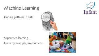
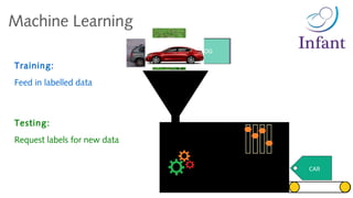
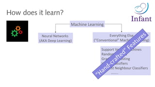
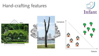
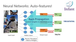
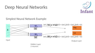
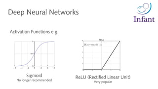
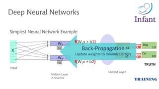
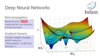
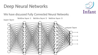
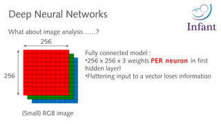
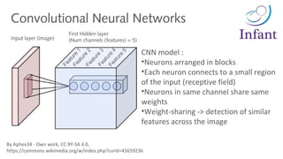
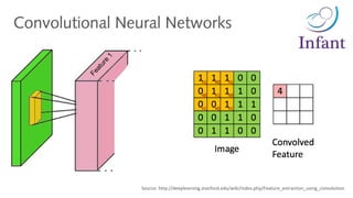
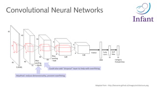
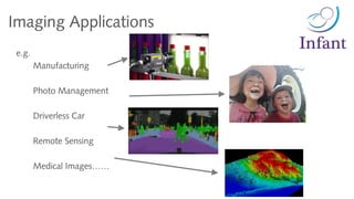
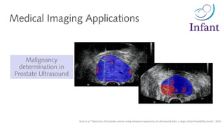
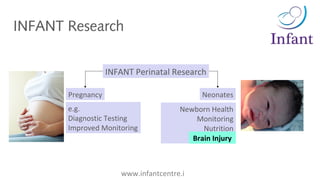
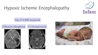
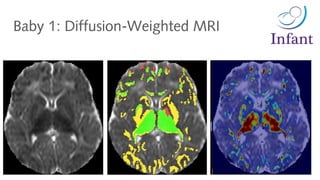
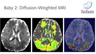
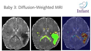
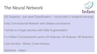
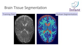
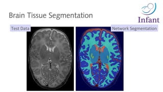
The document discusses deep learning for medical image analysis, focusing on applications in neonatal medical imaging. It provides an overview of deep learning and convolutional neural networks, including examples of their use for tasks like brain tissue segmentation in MRI scans of newborns. The presenter describes their research using deep learning for segmentation and diagnosis of hypoxic ischemic encephalopathy in MRI scans of newborns.


![# Code Snippet
model = Sequential()
model.add(Conv2D(32, kernel_size=(3, 3), activation='relu', input_shape=input_shape))
model.add(Conv2D(64, (3, 3), activation='relu'))
model.add(MaxPooling2D(pool_size=(2, 2)))
model.add(Dropout(0.25))
model.add(Flatten())
model.add(Dense(128, activation='relu'))
model.add(Dropout(0.5))
model.add(Dense(num_classes, activation='softmax'))
model.compile(loss=keras.losses.categorical_crossentropy,
optimizer=keras.optimizers.Adadelta(),
metrics=['accuracy'])
model.fit(x_train, y_train,
batch_size=batch_size,
epochs=epochs,
verbose=1,
validation_data=(x_test, y_test))
score = model.evaluate(x_test, y_test, verbose=0)
Convolutional Neural Networks
https://github.com/fchollet/keras/blob/master/examples/mnist_cnn.py](https://image.slidesharecdn.com/20170703meetupd-170704122730/85/2017-07-03_meetup_d-27-320.jpg)


![[台灣人工智慧學校] 主題演講 - 張智威總經理 (President of HTC DeepQ)](https://cdn.slidesharecdn.com/ss_thumbnails/aischoolhsinzhukeynoteedwardchang-180725074840-thumbnail.jpg?width=640&height=640&fit=bounds)


