This study evaluated different methods of gene therapy to treat infantile Batten disease (INCL) in a mouse model. Intrathecal injection targeting the spinal cord alone extended lifespan by 3 months and showed motor deficits at 7 months while decreasing disease markers in the spinal cord but not brain. Intracranial injection targeting the brain alone extended lifespan by 5.3 months and showed motor deficits at 9 months while decreasing markers in the brain but not spinal cord. Combination intrathecal and intracranial injection provided the most effective treatment, extending lifespan by 11.5 months and delaying motor deficits until 15 months by decreasing disease markers in both the brain and spinal cord. Targeting both areas simultaneously provided dramatic improvement over targeting
Expert Recommendations for the Laboratory Diagnosis of Neuronal Ceroid Lipofuscinosis Type 2 (CLN2 disease): Diagnostic Algorithm and Best Practice Guidelines for a Timely Diagnosis
Role of pro-brain-derived neurotrophic factor (proBDNF)to ma.docxjoellemurphey
Role of pro-brain-derived neurotrophic factor (proBDNF)
to mature BDNF conversion in activity-dependent
competition at developing neuromuscular synapses
H. Shawn Jea,b,c,1, Feng Yanga,b,d,1, Yuanyuan Jia,e, Guhan Nagappana,e, Barbara L. Hempsteadf, and Bai Lua,b,e,2
aSection on Neural Development and Plasticity, National Institute of Child Health and Human Development, Bethesda, MD 20892-3714; bGenes, Cognition,
and Psychosis Program (GCAP), National Institute of Mental Health, Bethesda, MD 20892-3714; cProgram in Neuroscience and Behavioral Disorders, Duke–
National University of Singapore (Duke-NUS) Graduate Medical School, 169857, Singapore; dLieber Institute for Brain Development, The Johns Hopkins
University Medical Campus, Baltimore, MD 21205; eR&D China, GlaxoSmithKline, Pudong, Shanghai 201203, China; and fDivision of Hematology, Department
of Medicine, Weill Medical College, Cornell University, New York, NY 10021
Edited* by Richard L. Huganir, The Johns Hopkins University School of Medicine, Baltimore, MD, and approved August 20, 2012 (received for review May
10, 2012)
Formation of specific neuronal connections often involves compe-
tition between adjacent axons, leading to stabilization of the active
terminal, while retraction of the less active ones. The underlying
molecular mechanisms remain unknown. We show that activity-
dependent conversion of pro–brain-derived neurotrophic factor
(proBDNF) to mature (m)BDNF mediates synaptic competition. Stim-
ulation of motoneurons triggers proteolytic conversion of proBDNF
to mBDNF at nerve terminals. In Xenopus nerve–muscle cocultures,
in which two motoneurons innervate one myocyte, proBDNF-
p75NTR signaling promotes retraction of the less active terminal,
whereas mBDNF–tyrosine-related kinase B (TrkB) p75NTR (p75 neu-
rotrophin receptor) facilitates stabilization of the active one. Thus,
proBDNF and mBDNF may serve as potential “punishment” and “re-
ward” signals for inactive and active terminals, respectively, and
activity-dependent conversion of proBDNF to mBDNF may regulate
synapse elimination.
neuromuscular junction | pro-neurotrophin | synapse competition
The nervous system responds to experience by altering thenumber and strength of synaptic connections (1). Activity-
dependent synaptic competition, a general process seen in many
parts of the developing nervous system, plays a critical role in
shaping patterns of neuronal connections (2–7). At the neuro-
muscular junction (NMJ), for example, multiple axons compete
for the same postsynaptic muscle cell during early postnatal life
until all but one is eliminated (8–10). Extensive experimental
data support the view that the more active terminal or “cartel”
gets stabilized, whereas less active ones withdraw, resulting in
canonical elimination of polyneuronal innervation (8, 11). It is
generally believed that this synaptic competition is mediated by
a “punishment” or “elimination” signal, produced by the post-
synaptic cell, that cau ...
Suac Syndrome is an autoimmune endotheliopathy with about 304 cases described until 2013. It charaterized by the triad of E-H-V (Encephalopathy, Hearing loss and Vision - branch retinal artery occlusions/BRAO [3]. The case report early-onset autoimmune neuropsychiatric disorder in a pre-pubertal 12 year old girl innitialy presenting with behavioral and emotional manifestations
Similar to 2016 BDSRA Shyng, Nelvagal, Dearborn, Cooper, Sands CLN1 (20)
Title: Sense of Taste
Presenter: Dr. Faiza, Assistant Professor of Physiology
Qualifications:
MBBS (Best Graduate, AIMC Lahore)
FCPS Physiology
ICMT, CHPE, DHPE (STMU)
MPH (GC University, Faisalabad)
MBA (Virtual University of Pakistan)
Learning Objectives:
Describe the structure and function of taste buds.
Describe the relationship between the taste threshold and taste index of common substances.
Explain the chemical basis and signal transduction of taste perception for each type of primary taste sensation.
Recognize different abnormalities of taste perception and their causes.
Key Topics:
Significance of Taste Sensation:
Differentiation between pleasant and harmful food
Influence on behavior
Selection of food based on metabolic needs
Receptors of Taste:
Taste buds on the tongue
Influence of sense of smell, texture of food, and pain stimulation (e.g., by pepper)
Primary and Secondary Taste Sensations:
Primary taste sensations: Sweet, Sour, Salty, Bitter, Umami
Chemical basis and signal transduction mechanisms for each taste
Taste Threshold and Index:
Taste threshold values for Sweet (sucrose), Salty (NaCl), Sour (HCl), and Bitter (Quinine)
Taste index relationship: Inversely proportional to taste threshold
Taste Blindness:
Inability to taste certain substances, particularly thiourea compounds
Example: Phenylthiocarbamide
Structure and Function of Taste Buds:
Composition: Epithelial cells, Sustentacular/Supporting cells, Taste cells, Basal cells
Features: Taste pores, Taste hairs/microvilli, and Taste nerve fibers
Location of Taste Buds:
Found in papillae of the tongue (Fungiform, Circumvallate, Foliate)
Also present on the palate, tonsillar pillars, epiglottis, and proximal esophagus
Mechanism of Taste Stimulation:
Interaction of taste substances with receptors on microvilli
Signal transduction pathways for Umami, Sweet, Bitter, Sour, and Salty tastes
Taste Sensitivity and Adaptation:
Decrease in sensitivity with age
Rapid adaptation of taste sensation
Role of Saliva in Taste:
Dissolution of tastants to reach receptors
Washing away the stimulus
Taste Preferences and Aversions:
Mechanisms behind taste preference and aversion
Influence of receptors and neural pathways
Impact of Sensory Nerve Damage:
Degeneration of taste buds if the sensory nerve fiber is cut
Abnormalities of Taste Detection:
Conditions: Ageusia, Hypogeusia, Dysgeusia (parageusia)
Causes: Nerve damage, neurological disorders, infections, poor oral hygiene, adverse drug effects, deficiencies, aging, tobacco use, altered neurotransmitter levels
Neurotransmitters and Taste Threshold:
Effects of serotonin (5-HT) and norepinephrine (NE) on taste sensitivity
Supertasters:
25% of the population with heightened sensitivity to taste, especially bitterness
Increased number of fungiform papillae
Title: Sense of Smell
Presenter: Dr. Faiza, Assistant Professor of Physiology
Qualifications:
MBBS (Best Graduate, AIMC Lahore)
FCPS Physiology
ICMT, CHPE, DHPE (STMU)
MPH (GC University, Faisalabad)
MBA (Virtual University of Pakistan)
Learning Objectives:
Describe the primary categories of smells and the concept of odor blindness.
Explain the structure and location of the olfactory membrane and mucosa, including the types and roles of cells involved in olfaction.
Describe the pathway and mechanisms of olfactory signal transmission from the olfactory receptors to the brain.
Illustrate the biochemical cascade triggered by odorant binding to olfactory receptors, including the role of G-proteins and second messengers in generating an action potential.
Identify different types of olfactory disorders such as anosmia, hyposmia, hyperosmia, and dysosmia, including their potential causes.
Key Topics:
Olfactory Genes:
3% of the human genome accounts for olfactory genes.
400 genes for odorant receptors.
Olfactory Membrane:
Located in the superior part of the nasal cavity.
Medially: Folds downward along the superior septum.
Laterally: Folds over the superior turbinate and upper surface of the middle turbinate.
Total surface area: 5-10 square centimeters.
Olfactory Mucosa:
Olfactory Cells: Bipolar nerve cells derived from the CNS (100 million), with 4-25 olfactory cilia per cell.
Sustentacular Cells: Produce mucus and maintain ionic and molecular environment.
Basal Cells: Replace worn-out olfactory cells with an average lifespan of 1-2 months.
Bowman’s Gland: Secretes mucus.
Stimulation of Olfactory Cells:
Odorant dissolves in mucus and attaches to receptors on olfactory cilia.
Involves a cascade effect through G-proteins and second messengers, leading to depolarization and action potential generation in the olfactory nerve.
Quality of a Good Odorant:
Small (3-20 Carbon atoms), volatile, water-soluble, and lipid-soluble.
Facilitated by odorant-binding proteins in mucus.
Membrane Potential and Action Potential:
Resting membrane potential: -55mV.
Action potential frequency in the olfactory nerve increases with odorant strength.
Adaptation Towards the Sense of Smell:
Rapid adaptation within the first second, with further slow adaptation.
Psychological adaptation greater than receptor adaptation, involving feedback inhibition from the central nervous system.
Primary Sensations of Smell:
Camphoraceous, Musky, Floral, Pepperminty, Ethereal, Pungent, Putrid.
Odor Detection Threshold:
Examples: Hydrogen sulfide (0.0005 ppm), Methyl-mercaptan (0.002 ppm).
Some toxic substances are odorless at lethal concentrations.
Characteristics of Smell:
Odor blindness for single substances due to lack of appropriate receptor protein.
Behavioral and emotional influences of smell.
Transmission of Olfactory Signals:
From olfactory cells to glomeruli in the olfactory bulb, involving lateral inhibition.
Primitive, less old, and new olfactory systems with different path
Ethanol (CH3CH2OH), or beverage alcohol, is a two-carbon alcohol
that is rapidly distributed in the body and brain. Ethanol alters many
neurochemical systems and has rewarding and addictive properties. It
is the oldest recreational drug and likely contributes to more morbidity,
mortality, and public health costs than all illicit drugs combined. The
5th edition of the Diagnostic and Statistical Manual of Mental Disorders
(DSM-5) integrates alcohol abuse and alcohol dependence into a single
disorder called alcohol use disorder (AUD), with mild, moderate,
and severe subclassifications (American Psychiatric Association, 2013).
In the DSM-5, all types of substance abuse and dependence have been
combined into a single substance use disorder (SUD) on a continuum
from mild to severe. A diagnosis of AUD requires that at least two of
the 11 DSM-5 behaviors be present within a 12-month period (mild
AUD: 2–3 criteria; moderate AUD: 4–5 criteria; severe AUD: 6–11 criteria).
The four main behavioral effects of AUD are impaired control over
drinking, negative social consequences, risky use, and altered physiological
effects (tolerance, withdrawal). This chapter presents an overview
of the prevalence and harmful consequences of AUD in the U.S.,
the systemic nature of the disease, neurocircuitry and stages of AUD,
comorbidities, fetal alcohol spectrum disorders, genetic risk factors, and
pharmacotherapies for AUD.
- Video recording of this lecture in English language: https://youtu.be/lK81BzxMqdo
- Video recording of this lecture in Arabic language: https://youtu.be/Ve4P0COk9OI
- Link to download the book free: https://nephrotube.blogspot.com/p/nephrotube-nephrology-books.html
- Link to NephroTube website: www.NephroTube.com
- Link to NephroTube social media accounts: https://nephrotube.blogspot.com/p/join-nephrotube-on-social-media.html
These simplified slides by Dr. Sidra Arshad present an overview of the non-respiratory functions of the respiratory tract.
Learning objectives:
1. Enlist the non-respiratory functions of the respiratory tract
2. Briefly explain how these functions are carried out
3. Discuss the significance of dead space
4. Differentiate between minute ventilation and alveolar ventilation
5. Describe the cough and sneeze reflexes
Study Resources:
1. Chapter 39, Guyton and Hall Textbook of Medical Physiology, 14th edition
2. Chapter 34, Ganong’s Review of Medical Physiology, 26th edition
3. Chapter 17, Human Physiology by Lauralee Sherwood, 9th edition
4. Non-respiratory functions of the lungs https://academic.oup.com/bjaed/article/13/3/98/278874
Ozempic: Preoperative Management of Patients on GLP-1 Receptor Agonists Saeid Safari
Preoperative Management of Patients on GLP-1 Receptor Agonists like Ozempic and Semiglutide
ASA GUIDELINE
NYSORA Guideline
2 Case Reports of Gastric Ultrasound
Report Back from SGO 2024: What’s the Latest in Cervical Cancer?bkling
Are you curious about what’s new in cervical cancer research or unsure what the findings mean? Join Dr. Emily Ko, a gynecologic oncologist at Penn Medicine, to learn about the latest updates from the Society of Gynecologic Oncology (SGO) 2024 Annual Meeting on Women’s Cancer. Dr. Ko will discuss what the research presented at the conference means for you and answer your questions about the new developments.
Lung Cancer: Artificial Intelligence, Synergetics, Complex System Analysis, S...Oleg Kshivets
RESULTS: Overall life span (LS) was 2252.1±1742.5 days and cumulative 5-year survival (5YS) reached 73.2%, 10 years – 64.8%, 20 years – 42.5%. 513 LCP lived more than 5 years (LS=3124.6±1525.6 days), 148 LCP – more than 10 years (LS=5054.4±1504.1 days).199 LCP died because of LC (LS=562.7±374.5 days). 5YS of LCP after bi/lobectomies was significantly superior in comparison with LCP after pneumonectomies (78.1% vs.63.7%, P=0.00001 by log-rank test). AT significantly improved 5YS (66.3% vs. 34.8%) (P=0.00000 by log-rank test) only for LCP with N1-2. Cox modeling displayed that 5YS of LCP significantly depended on: phase transition (PT) early-invasive LC in terms of synergetics, PT N0—N12, cell ratio factors (ratio between cancer cells- CC and blood cells subpopulations), G1-3, histology, glucose, AT, blood cell circuit, prothrombin index, heparin tolerance, recalcification time (P=0.000-0.038). Neural networks, genetic algorithm selection and bootstrap simulation revealed relationships between 5YS and PT early-invasive LC (rank=1), PT N0—N12 (rank=2), thrombocytes/CC (3), erythrocytes/CC (4), eosinophils/CC (5), healthy cells/CC (6), lymphocytes/CC (7), segmented neutrophils/CC (8), stick neutrophils/CC (9), monocytes/CC (10); leucocytes/CC (11). Correct prediction of 5YS was 100% by neural networks computing (area under ROC curve=1.0; error=0.0).
CONCLUSIONS: 5YS of LCP after radical procedures significantly depended on: 1) PT early-invasive cancer; 2) PT N0--N12; 3) cell ratio factors; 4) blood cell circuit; 5) biochemical factors; 6) hemostasis system; 7) AT; 8) LC characteristics; 9) LC cell dynamics; 10) surgery type: lobectomy/pneumonectomy; 11) anthropometric data. Optimal diagnosis and treatment strategies for LC are: 1) screening and early detection of LC; 2) availability of experienced thoracic surgeons because of complexity of radical procedures; 3) aggressive en block surgery and adequate lymph node dissection for completeness; 4) precise prediction; 5) adjuvant chemoimmunoradiotherapy for LCP with unfavorable prognosis.
TEST BANK for Operations Management, 14th Edition by William J. Stevenson, Ve...kevinkariuki227
TEST BANK for Operations Management, 14th Edition by William J. Stevenson, Verified Chapters 1 - 19, Complete Newest Version.pdf
Pulmonary Thromboembolism - etilogy, types, medical- Surgical and nursing man...VarunMahajani
Disruption of blood supply to lung alveoli due to blockage of one or more pulmonary blood vessels is called as Pulmonary thromboembolism. In this presentation we will discuss its causes, types and its management in depth.
These lecture slides, by Dr Sidra Arshad, offer a quick overview of physiological basis of a normal electrocardiogram.
Learning objectives:
1. Define an electrocardiogram (ECG) and electrocardiography
2. Describe how dipoles generated by the heart produce the waveforms of the ECG
3. Describe the components of a normal electrocardiogram of a typical bipolar leads (limb II)
4. Differentiate between intervals and segments
5. Enlist some common indications for obtaining an ECG
Study Resources:
1. Chapter 11, Guyton and Hall Textbook of Medical Physiology, 14th edition
2. Chapter 9, Human Physiology - From Cells to Systems, Lauralee Sherwood, 9th edition
3. Chapter 29, Ganong’s Review of Medical Physiology, 26th edition
4. Electrocardiogram, StatPearls - https://www.ncbi.nlm.nih.gov/books/NBK549803/
5. ECG in Medical Practice by ABM Abdullah, 4th edition
6. ECG Basics, http://www.nataliescasebook.com/tag/e-c-g-basics
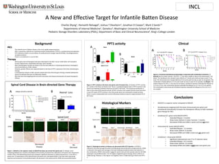
1. Spinal Cord Disease in Brain-directed Gene Therapy
Figure 1. AAV2/9 is a far superior vector to AAV2/5 but does not correct the spinal cord. A) Lifespan curve
comparing AAV2/9 (13.7 months) and AAV2/5 (10.6 months). B) Axonal loss observed in the spinal cord. Fluorescent
markers (Thy1-YFP) delineating axonal tracts showed a significant decrease in relative fluorescence in the PPT1-/-
spinal cord. C) Examination of the AAV2/9 brain and spinal cord. Decreased AFSM in the brain with intracranial
injection and resolution of neuroinflammation. No decrease in the spinal cord for AFSM and neuroinflammation
A New and Effective Target for Infantile Batten Disease
Charles Shyng1, Hemanth Nelvagal2, Joshua T Dearborn1, Jonathan D Cooper2, Mark S Sands1,3
Departments of Internal Medicine1, Genetics3; Washington University School of Medicine
Pediatric Storage Disorders Laboratory (PSDL), Department of Basic and Clinical Neuroscience2, King's College London
Background
INCL
• The infantile form of Batten disease is the most rapidly progressing form
• INCL is caused by a deficiency in palmitoyl-protein thioeseterase-1 (PPT1), a soluble lysosomal hydrolase
• PPT1 deficiency leads to accumulation of autofluorescent storage material (AFSM), neurodegeneration,
and glial activation
• There is no treatment or cure for INCL
Therapy
• Various gene and cell therapies have been attempted in the INCL mouse model (Stem cell transplant,
Enzyme replacement, Small Molecule Drugs, Gene Transfer)
• Brain-directed gene transfer has shown to be the most effective in improving biochemical, histological,
and clinical features of INCL disease
• However, greater efficacy was expected based on the level of PPT1 expression from brain-directed gene
therapy
• A retrospective analysis of INCL animals treated with brain-directed gene therapy revealed widespread
spinal cord disease that was not effectively treated.
• We show here that targeting both the brain and spinal cord disease dramatically increased therapeutic
efficacy.
PPT1 activity
Figure 2. PPT1 activity in the brain and spinal cord of treated mice. A) Analysis of PPT1 activity
at 1 month in the Brain and Spinal cord. There is supraphysiological levels of PPT1 activity in the
spinal cord following intrathecal injection but little in the brain. The reciprocal distribution is
seen in the intracranial injected animals. B) PPT1 activity in the treated animals show that there
is near WT levels of PPT1 activity in the intracranial alone and combination-treated animals.
There is nearly undetectable brain activity following intrathecal injection (approximately 5% of
WT levels)
Histological Markers
Figure 3. Histological markers of disease are decreased with IC/IT injection. A) AFSM is
reduced in the brain but not the spinal cord following intracranial alone (orange asterick). AFSM
is reduced in the spinal cord and not in the brain following intrathecal alone (red asterick). In
the combination, AFSM is decreased in both the spinal cord and brain (purple asterick). B) CD68
staining for neuroinfllammation. For the regions targeted by gene therapy, there is little to no
CD68 staining. However, in the regions not targeted, there is CD68 staining at levels near PPT1-
/-.
Clinical
Figure 4. Functional and behavioral phenotype is improved with combination treatment. A)
Rotarod test for motor function. The PPT1 -/- mice were unable to stay on the rotarod past 7 mo
(blue). The intrathecal mice showed motor deficits at 7 month and could not stay on past 11 mo
(red). The intracranial mice showed deficits at 9 mo and could not stay on past 13 mo (green). The
combination showed deficits at 15 mo and could not stay on past 19 mo (purple). B) Lifespan. The
median lifespan for PPT1-/- mice was 8.4 mo (blue). Intrathecal injection extended the lifespan to
11.3 mo (+3 mo, red). Intracranial injection extended the lifespan to 13.7 mo (+5.3 mo, green).
The combination intracranial and intrathecal injection extended the lifespan to 19.5 mo (+11.5
mo, purple).
Conclusions
• AAV2/9 is a superior vector compared to AAV2/5
• Simultaneously targeting both the brain (intracranial) and spinal cord
(intrathecal) dramatically increases the therapeutic efficacy of AAV-mediated
gene therapy for INCL
• Intrathecal (IT, spinal cord) AAV2/9-hPPT1
• Extended lifespan: 3 months
• Show motor deficits in motor function: 7 months.
• Decreased AFSM and CD68 in spinal cord but not brain
• Intracranial (IC, brain) AAV2/9-hPPT1
• Extended lifespan: 5.3 months
• Show motor deficits: 9 months.
• Decreased AFSM and CD68 in brain but not spinal cord
• Intrathecal + Intracranial AAV2/9
• significant improvement over IT or IC injection alone
• Extended lifespan: 19.5 months
• Show motor deficits: 15 months
• Decreased AFSM and CD68 in both brain and spinal cord
Brain
Spinal Cord
Intracranial IntrathecalWildtype IC/ITPpt1-/-
IC/IT
Brain
Spinal Cord
Intracranial IntrathecalWildtype Ppt1-/-
Brain
IntracranialWildtype Ppt1-/-
Spinal Cord
IntracranialWildtype Ppt1-/-
AFSM
CD68
Decreased
Disease
No
Decrease
A B
A
B
B
A
BA
C
Axonal Loss
*
*
*
*
*
*
*
*
*
*
*
*
INCL