SlideShare a Scribd company logo
Targeting Brain Endothelial cells for JNCL therapy 
Colleen S Stein1, Mark Schultz2, Luis Tecedor3, and Beverly L Davidson3, 1Dept. of Internal Medicine, Carver College of Medicine, University of Iowa; 2Dept. 
of Pathology, University of Michigan Medical School, Ann Arbor, MI: 3Children's Hospital of Philadelphia, Philadelphia, PA. contact e-mail: colleen-stein@uiowa.edu 
CLN3 gene 
2) Intravenous injection 
For gene therapy, AAV-CLN3 
particles will be injected 
intravenously into JNCL mice. 
AAV-CLN3 particles will bind 
and deliver the CLN3 gene to 
endothelial cells lining the blood 
vessels of the brain. With a 
good copy of the CLN3 gene, 
the endothelial cells can then 
make CLN3 protein. 
3) Therapeutic effect? 
JNCL mice injected with 
AAV-CLN3 will be 
monitored to determine if 
they exhibit superior 
performance on motor tasks 
and reduced brain 
pathology compared to 
untreated JNCL mice. 
Mice will be tested for ability to 
maintain balance on a rotating 
rod. Image adapted from Nature 
Neuroscience 16: 658, 2013. 
WHAT THIS MEANS FOR JNCL 
THERAPY 
Endothelial cells from JNCL mouse brain 
have trouble internalizing molecules 
CLN3 positive 
endothelial 
cells 
outside of cell 
plasma membrane 
inside of cell 
caveolin-1 
CLN3 negative 
endothelial 
cells 
TWO OF THREE TYPES OF ENDOCYTOSIS ARE DEFECTIVE IN JNCL MOUSE BRAIN 
ENDOTHELIAL CELLS. Cells internalize extracellular molecules by a process called endocytosis, in 
which a portion of the outer membrane of the cell buds into the cell and pinches off, bringing in a 
"mouthful" of molecules. Three main forms of endocytosis are depicted in the drawing: clathrin-dependent, 
caveolar, and fluid-phase endocytosis. Red-tagged transferrin, green-tagged albumin, and 
green-tagged dextran are used to examine these forms of endocytosis, respectively. In this experiment 
we prepared cultures of endothelial cells harvested from unaffected and JNCL mouse brains and 
examined internalization of the red- or green-tagged molecules. Interestingly, we found that both caveolar 
and fluid-phase endocytic pathways are impaired in the JNCL endothelial cells, while clathrin-dependent 
endocytosis is intact. 
Acknowledgements 
• These studies are funded by the BDSRA, 
Beyond Batten Disease Foundation 
(BBDF), and the NIH. 
• We thank the Gene Transfer Vector Core 
and the Central Microscopy Research 
Facility at the University of Iowa. 
1 
Art by Mark L Schultz 
INTRODUCTION 
JNCL 
Juvenile Batten disease (JNCL) is caused by mutations in the CLN3 gene, creating 
a deficiency or defect in the CLN3 protein. CLN3 deficiency leads to JNCL 
symptoms, which include loss of vision, seizures, and progressive decline in motor 
and mental capabilities. 
CLN3 
CLN3 is situated within lipid membrane regions inside cells, and is not secreted. We 
don't know the exact function of CLN3, but it appears to play a basic role in the 
normal transport of proteins and lipids inside cells. 
While CLN3 is found at some level in all cells throughout the body, JNCL is a 
disease of central nervous system. So it seems that CLN3 has a critical role in cells 
that control the function of the brain and eye. One important type of cell that is often 
overlooked in studies of neurological diseases is the endothelial cell. Endothelial 
cells form the inside layer of blood vessels. 
The Blood-Brain Barrier (BBB) 
In the blood vessels (vasculature) of the brain, endothelial cells are specialized to 
protect and feed the brain. Here endothelial cells butt up against eachother tightly, 
eliminating spaces between cells. This creates a physical barrier between the blood 
and the brain, aptly called the blood-brain barrier (BBB). The BBB protects the brain 
from entry of microbes, toxic substances, and destructive immune cells. Nutrients 
are selectively taken into and transported through the endothelial cells. Thus the 
underlying brain cells (neurons and glial cells), rely on endothelial cells for nutrient 
supply for optimal neural performance and viability. 
In our previous work, our CLN3 reporter mouse indicated that CLN3 is made at 
relatively high level in endothelial cells throughout the brain vasculature. Thus it is 
possible that CLN3 is critical to BBB functions, which dictates brain health. 
Dense vasculature in the brain: 
Endothelial cells that line the blood vessels regulate nutrient 
supply from the blood to the brain. 
Astrocyte 
extends 
processes onto 
micro-vessels 
Neurons 
Mary Moye-Rowley, Iowa City, IA 
Cross-sectional view of 
a microvessel illustrates 
extensive cellular 
communication 
Adapted from Abbott et al., Nature Reviews, Neuroscience, 2006 
The brain is the most densely vascularized organ in the body. This density is necessary to meet the oxygen and 
nutrient demands of intense neuronal activity. Every neuron and glial cell in the brain contacts or is in close 
proximity to a blood vessel. The endothelial cells that line the blood vessels in the brain form tight cell-to-cell 
junctions with each other, creating a physical barrier between the blood and the brain, termed the blood-brain 
barrier (BBB). Most nutrients gain entry into the brain by passing though the endothelial cells. Endothelial cells at 
the BBB are equipped with numerous receptors and channels, and act as gatekeepers, regulating selective 
transport into and out of the brain. Thus neuronal health is dependent upon endothelial cell health. 
We propose that endothelial cell dysfunction is a critical event leading to JNCL symptoms, and hypothesize that 
restoring CLN3 to endothelial cells will protect against JNCL symptoms. 
Does restoration of CLN3 to endothelial 
cells prevent JNCL? 
2 
USE OF GENTICALLY ENGINEERED MICE TO TEST THE HYPOTHESIS. It is conceivable that vascular 
endothelial cell impairment is a primary event in JNCL pathogenesis. Thus, we may be able to avert disease 
by providing normal CLN3 exclusively to brain endothelial cells. To test this concept, we are using transgenic 
mice. A transgenic mouse strain is one that has been genetically altered to carry an extra gene, referred to 
as a transgene. 
For this project we are using JNCL mice that have been genetically engineered to carry an inducible CLN3 
transgene in their genome. By breeding this mouse to a Tie2-Cre inducer mouse, we will trigger CLN3 
expression in the endothelial cells of progeny mice. Progeny mice will be tested in motor and behavioral 
assays to assess whether they develop JNCL symptoms, or remain symptom-free or show milder disease. 
3 
Gene therapy to restore CLN3 to BBB 
endothelial cells 
1) Production of AAV-CLN3 
AAV particles are microscopic virus-like 
particles used to delivery genes 
into cells. For gene therapy, we are 
designing AAV particles that will 
effectively attach to and deliver 
genes to brain endothelium. We will 
package the CLN3 gene into these 
AAV particles. 
AAV-CLN3 GENE TRANSFER TO BBB ENDOTHELIAL CELLS. Apart from the transgenic 
mouse approach, another way to determine whether providing CLN3 to endothelial cells will 
protect against JNCL symptoms is to do Gene Therapy. Here we plan to utilize AAV virus-like-particles 
to transfer the CLN3 gene into endothelial cells of JNCL mice. 
These studies will tell us whether blood vessel 
endothelium is a worthwhile target for CLN3 gene 
transfer in JNCL patients: 
If we find that providing CLN3 to endothelial cells of the 
brain vasculature in the JNCL mouse, by either genetic 
manipulation (Fig. 2) or by gene transfer (Fig. 3) results 
in milder disease, this would support advancement of 
similar gene transfer strategies for clinical application to 
JNCL patients 
This gene transfer approach would involve i.v. injection to 
achieve widespread gene transfer throughout the brain 
vasculature, and would not require direct injection into 
the brain.

More Related Content

What's hot

CLN8 Pesaola 2013
CLN8 Pesaola 2013CLN8 Pesaola 2013
Poster-Kelly Berger
Poster-Kelly BergerPoster-Kelly Berger
Poster-Kelly BergerKelly Berger
 
BDSRA 2015 CLN1 CLN2 CLN3 Sleat, Lobel
BDSRA 2015 CLN1 CLN2 CLN3 Sleat, Lobel BDSRA 2015 CLN1 CLN2 CLN3 Sleat, Lobel
BDSRA 2015 CLN1 CLN2 CLN3 Sleat, Lobel
Batten Disease Support and Research Association
 
2013 Vlincl lehesjoki
2013 Vlincl lehesjoki2013 Vlincl lehesjoki
Faithful cell division requires tightly controlled protein placement
Faithful cell division requires tightly controlled protein placementFaithful cell division requires tightly controlled protein placement
Faithful cell division requires tightly controlled protein placement
Saris Arango
 
Mark Daly - Finding risk genes in psychiatric disorders
Mark Daly - Finding risk genes in psychiatric disordersMark Daly - Finding risk genes in psychiatric disorders
Mark Daly - Finding risk genes in psychiatric disorders
wef
 
SCHIZOPHRENIA RESEARCH FORUM - LIVE WEBINAR June 2017 Kristen Brennand
SCHIZOPHRENIA RESEARCH FORUM - LIVE WEBINAR June 2017   Kristen BrennandSCHIZOPHRENIA RESEARCH FORUM - LIVE WEBINAR June 2017   Kristen Brennand
SCHIZOPHRENIA RESEARCH FORUM - LIVE WEBINAR June 2017 Kristen Brennand
wef
 
2016 BDSRA Cotman, Chandrachud, Hillje, Ilo, Nowell, Oh CLN2, CLN3, CLN6, Adu...
2016 BDSRA Cotman, Chandrachud, Hillje, Ilo, Nowell, Oh CLN2, CLN3, CLN6, Adu...2016 BDSRA Cotman, Chandrachud, Hillje, Ilo, Nowell, Oh CLN2, CLN3, CLN6, Adu...
2016 BDSRA Cotman, Chandrachud, Hillje, Ilo, Nowell, Oh CLN2, CLN3, CLN6, Adu...
Batten Disease Support and Research Association
 
Treatment for lysosomal storage diseases using crispr cas9
Treatment for lysosomal storage diseases using crispr cas9Treatment for lysosomal storage diseases using crispr cas9
Treatment for lysosomal storage diseases using crispr cas9
limchloe
 
Dougherty Reeves Lucas Gamble Lesort Cowell 2012
Dougherty Reeves Lucas Gamble Lesort Cowell 2012Dougherty Reeves Lucas Gamble Lesort Cowell 2012
Dougherty Reeves Lucas Gamble Lesort Cowell 2012Elizabeth Lucas
 
2016 BDSRA Mole CLN1, CLN2, CLN3, CLN5, CLN6, CLN7,
2016 BDSRA Mole CLN1, CLN2, CLN3, CLN5, CLN6, CLN7,2016 BDSRA Mole CLN1, CLN2, CLN3, CLN5, CLN6, CLN7,
2016 BDSRA Mole CLN1, CLN2, CLN3, CLN5, CLN6, CLN7,
Batten Disease Support and Research Association
 
Stem Cell Therapy of Spinal Cord Injury
Stem Cell Therapy of Spinal Cord InjuryStem Cell Therapy of Spinal Cord Injury
Stem Cell Therapy of Spinal Cord Injury
wiseyou
 
Schizophrenia Research Forum Live Webinar - June 28, 2017 - Rusty Gage
Schizophrenia Research Forum Live Webinar - June 28, 2017 - Rusty Gage Schizophrenia Research Forum Live Webinar - June 28, 2017 - Rusty Gage
Schizophrenia Research Forum Live Webinar - June 28, 2017 - Rusty Gage
wef
 
Directed research spring 2016 Daniel Svedberg
Directed research spring 2016 Daniel SvedbergDirected research spring 2016 Daniel Svedberg
Directed research spring 2016 Daniel SvedbergDan Svedberg
 
Altered proliferation and networks in neural cells derived from idiopathic au...
Altered proliferation and networks in neural cells derived from idiopathic au...Altered proliferation and networks in neural cells derived from idiopathic au...
Altered proliferation and networks in neural cells derived from idiopathic au...
Masuma Sani
 
MOLECULAR BIOLOGY FOLDING
MOLECULAR BIOLOGY FOLDINGMOLECULAR BIOLOGY FOLDING
MOLECULAR BIOLOGY FOLDINGAndreaFacioG
 

What's hot (20)

CLN8 Pesaola 2013
CLN8 Pesaola 2013CLN8 Pesaola 2013
CLN8 Pesaola 2013
 
Poster-Kelly Berger
Poster-Kelly BergerPoster-Kelly Berger
Poster-Kelly Berger
 
2014 BDSRA Storch CLN7
2014 BDSRA Storch CLN72014 BDSRA Storch CLN7
2014 BDSRA Storch CLN7
 
BDSRA 2015 CLN1 CLN2 CLN3 Sleat, Lobel
BDSRA 2015 CLN1 CLN2 CLN3 Sleat, Lobel BDSRA 2015 CLN1 CLN2 CLN3 Sleat, Lobel
BDSRA 2015 CLN1 CLN2 CLN3 Sleat, Lobel
 
2013 Vlincl lehesjoki
2013 Vlincl lehesjoki2013 Vlincl lehesjoki
2013 Vlincl lehesjoki
 
Faithful cell division requires tightly controlled protein placement
Faithful cell division requires tightly controlled protein placementFaithful cell division requires tightly controlled protein placement
Faithful cell division requires tightly controlled protein placement
 
Vlincl tuxworth
Vlincl tuxworthVlincl tuxworth
Vlincl tuxworth
 
Mark Daly - Finding risk genes in psychiatric disorders
Mark Daly - Finding risk genes in psychiatric disordersMark Daly - Finding risk genes in psychiatric disorders
Mark Daly - Finding risk genes in psychiatric disorders
 
SCHIZOPHRENIA RESEARCH FORUM - LIVE WEBINAR June 2017 Kristen Brennand
SCHIZOPHRENIA RESEARCH FORUM - LIVE WEBINAR June 2017   Kristen BrennandSCHIZOPHRENIA RESEARCH FORUM - LIVE WEBINAR June 2017   Kristen Brennand
SCHIZOPHRENIA RESEARCH FORUM - LIVE WEBINAR June 2017 Kristen Brennand
 
2016 BDSRA Cotman, Chandrachud, Hillje, Ilo, Nowell, Oh CLN2, CLN3, CLN6, Adu...
2016 BDSRA Cotman, Chandrachud, Hillje, Ilo, Nowell, Oh CLN2, CLN3, CLN6, Adu...2016 BDSRA Cotman, Chandrachud, Hillje, Ilo, Nowell, Oh CLN2, CLN3, CLN6, Adu...
2016 BDSRA Cotman, Chandrachud, Hillje, Ilo, Nowell, Oh CLN2, CLN3, CLN6, Adu...
 
Treatment for lysosomal storage diseases using crispr cas9
Treatment for lysosomal storage diseases using crispr cas9Treatment for lysosomal storage diseases using crispr cas9
Treatment for lysosomal storage diseases using crispr cas9
 
Dougherty Reeves Lucas Gamble Lesort Cowell 2012
Dougherty Reeves Lucas Gamble Lesort Cowell 2012Dougherty Reeves Lucas Gamble Lesort Cowell 2012
Dougherty Reeves Lucas Gamble Lesort Cowell 2012
 
2016 BDSRA Mole CLN1, CLN2, CLN3, CLN5, CLN6, CLN7,
2016 BDSRA Mole CLN1, CLN2, CLN3, CLN5, CLN6, CLN7,2016 BDSRA Mole CLN1, CLN2, CLN3, CLN5, CLN6, CLN7,
2016 BDSRA Mole CLN1, CLN2, CLN3, CLN5, CLN6, CLN7,
 
Lrrk2 nww-wood
Lrrk2 nww-woodLrrk2 nww-wood
Lrrk2 nww-wood
 
Stem Cell Therapy of Spinal Cord Injury
Stem Cell Therapy of Spinal Cord InjuryStem Cell Therapy of Spinal Cord Injury
Stem Cell Therapy of Spinal Cord Injury
 
Schizophrenia Research Forum Live Webinar - June 28, 2017 - Rusty Gage
Schizophrenia Research Forum Live Webinar - June 28, 2017 - Rusty Gage Schizophrenia Research Forum Live Webinar - June 28, 2017 - Rusty Gage
Schizophrenia Research Forum Live Webinar - June 28, 2017 - Rusty Gage
 
Directed research spring 2016 Daniel Svedberg
Directed research spring 2016 Daniel SvedbergDirected research spring 2016 Daniel Svedberg
Directed research spring 2016 Daniel Svedberg
 
Altered proliferation and networks in neural cells derived from idiopathic au...
Altered proliferation and networks in neural cells derived from idiopathic au...Altered proliferation and networks in neural cells derived from idiopathic au...
Altered proliferation and networks in neural cells derived from idiopathic au...
 
MOLECULAR BIOLOGY FOLDING
MOLECULAR BIOLOGY FOLDINGMOLECULAR BIOLOGY FOLDING
MOLECULAR BIOLOGY FOLDING
 
Corrected Review Paper
Corrected Review PaperCorrected Review Paper
Corrected Review Paper
 

Similar to 2014 BDSRA Stein and Davidson JNCL

The nano skeleton in the closet
The nano skeleton in the closetThe nano skeleton in the closet
The nano skeleton in the closetAdonis Sfera, MD
 
2 and 3-D Human Cell-Based Assays
2 and 3-D Human Cell-Based Assays2 and 3-D Human Cell-Based Assays
2 and 3-D Human Cell-Based Assays
Pete Shuster
 
stem cell and Traumatic brain injury
stem cell and Traumatic brain injurystem cell and Traumatic brain injury
stem cell and Traumatic brain injury
aishudiva
 
role of stem cells in CNS repair-Maha Hammady.pptx
role of stem cells in CNS repair-Maha Hammady.pptxrole of stem cells in CNS repair-Maha Hammady.pptx
role of stem cells in CNS repair-Maha Hammady.pptx
Maha Hammady
 
Stem Cell Transplantation for Stroke
Stem Cell Transplantation for StrokeStem Cell Transplantation for Stroke
Stem Cell Transplantation for Stroke
Ankita-rastogi
 
Umbilical Cord Blood Cells and Brain Stroke Injury - Cryoviva India
Umbilical Cord Blood Cells and Brain Stroke Injury - Cryoviva IndiaUmbilical Cord Blood Cells and Brain Stroke Injury - Cryoviva India
Umbilical Cord Blood Cells and Brain Stroke Injury - Cryoviva India
Ankita-rastogi
 
The central nervous system
The central nervous systemThe central nervous system
The central nervous systemraj kumar
 
Stem cell therapy in neurological diseases
Stem cell therapy in neurological diseasesStem cell therapy in neurological diseases
Stem cell therapy in neurological diseasesNeurologyKota
 
Intra Cranial Stem Cell Transplant For Npc.Ppt 2
Intra Cranial Stem Cell Transplant For Npc.Ppt 2Intra Cranial Stem Cell Transplant For Npc.Ppt 2
Intra Cranial Stem Cell Transplant For Npc.Ppt 2
Duriya Lakdawala
 
Stem cell therapy in neurological disorder
Stem cell therapy in neurological disorderStem cell therapy in neurological disorder
Stem cell therapy in neurological disorder
NeurologyKota
 
Stem cell therapy in neurological disorder
Stem cell therapy in neurological disorder  Stem cell therapy in neurological disorder
Stem cell therapy in neurological disorder
NeurologyKota
 
ChowetalTINSreview.pdf
ChowetalTINSreview.pdfChowetalTINSreview.pdf
ChowetalTINSreview.pdf
alaaaltaee3
 
Neuromics presentation november 2021
Neuromics presentation november 2021Neuromics presentation november 2021
Neuromics presentation november 2021
Pete Shuster
 
Myelination
MyelinationMyelination
Myelination
Aman Kumar Naik
 
Stem cells Used to Develop Mini Human Brain & Stem Cells for Spinal Cord Inju...
Stem cells Used to Develop Mini Human Brain & Stem Cells for Spinal Cord Inju...Stem cells Used to Develop Mini Human Brain & Stem Cells for Spinal Cord Inju...
Stem cells Used to Develop Mini Human Brain & Stem Cells for Spinal Cord Inju...
Ankita-rastogi
 
Grab Chakravorty Heyde and STINS microbial interactions neuronal function 2011
Grab Chakravorty Heyde and STINS microbial interactions neuronal function 2011Grab Chakravorty Heyde and STINS microbial interactions neuronal function 2011
Grab Chakravorty Heyde and STINS microbial interactions neuronal function 2011Monique Stins, M.A.T., PhD.
 
Diapos seminario microglía
Diapos seminario microglía Diapos seminario microglía
Diapos seminario microglía
IsabellaMadrigalAlva
 
Diapositivas seminario biomol
Diapositivas seminario biomolDiapositivas seminario biomol
Diapositivas seminario biomol
IsabellaMadrigalAlva
 
FOLDING (Central dogma of genetics)
FOLDING (Central dogma of genetics) FOLDING (Central dogma of genetics)
FOLDING (Central dogma of genetics)
Maria Giraldo
 

Similar to 2014 BDSRA Stein and Davidson JNCL (20)

The nano skeleton in the closet
The nano skeleton in the closetThe nano skeleton in the closet
The nano skeleton in the closet
 
2 and 3-D Human Cell-Based Assays
2 and 3-D Human Cell-Based Assays2 and 3-D Human Cell-Based Assays
2 and 3-D Human Cell-Based Assays
 
stem cell and Traumatic brain injury
stem cell and Traumatic brain injurystem cell and Traumatic brain injury
stem cell and Traumatic brain injury
 
role of stem cells in CNS repair-Maha Hammady.pptx
role of stem cells in CNS repair-Maha Hammady.pptxrole of stem cells in CNS repair-Maha Hammady.pptx
role of stem cells in CNS repair-Maha Hammady.pptx
 
Stem Cell Transplantation for Stroke
Stem Cell Transplantation for StrokeStem Cell Transplantation for Stroke
Stem Cell Transplantation for Stroke
 
Umbilical Cord Blood Cells and Brain Stroke Injury - Cryoviva India
Umbilical Cord Blood Cells and Brain Stroke Injury - Cryoviva IndiaUmbilical Cord Blood Cells and Brain Stroke Injury - Cryoviva India
Umbilical Cord Blood Cells and Brain Stroke Injury - Cryoviva India
 
PNAS-2013-Nikolakopoulou-8714-9
PNAS-2013-Nikolakopoulou-8714-9PNAS-2013-Nikolakopoulou-8714-9
PNAS-2013-Nikolakopoulou-8714-9
 
The central nervous system
The central nervous systemThe central nervous system
The central nervous system
 
Stem cell therapy in neurological diseases
Stem cell therapy in neurological diseasesStem cell therapy in neurological diseases
Stem cell therapy in neurological diseases
 
Intra Cranial Stem Cell Transplant For Npc.Ppt 2
Intra Cranial Stem Cell Transplant For Npc.Ppt 2Intra Cranial Stem Cell Transplant For Npc.Ppt 2
Intra Cranial Stem Cell Transplant For Npc.Ppt 2
 
Stem cell therapy in neurological disorder
Stem cell therapy in neurological disorderStem cell therapy in neurological disorder
Stem cell therapy in neurological disorder
 
Stem cell therapy in neurological disorder
Stem cell therapy in neurological disorder  Stem cell therapy in neurological disorder
Stem cell therapy in neurological disorder
 
ChowetalTINSreview.pdf
ChowetalTINSreview.pdfChowetalTINSreview.pdf
ChowetalTINSreview.pdf
 
Neuromics presentation november 2021
Neuromics presentation november 2021Neuromics presentation november 2021
Neuromics presentation november 2021
 
Myelination
MyelinationMyelination
Myelination
 
Stem cells Used to Develop Mini Human Brain & Stem Cells for Spinal Cord Inju...
Stem cells Used to Develop Mini Human Brain & Stem Cells for Spinal Cord Inju...Stem cells Used to Develop Mini Human Brain & Stem Cells for Spinal Cord Inju...
Stem cells Used to Develop Mini Human Brain & Stem Cells for Spinal Cord Inju...
 
Grab Chakravorty Heyde and STINS microbial interactions neuronal function 2011
Grab Chakravorty Heyde and STINS microbial interactions neuronal function 2011Grab Chakravorty Heyde and STINS microbial interactions neuronal function 2011
Grab Chakravorty Heyde and STINS microbial interactions neuronal function 2011
 
Diapos seminario microglía
Diapos seminario microglía Diapos seminario microglía
Diapos seminario microglía
 
Diapositivas seminario biomol
Diapositivas seminario biomolDiapositivas seminario biomol
Diapositivas seminario biomol
 
FOLDING (Central dogma of genetics)
FOLDING (Central dogma of genetics) FOLDING (Central dogma of genetics)
FOLDING (Central dogma of genetics)
 

More from Batten Disease Support and Research Association

2018 BDSRA Gayko
2018 BDSRA Gayko2018 BDSRA Gayko
2018 BDSRA Whiting CLN2
2018 BDSRA Whiting CLN22018 BDSRA Whiting CLN2
2018 BDSRA Vierhile
2018 BDSRA Vierhile2018 BDSRA Vierhile
2018 BDSRA Storch CLN7
2018 BDSRA Storch CLN72018 BDSRA Storch CLN7
2018 BDSRA Gray CLN1
2018 BDSRA Gray CLN12018 BDSRA Gray CLN1
2018 BDSRA Hughes CLN6
2018 BDSRA Hughes CLN62018 BDSRA Hughes CLN6
2018 BDSRA Roine CLN3
2018 BDSRA Roine CLN32018 BDSRA Roine CLN3
2018 BDSRA Roberge
2018 BDSRA Roberge 2018 BDSRA Roberge
2018 BDSRA Circumvent Pharmaceuticals CLN1
2018 BDSRA Circumvent Pharmaceuticals CLN12018 BDSRA Circumvent Pharmaceuticals CLN1
2018 BDSRA Circumvent Pharmaceuticals CLN1
Batten Disease Support and Research Association
 
2018 BDSRA Cooper, Nelvagal
2018 BDSRA Cooper, Nelvagal2018 BDSRA Cooper, Nelvagal
2018 BDSRA Feuerborn
2018 BDSRA Feuerborn2018 BDSRA Feuerborn
2018 BDSRA Cotman MGH Center for Genomic Medicine
2018 BDSRA Cotman MGH Center for Genomic Medicine2018 BDSRA Cotman MGH Center for Genomic Medicine
2018 BDSRA Cotman MGH Center for Genomic Medicine
Batten Disease Support and Research Association
 
2018 BDSRA Cotman CLN3
2018 BDSRA Cotman CLN32018 BDSRA Cotman CLN3
2018 BDSRA Dang Do CLN3
2018 BDSRA Dang Do CLN32018 BDSRA Dang Do CLN3
2017 BDSRA Whiting and Katz CLN2
2017 BDSRA Whiting and Katz CLN22017 BDSRA Whiting and Katz CLN2
2017 BDSRA Whiting and Katz CLN2
Batten Disease Support and Research Association
 
2017 BDSRA Tammen, Grupen, James and Delerue CLN6 CLN7
2017 BDSRA Tammen, Grupen, James and Delerue CLN6 CLN72017 BDSRA Tammen, Grupen, James and Delerue CLN6 CLN7
2017 BDSRA Tammen, Grupen, James and Delerue CLN6 CLN7
Batten Disease Support and Research Association
 
2017 BDSRA Storch and Danyukova CLN7
2017 BDSRA Storch and Danyukova CLN72017 BDSRA Storch and Danyukova CLN7
2017 BDSRA Storch and Danyukova CLN7
Batten Disease Support and Research Association
 
2017 BDSRA Stehr and van der Putten, CLN3
2017 BDSRA Stehr and van der Putten, CLN32017 BDSRA Stehr and van der Putten, CLN3
2017 BDSRA Stehr and van der Putten, CLN3
Batten Disease Support and Research Association
 
2017 BDSRA Autti, U. Roine, T. Roine, Aberg, Tokola, Balk, Hakkarainen, Manne...
2017 BDSRA Autti, U. Roine, T. Roine, Aberg, Tokola, Balk, Hakkarainen, Manne...2017 BDSRA Autti, U. Roine, T. Roine, Aberg, Tokola, Balk, Hakkarainen, Manne...
2017 BDSRA Autti, U. Roine, T. Roine, Aberg, Tokola, Balk, Hakkarainen, Manne...
Batten Disease Support and Research Association
 
2017 BDSRA Trometer, Potier, Cournoyer, and Schermer
2017 BDSRA Trometer, Potier, Cournoyer, and Schermer2017 BDSRA Trometer, Potier, Cournoyer, and Schermer
2017 BDSRA Trometer, Potier, Cournoyer, and Schermer
Batten Disease Support and Research Association
 

More from Batten Disease Support and Research Association (20)

2018 BDSRA Gayko
2018 BDSRA Gayko2018 BDSRA Gayko
2018 BDSRA Gayko
 
2018 BDSRA Whiting CLN2
2018 BDSRA Whiting CLN22018 BDSRA Whiting CLN2
2018 BDSRA Whiting CLN2
 
2018 BDSRA Vierhile
2018 BDSRA Vierhile2018 BDSRA Vierhile
2018 BDSRA Vierhile
 
2018 BDSRA Storch CLN7
2018 BDSRA Storch CLN72018 BDSRA Storch CLN7
2018 BDSRA Storch CLN7
 
2018 BDSRA Gray CLN1
2018 BDSRA Gray CLN12018 BDSRA Gray CLN1
2018 BDSRA Gray CLN1
 
2018 BDSRA Hughes CLN6
2018 BDSRA Hughes CLN62018 BDSRA Hughes CLN6
2018 BDSRA Hughes CLN6
 
2018 BDSRA Roine CLN3
2018 BDSRA Roine CLN32018 BDSRA Roine CLN3
2018 BDSRA Roine CLN3
 
2018 BDSRA Roberge
2018 BDSRA Roberge 2018 BDSRA Roberge
2018 BDSRA Roberge
 
2018 BDSRA Circumvent Pharmaceuticals CLN1
2018 BDSRA Circumvent Pharmaceuticals CLN12018 BDSRA Circumvent Pharmaceuticals CLN1
2018 BDSRA Circumvent Pharmaceuticals CLN1
 
2018 BDSRA Cooper, Nelvagal
2018 BDSRA Cooper, Nelvagal2018 BDSRA Cooper, Nelvagal
2018 BDSRA Cooper, Nelvagal
 
2018 BDSRA Feuerborn
2018 BDSRA Feuerborn2018 BDSRA Feuerborn
2018 BDSRA Feuerborn
 
2018 BDSRA Cotman MGH Center for Genomic Medicine
2018 BDSRA Cotman MGH Center for Genomic Medicine2018 BDSRA Cotman MGH Center for Genomic Medicine
2018 BDSRA Cotman MGH Center for Genomic Medicine
 
2018 BDSRA Cotman CLN3
2018 BDSRA Cotman CLN32018 BDSRA Cotman CLN3
2018 BDSRA Cotman CLN3
 
2018 BDSRA Dang Do CLN3
2018 BDSRA Dang Do CLN32018 BDSRA Dang Do CLN3
2018 BDSRA Dang Do CLN3
 
2017 BDSRA Whiting and Katz CLN2
2017 BDSRA Whiting and Katz CLN22017 BDSRA Whiting and Katz CLN2
2017 BDSRA Whiting and Katz CLN2
 
2017 BDSRA Tammen, Grupen, James and Delerue CLN6 CLN7
2017 BDSRA Tammen, Grupen, James and Delerue CLN6 CLN72017 BDSRA Tammen, Grupen, James and Delerue CLN6 CLN7
2017 BDSRA Tammen, Grupen, James and Delerue CLN6 CLN7
 
2017 BDSRA Storch and Danyukova CLN7
2017 BDSRA Storch and Danyukova CLN72017 BDSRA Storch and Danyukova CLN7
2017 BDSRA Storch and Danyukova CLN7
 
2017 BDSRA Stehr and van der Putten, CLN3
2017 BDSRA Stehr and van der Putten, CLN32017 BDSRA Stehr and van der Putten, CLN3
2017 BDSRA Stehr and van der Putten, CLN3
 
2017 BDSRA Autti, U. Roine, T. Roine, Aberg, Tokola, Balk, Hakkarainen, Manne...
2017 BDSRA Autti, U. Roine, T. Roine, Aberg, Tokola, Balk, Hakkarainen, Manne...2017 BDSRA Autti, U. Roine, T. Roine, Aberg, Tokola, Balk, Hakkarainen, Manne...
2017 BDSRA Autti, U. Roine, T. Roine, Aberg, Tokola, Balk, Hakkarainen, Manne...
 
2017 BDSRA Trometer, Potier, Cournoyer, and Schermer
2017 BDSRA Trometer, Potier, Cournoyer, and Schermer2017 BDSRA Trometer, Potier, Cournoyer, and Schermer
2017 BDSRA Trometer, Potier, Cournoyer, and Schermer
 

2014 BDSRA Stein and Davidson JNCL

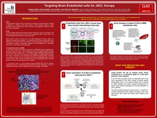
  • 1. Targeting Brain Endothelial cells for JNCL therapy Colleen S Stein1, Mark Schultz2, Luis Tecedor3, and Beverly L Davidson3, 1Dept. of Internal Medicine, Carver College of Medicine, University of Iowa; 2Dept. of Pathology, University of Michigan Medical School, Ann Arbor, MI: 3Children's Hospital of Philadelphia, Philadelphia, PA. contact e-mail: colleen-stein@uiowa.edu CLN3 gene 2) Intravenous injection For gene therapy, AAV-CLN3 particles will be injected intravenously into JNCL mice. AAV-CLN3 particles will bind and deliver the CLN3 gene to endothelial cells lining the blood vessels of the brain. With a good copy of the CLN3 gene, the endothelial cells can then make CLN3 protein. 3) Therapeutic effect? JNCL mice injected with AAV-CLN3 will be monitored to determine if they exhibit superior performance on motor tasks and reduced brain pathology compared to untreated JNCL mice. Mice will be tested for ability to maintain balance on a rotating rod. Image adapted from Nature Neuroscience 16: 658, 2013. WHAT THIS MEANS FOR JNCL THERAPY Endothelial cells from JNCL mouse brain have trouble internalizing molecules CLN3 positive endothelial cells outside of cell plasma membrane inside of cell caveolin-1 CLN3 negative endothelial cells TWO OF THREE TYPES OF ENDOCYTOSIS ARE DEFECTIVE IN JNCL MOUSE BRAIN ENDOTHELIAL CELLS. Cells internalize extracellular molecules by a process called endocytosis, in which a portion of the outer membrane of the cell buds into the cell and pinches off, bringing in a "mouthful" of molecules. Three main forms of endocytosis are depicted in the drawing: clathrin-dependent, caveolar, and fluid-phase endocytosis. Red-tagged transferrin, green-tagged albumin, and green-tagged dextran are used to examine these forms of endocytosis, respectively. In this experiment we prepared cultures of endothelial cells harvested from unaffected and JNCL mouse brains and examined internalization of the red- or green-tagged molecules. Interestingly, we found that both caveolar and fluid-phase endocytic pathways are impaired in the JNCL endothelial cells, while clathrin-dependent endocytosis is intact. Acknowledgements • These studies are funded by the BDSRA, Beyond Batten Disease Foundation (BBDF), and the NIH. • We thank the Gene Transfer Vector Core and the Central Microscopy Research Facility at the University of Iowa. 1 Art by Mark L Schultz INTRODUCTION JNCL Juvenile Batten disease (JNCL) is caused by mutations in the CLN3 gene, creating a deficiency or defect in the CLN3 protein. CLN3 deficiency leads to JNCL symptoms, which include loss of vision, seizures, and progressive decline in motor and mental capabilities. CLN3 CLN3 is situated within lipid membrane regions inside cells, and is not secreted. We don't know the exact function of CLN3, but it appears to play a basic role in the normal transport of proteins and lipids inside cells. While CLN3 is found at some level in all cells throughout the body, JNCL is a disease of central nervous system. So it seems that CLN3 has a critical role in cells that control the function of the brain and eye. One important type of cell that is often overlooked in studies of neurological diseases is the endothelial cell. Endothelial cells form the inside layer of blood vessels. The Blood-Brain Barrier (BBB) In the blood vessels (vasculature) of the brain, endothelial cells are specialized to protect and feed the brain. Here endothelial cells butt up against eachother tightly, eliminating spaces between cells. This creates a physical barrier between the blood and the brain, aptly called the blood-brain barrier (BBB). The BBB protects the brain from entry of microbes, toxic substances, and destructive immune cells. Nutrients are selectively taken into and transported through the endothelial cells. Thus the underlying brain cells (neurons and glial cells), rely on endothelial cells for nutrient supply for optimal neural performance and viability. In our previous work, our CLN3 reporter mouse indicated that CLN3 is made at relatively high level in endothelial cells throughout the brain vasculature. Thus it is possible that CLN3 is critical to BBB functions, which dictates brain health. Dense vasculature in the brain: Endothelial cells that line the blood vessels regulate nutrient supply from the blood to the brain. Astrocyte extends processes onto micro-vessels Neurons Mary Moye-Rowley, Iowa City, IA Cross-sectional view of a microvessel illustrates extensive cellular communication Adapted from Abbott et al., Nature Reviews, Neuroscience, 2006 The brain is the most densely vascularized organ in the body. This density is necessary to meet the oxygen and nutrient demands of intense neuronal activity. Every neuron and glial cell in the brain contacts or is in close proximity to a blood vessel. The endothelial cells that line the blood vessels in the brain form tight cell-to-cell junctions with each other, creating a physical barrier between the blood and the brain, termed the blood-brain barrier (BBB). Most nutrients gain entry into the brain by passing though the endothelial cells. Endothelial cells at the BBB are equipped with numerous receptors and channels, and act as gatekeepers, regulating selective transport into and out of the brain. Thus neuronal health is dependent upon endothelial cell health. We propose that endothelial cell dysfunction is a critical event leading to JNCL symptoms, and hypothesize that restoring CLN3 to endothelial cells will protect against JNCL symptoms. Does restoration of CLN3 to endothelial cells prevent JNCL? 2 USE OF GENTICALLY ENGINEERED MICE TO TEST THE HYPOTHESIS. It is conceivable that vascular endothelial cell impairment is a primary event in JNCL pathogenesis. Thus, we may be able to avert disease by providing normal CLN3 exclusively to brain endothelial cells. To test this concept, we are using transgenic mice. A transgenic mouse strain is one that has been genetically altered to carry an extra gene, referred to as a transgene. For this project we are using JNCL mice that have been genetically engineered to carry an inducible CLN3 transgene in their genome. By breeding this mouse to a Tie2-Cre inducer mouse, we will trigger CLN3 expression in the endothelial cells of progeny mice. Progeny mice will be tested in motor and behavioral assays to assess whether they develop JNCL symptoms, or remain symptom-free or show milder disease. 3 Gene therapy to restore CLN3 to BBB endothelial cells 1) Production of AAV-CLN3 AAV particles are microscopic virus-like particles used to delivery genes into cells. For gene therapy, we are designing AAV particles that will effectively attach to and deliver genes to brain endothelium. We will package the CLN3 gene into these AAV particles. AAV-CLN3 GENE TRANSFER TO BBB ENDOTHELIAL CELLS. Apart from the transgenic mouse approach, another way to determine whether providing CLN3 to endothelial cells will protect against JNCL symptoms is to do Gene Therapy. Here we plan to utilize AAV virus-like-particles to transfer the CLN3 gene into endothelial cells of JNCL mice. These studies will tell us whether blood vessel endothelium is a worthwhile target for CLN3 gene transfer in JNCL patients: If we find that providing CLN3 to endothelial cells of the brain vasculature in the JNCL mouse, by either genetic manipulation (Fig. 2) or by gene transfer (Fig. 3) results in milder disease, this would support advancement of similar gene transfer strategies for clinical application to JNCL patients This gene transfer approach would involve i.v. injection to achieve widespread gene transfer throughout the brain vasculature, and would not require direct injection into the brain.