Respiration in Anesthetized Mice: Evidence-Based Recommendations for Improved...InsideScientific
Join Dr. James Marx as he discusses improving the care and use of mice in biomedical research, with a focus on the pharmacology behind the effects of anesthetics on respiration.
Essentially, all anesthetic drugs suppress respiration in animals. This fact, combined with the high metabolic rate of mice, means that monitoring and respiratory support are critical for ensuring successful anesthesia of mice. Unfortunately, the monitoring and support for anesthetized mice lags well behind the care we give our other species. Recent publications and advances in equipment have given us the opportunity to make significant improvements in the care we provide anesthetized mice.
In this talk, Dr. Marx will briefly discuss the pharmacology behind the effects of anesthetics on respiration. He will then use evidence-based medicine to give practical recommendations for monitoring and support of respiration. An important part of this will include discussing normal physiologic values for respiratory parameters and highlighting differences between anesthetics and their effects on respiration. The recommendations in this discussion will be based on evidence of treatable or preventable issues. Lastly, he will briefly discuss technologies which are commonly used in other species and may be applied in mice in the near future.
Key Topics Include:
- The significant effects of anesthesia on respiration in mice
- Practical methods of monitoring and providing support to anesthetized mice
- Discussing equipment that will provide valuable information while addressing the depth of anesthesia in mice
Studying Epilepsy in Awake Head-Fixed Mice Using Microscopy, Electrophysiolog...InsideScientific
Epilepsy research employs sophisticated research methods such as fluorescence optical imaging and optogenetics, as well as novel electrophysiological techniques, to address unresolved questions about seizure generation and propagation on the cellular and circuitry levels. Since epilepsy research is most relevant when performed in non-anesthetized mice, it requires specialized tools that ensure stable head fixation during high-precision imaging and recordings.
In this webinar, Dr. Anthony Umpierre (Prof. LongJun Wu group, Mayo Clinic, USA) and Prof. Rob Wykes (UCL, UK) present their research on microglial calcium signaling and epileptic networks carried out in awake head-fixed mice. In addition to sharing exciting new findings, the presenters address the challenges of working with awake mice.
Key topics will include…
- Mesoscopic investigations of seizure dynamics and propagation using widefield calcium imaging
- Generating full-bandwidth electrophysiological recordings enabled by graphene micro-transistors to detect spreading depolarizations and seizures
- On-demand optogenetic induction of spreading depolarizations to investigate pharmacological suppression in the awake brain
- The impact of acute versus chronic window preparations on microglial calcium activity
- The use of genetically encoded calcium indicators to study calcium dynamics in microglia
- The effects of bi-directional shifts in neuronal activity caused by kainate-triggered status epilepticus and isoflurane anesthesia on microglial calcium
Functional Ultrasound (fUS) Imaging in the Brain of Awake Behaving MiceInsideScientific
To watch the webinar, visit:
https://insidescientific.com/webinar/functional-ultrasound-imaging-brain-awake-behaving-mice-neurotar-iconeus
Functional ultrasound (fUS) imaging is a new kid on the block in neuroimaging. It combines high spaciotemporal resolution with deep tissue penetration, which enables non-invasive whole-brain imaging in mice.
This exciting new technology complements and extends classical imaging modalities: it enables more straightforward, unobstructed and non-invasive functional measurements in mouse models of CNS diseases. Sensitive to changes in cerebral blood volume, fUS imaging is used to characterize brain networks with functional connectivity analysis and to measure the responses to sensory stimuli and pharmacological challenges.
fUS imaging performed in the brain of awake mice removes the biases and artifacts associated with the use of general anesthesia, which is no longer a “necessary evil” of translational imaging. Besides that: fUS imaging in awake mice allows integrating functional imaging with behavioral readouts starting from open field locomotion tracking to maze navigation and sociability studies.
In this webinar, you will learn:
– Functional ultrasound (fUS) imaging methodology
– How translational fUS neuroimaging helps to advance basic neuroscience research and preclinical drug discovery
– The main advantages and limitations of using functional ultrasound compared to other techniques such as BOLD fMRI
– The benefits of imaging in awake, head-restrained but otherwise freely moving mice
– Imaging functional activation, connectivity and pharmacologically-induced changes in awake and behaving mice
– How to combine fUS imaging with behavioral observation
Single-Cell Electrophysiology and 2-Photon Imaging in Awake Mice with 2D-Loco...InsideScientific
In this webinar sponsored by Neurotar, experts present their research utilizing the Mobile HomeCage®, an experimental tool which ensures the stability required for high-precision neurophysiological techniques while allowing mice to navigate and explore their environment.
Case Study #1:
Dr. Sarah Stuart and Dr. Jon Palacios-Filardo of the University of Bristol present their studies combining analysis of goal-directed behavior with whole-cell recordings from the hippocampus of awake mice. The researchers share useful tips for the surgery protocol and for adjusting the head fixation angle in order to facilitate mouse motility and exploratory behavior.
Case Study #2:
Dr. Alexander Dityatev and Weilun Sun from the German Center for Neurodegenerative Diseases (DZNE) discuss 2-photon imaging of fluorescently labeled microglia in vivo in the context of neurodegenerative disease. They also present their recent data on the effects of different anesthetics on the microglial response to localized laser injury.
Case Study #3:
Dr. Norbert Hájos from the Hungarian Academy of Sciences presents his lab’s research into the amygdala’s role in reward-driven behavior. He shares the challenges of making single-unit recordings using silicon probes during mouse locomotion and subsequent morphological identification of active neurons in the amygdala.
Key topics covered during this webinar include:
- Requirements for stable single-cell recordings and 2-photon imaging in behaving mice
- Challenges of combining high-precision techniques with behavioral research
- Methodological considerations for improving exploratory behavior in head-fixed mice
- Quantitative analysis of microglial function using 2-photon microscopy in awake mice
- Recording neuronal activity in the amygdala of awake mice followed by morphological identification of recorded neurons
A characteristic of the developing mammalian visual system is a brief interval of plasticity, termed the “critical period,” when the circuitry of
primary visual cortex is most sensitive to perturbation of visual experience. Depriving one eye of vision (monocular deprivation [MD]) during
the critical period alters ocular dominance (OD) by shifting the responsiveness of neurons in visual cortex to favor the nondeprived eye. A
disinhibitory microcircuit involving parvalbumin-expressing (PV) interneurons initiates this OD plasticity. The gene encoding the neuronal
nogo-66-receptor1(ngr1/rtn4r) is required to close the critical period.Herewecombinedmousegenetics, electrophysiology,andcircuitmapping
with laser-scanning photostimulation to investigate whether disinhibition is confined to the critical period by ngr1.We demonstrate that ngr1
mutant mice retain plasticity characteristic of the critical period as adults, and that ngr1 operates within PV interneurons to restrict the loss of
intracortical excitatory synaptic input following MD in adult mice, and this disinhibition induces a “lower PV network configuration” in both
critical-period wild-type miceandadult ngr1/mice.Wepropose that ngr1 limits disinhibition to close the critical period forODplasticityand
that a decrease in PV expression levels reports the diminished recent cumulative activity of these interneurons.
Sensorimotor Network Development During Early Postnatal Life in the Awake and...InsideScientific
In the last decades, electrophysiological and imaging-based approaches provided significant new insights into the mechanisms of neuronal development. Nevertheless, many important questions remain unanswered. How does the fine control of a motor output develop? How does sensorimotor integration in the early and subsequent phases of brain development shape behavior? How does sensorimotor development evolve in awake and sleeping states? What role do myoclonic twitches play in this process?
Answering these questions requires performing high-precision tests in the brain of non-anesthetized animals across sleep and wake during the early stages of their postnatal development. Such tests require head-fixation apparatus suitable for neonatal and juvenile rodents. The Mobile HomeCage combines a stable head-fixation with an air-lifted cage that closely resembles laboratory rodents’ natural habitat – an optimal platform for studying early postnatal brain development.
In this webinar, Dr. Cavaccini (Prof. Karayannis’s lab at the Brain Research Institute, University of Zurich) and Dr. Dooley (Prof. Blumberg’s lab at the University of Iowa), share their insights into the development of rodent sensorimotor neuronal circuits during early postnatal life. They elucidate the cortical and subcortical mechanisms involved in the development of sensorimotor circuitry during wakefulness (in a mouse model) and REM sleep (in a rat model).
Key Takeaways
Dr. Anna Cavaccini:
- Anatomical and functional changes occur at the striatal level before and after the onset of different sensory modalities
- Locomotor activity changes throughout early development, and it correlates with striatal function
- Sensory information coming from whiskers affects locomotion and striatal function before and after the onset of different sensory modalities
Dr. James Dooley:
- Myoclonic twitches in REM sleep continue to trigger cortical and thalamic activity beyond the early postnatal period
- Twitch-related thalamic activity is spatiotemporally refined by the third postnatal week
- Motor thalamus activity reflects an internal model of movement produced by twitches and is dependent on the cerebellar output
Electrophysiology of Human Native Receptors in Neurological and Mental DisordersInsideScientific
Dr. Agenor Limon presents research integrating functional metrics with large anatomical, transcriptomic, and proteomic datasets to evaluate the relationship between synaptic E/I ratio and behavioral abnormalities across postmortem intervals and brain banks.
Alterations in synaptic function have been found in transcriptomic, genetic, and proteomic studies of neurological and mental disorders. Clinical and preclinical studies suggest that synaptic dysfunction and behavioral abnormalities in disorders like Alzheimer’s disease and schizophrenia may be mechanistically linked to the emergence of imbalances between excitatory (E) and inhibitory (I) receptors. However, until recently, the electrophysiological E/I synaptic ratio had only been measured in animal models.
Using pioneering methods developed in the lab including reactivation and microtransplantation of synaptic receptors from frozen human brains, Dr. Agenor Limon’s research team has obtained electrophysiological metrics of global synaptic E/I ratios in cortical brain regions of subjects that were affected by Alzheimer’s Disease and synaptic measurements in schizophrenia.
In this webinar, Dr. Agenor Limon will present recent research integrating functional metrics with large anatomical, transcriptomic, and proteomic datasets to evaluate the relationship between synaptic E/I ratio and behavioral abnormalities across postmortem intervals and brain banks.
Key Topics Include:
- Understand the global synaptic excitation to inhibition ratio between excitatory and inhibitory synaptic receptors determined from reactivated frozen human brain tissue
- Understand the relationship between electrophysiological metrics of receptor function and multi-omic data in neurodegenerative disorders
- Understand the role of deviations of the excitation to inhibition ratio with clinical presentation in Alzheimer’s disease
From “Artificial” to “Real”: What 24/7 Home Cage Monitoring Teaches Us In Pre...InsideScientific
In this webinar Dr. Stefano Gaburro, Scientific Director at Tecniplast, will present an innovative non-invasive and scalable technique called Digital Ventilated Cage (DVC) that is meant to perform longitudinal studies for neurodegenerative disease models using long term monitoring of mice in a stress-free environment.
In the second part of the webinar, Dr. Brun Ulfhake from Karolinska Institutet will show how this technique can be used to study biorhythmicity (circadian and circannual) of small rodents and approaches to characterize and extract metrics of the spontaneous home-cage way of life for mice. These metrics may translate better to behavioral observations made in humans.
The Brain as a Whole: Executive Neurons and Sustaining Homeostatic GliaInsideScientific
Carl Petersen and Alexei Verkhratsky share their research on homeostatic neuroglia and imaging of neuronal network function. This webinar is brought to you by APS’ new journal, Function, and part of their Physiology in Focus learning series.
During this exclusive live webinar, Carl Petersen and Alexei Verkhratsky discuss astrocyte-mediated homeostatic control of the central nervous system, and how optical and 2-photon microscopy can be used for functional neuroimaging.
Imaging Neuronal Function
Carl Petersen, PhD
Highly dynamic and spatially distributed neuronal circuits in the brain control mammalian behavior. Through technological advances, optical measurements of neuronal function can now be carried out in behaving mice at multiple scales. Wide-field imaging allows the dynamic interactions between different brain areas to be studied as sensory information is processed and transformed into behavioral output. Within a brain region, two-photon microscopy can be used to image the neuronal network activity with cellular resolution allowing different types of projection neurons to be distinguished. Together optical methods provide versatile tools for causal mechanistic understanding of neuronal network function in mice.
Astrocytes: indispensable neuronal supporters in sickness and in health
Alexei Verkhratsky, MD, PhD, DSc
The nervous system is composed of two arms: the executive neurons and the homeostatic neuroglia. The neurons require energy, support, and protection, all of which is provided by the neuroglia. Astrocytes, the principal homeostatic cells of the brain and spinal cord, are tightly integrated into the neural networks and act within the context of the neural tissue. As astrocytes control the homeostasis of the central nervous system at all levels of organization, from the molecular to the whole organ level, we can begin to define and understand brain vulnerabilities to aging and diseases.
Place Cell Mapping and Stress Monitoring in Head-Fixed Mice Navigating an Air...InsideScientific
In this webinar sponsored by Neurotar, experts present their research on 2-photon imaging of hippocampal place cells and on stress monitoring in head-fixed awake behaving mice. Dr. Konrad Juczewski from the National Institutes of Health (NIH)/National Institute on Alcohol Abuse and Alcoholism (NIAAA) discusses the impact of head fixation on animal’s stress, locomotion and performance in classical behavioral paradigms.
Dr. Mary Ann Go from the Laboratory of Neural Coding and Neurodegenerative Disease at Imperial College London led by Prof. Simon Schultz presents her research using 2-photon microscopy aimed at place cell mapping in the hippocampus during exploration and navigation of a circular linear track.
Key Discussion Topics Include:
- Stress reduction in head-fixed rodents
- Improving data reproducibility and translational value of the data acquired from head-fixed rodents
- Effects of head fixation on blood corticosterone concentration, locomotion patterns and performance in stress-associated behavioral tests
- Optimizing habituation protocol for head-fixed mice
- Monitoring neural activity and mapping of place cells using 2-photon microscopy during navigation and exploration behavior
- Automating the experiments using a closed-loop approach and behavior-triggered reward systems
Case Studies in Home Cage Monitoring: Rodent Behavior, Circadian Biology and ...InsideScientific
Automated home cage behavioral monitoring is receiving increasing attention from the scientific community because of its benefits with regards to translational research, data replicability and animal welfare. In this webinar, Kenneth Dyar (Helmholtz Diabetes Center) and Joanna Moore (GSK) discuss how home cage monitoring can be used to reduce animal stress, optimize methodology and guide physiology and animal behavior research.
Dr. Kenneth Dyar
Passive locomotor activity monitoring for real-time circadian study design
Circadian clocks are fundamental determinants of physiology, behavior and health. For skeletal muscle, the circadian clock promotes insulin sensitivity and orchestrates rhythms of glucose, lipid, and amino acid metabolism. Physical activity synchronizes circadian clocks by altering body temperature and through distribution of various hormones and metabolites. Research suggests that misalignment of the ‘muscle clock’ plays an important pathophysiological role in metabolic disease. In this webinar, Dr. Kenneth Dyar highlights some examples of how the DVC system can be used for locomotor activity monitoring in order to evaluate circadian alignment before, during or after various dietary and pharmacological interventions.
Dr. Joanna Moore
Using home-cage monitoring to determine the impact of timed mating on male mouse welfare
The use of sterile male mice to induce pseudopregnancy in female mice assigned for the implantation of embryos is a vital component in the production of Genetically Altered Animals (GAA). This process involves swapping a genetically sterile male’s female companion for a new female. In this presentation, Dr. Joanna Moore discusses the use of home cage activity monitoring to evaluate the potential impact of this procedure on the welfare of male mice and how the impact of this intervention may be reduced. All animal studies were ethically reviewed and carried out in accordance with the Animals (Scientific Procedures) Act 1986 and the GSK Policy on the Care, Welfare and Treatment of Animals.
Key topics will include…
- Using home cage activity as a readout for animal welfare
- Using locomotor activity to optimize methodology and validate study design in real-time
- Pre-study screening of cohorts for outliers
Self Head Fixation Training for the Study of Perceptual Decisions in MiceInsideScientific
In this webinar, Andrea Benucci, PhD will discuss a setup developed in his laboratory for high-throughput behavioral training of mice based on voluntary head fixation. He will describe its flexible use for behavioral training and concurrent neural recordings, delving into some technical considerations related to user-specific customizations as well.
In Andrea’s lab, they study the neural substrate of visual processing and vision-based decision making. To this end, they aim to define a research framework capable of linking neural architectures to the underlying computations. The solution they have developed is to integrate experimental methods for all-optical dissection of neuronal circuits with large-scale dynamical network models based on artificial neural networks (aNNs). The connectivity architecture of aNNs closely mirror that of biological neural networks, thus representing an effective theoretical framework to unify computational, algorithmic, and implementation levels of analysis.
Finally, Andrea will present some examples of unique research achievements made possible by the use of this setup.
ESP block - future direction and remaining questionsAmit Pawa
This Talk was delivered by Dr Pawa on 5th June 2021 as part of the ISURA 2021 hybrid conference held in Toronto.
The Future Direction of this block and remaining questions to be answered are covered here
This is a poster which describes an experiment which was executed by our group. A part was our own experiment (the i-Doser part) and the other part was classical.
We came up with the i-Doser experiment, which was voted as the winning experiment. Furthermore, our poster was chosen as the best poster out of all the other posters. (June 2010)
A monkey model of auditory scene analysisPradeepD32
My work impacts half the world who develop age-related hearing loss with difficulty understanding speech in noise. To understand how the brain solves the cocktail party problem, I need to record from neurons suitable only in animals. Monkeys are best suited for this given our similar auditory brains. I use sounds without semantics and employ fMRI to show that monkeys use similar brain regions as humans to separate overlapping sounds. This study is the first to show such evidence in any animal. Now, I can record from monkey neurons and generalize the results to humans!
Don't Miss a Beat: Understanding Continuous, Real Time Physiologic MonitoringInsideScientific
In vivo, preclinical research encompasses numerous study designs with various species and endpoints being monitored. Having access to all available study data allows the scientist to comprehensively understand the study paradigm and make informed research decisions. During Session 3 of our webseries "Biotelemetry For The Life Sciences", presenters discussed the importance of continuous, real-time monitoring in preclinical research. Case studies included using EEG as a biomarker for CNS activity and drug discovery and using telemetry for disease characterizations and and evaluation of vaccines in Biodefense research.
During this exclusive webinar sponsored by Data Sciences International, Steve Fox shares his experience from pharmaceutical development; discussing the importance of continuous EEG monitoring for sleep studies. Anna Honko explains the importance of having access to real-time, continuous data when studying infectious diseases in non-human primates in a Biodefense setting. In addition, Dusty Sarazan reviews how and why continuous, real-time monitoring has become a preferred and essential method for acquiring and studying physiology in today's preclinical research setting.
Key Topics:
EEG as a biomarker for CNS activity and a platform for pre-clincal drug discovery
Sleep/wake patterns and rhythms, and how qEEG signatures allow for accurate clinical predictions of efficacy and CNS adverse event screening
Considering the FDA Animal Rule
Basic disease characterizations and evaluation of vaccines and therapeutics
Non-human primate models of viral biodefense and emerging pathogens
Translating pre-clinical study findings to human, clinical populations
Guest Speakers:
Steve Fox, BS
Associate Principal Scientist,
Merck & Co., Inc.
Anna Honko, PhD
Staff Scientist,
NIH/NIAID Integrated Research Facility
R. Dustan Sarazan, DVM, PhD
Vice President & Chief Scientific Officer, Data Sciences International
Presentation by Jared Jageler, David Adler, Noelia Duchovny, and Evan Herrnstadt, analysts in CBO’s Microeconomic Studies and Health Analysis Divisions, at the Association of Environmental and Resource Economists Summer Conference.
What is the point of small housing associations.pptxPaul Smith
Given the small scale of housing associations and their relative high cost per home what is the point of them and how do we justify their continued existance
This session provides a comprehensive overview of the latest updates to the Uniform Administrative Requirements, Cost Principles, and Audit Requirements for Federal Awards (commonly known as the Uniform Guidance) outlined in the 2 CFR 200.
With a focus on the 2024 revisions issued by the Office of Management and Budget (OMB), participants will gain insight into the key changes affecting federal grant recipients. The session will delve into critical regulatory updates, providing attendees with the knowledge and tools necessary to navigate and comply with the evolving landscape of federal grant management.
Learning Objectives:
- Understand the rationale behind the 2024 updates to the Uniform Guidance outlined in 2 CFR 200, and their implications for federal grant recipients.
- Identify the key changes and revisions introduced by the Office of Management and Budget (OMB) in the 2024 edition of 2 CFR 200.
- Gain proficiency in applying the updated regulations to ensure compliance with federal grant requirements and avoid potential audit findings.
- Develop strategies for effectively implementing the new guidelines within the grant management processes of their respective organizations, fostering efficiency and accountability in federal grant administration.
ZGB - The Role of Generative AI in Government transformation.pdfSaeed Al Dhaheri
This keynote was presented during the the 7th edition of the UAE Hackathon 2024. It highlights the role of AI and Generative AI in addressing government transformation to achieve zero government bureaucracy
Many ways to support street children.pptxSERUDS INDIA
By raising awareness, providing support, advocating for change, and offering assistance to children in need, individuals can play a crucial role in improving the lives of street children and helping them realize their full potential
Donate Us
https://serudsindia.org/how-individuals-can-support-street-children-in-india/
#donatefororphan, #donateforhomelesschildren, #childeducation, #ngochildeducation, #donateforeducation, #donationforchildeducation, #sponsorforpoorchild, #sponsororphanage #sponsororphanchild, #donation, #education, #charity, #educationforchild, #seruds, #kurnool, #joyhome
A process server is a authorized person for delivering legal documents, such as summons, complaints, subpoenas, and other court papers, to peoples involved in legal proceedings.
Jennifer Schaus and Associates hosts a complimentary webinar series on The FAR in 2024. Join the webinars on Wednesdays and Fridays at noon, eastern.
Recordings are on YouTube and the company website.
https://www.youtube.com/@jenniferschaus/videos
Russian anarchist and anti-war movement in the third year of full-scale warAntti Rautiainen
Anarchist group ANA Regensburg hosted my online-presentation on 16th of May 2024, in which I discussed tactics of anti-war activism in Russia, and reasons why the anti-war movement has not been able to make an impact to change the course of events yet. Cases of anarchists repressed for anti-war activities are presented, as well as strategies of support for political prisoners, and modest successes in supporting their struggles.
Thumbnail picture is by MediaZona, you may read their report on anti-war arson attacks in Russia here: https://en.zona.media/article/2022/10/13/burn-map
Links:
Autonomous Action
http://Avtonom.org
Anarchist Black Cross Moscow
http://Avtonom.org/abc
Solidarity Zone
https://t.me/solidarity_zone
Memorial
https://memopzk.org/, https://t.me/pzk_memorial
OVD-Info
https://en.ovdinfo.org/antiwar-ovd-info-guide
RosUznik
https://rosuznik.org/
Uznik Online
http://uznikonline.tilda.ws/
Russian Reader
https://therussianreader.com/
ABC Irkutsk
https://abc38.noblogs.org/
Send mail to prisoners from abroad:
http://Prisonmail.online
YouTube: https://youtu.be/c5nSOdU48O8
Spotify: https://podcasters.spotify.com/pod/show/libertarianlifecoach/episodes/Russian-anarchist-and-anti-war-movement-in-the-third-year-of-full-scale-war-e2k8ai4
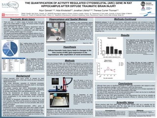
1. THE QUANTIFICATION OF ACTIVITY REGULATED CYTOSKELETAL (ARC) GENE IN RAT
HIPPOCAMPUS AFTER DIFFUSE TRAUMATIC BRAIN INJURY
1BASIS Chandler High School, Chandler, AZ, 2BARROW Neurological Institute at Phoenix Children’s Hospital- Phoenix, AZ, 3Department of Child Health, University of Arizona College of Medicine –
Phoenix, AZ, 4Phoenix VA Healthcare System- Phoenix, AZ, 5Department of Neuroscience, University of Strasbourg, France, 6Neuroscience Program, Arizona State University, Tempe, AZ,
References
Background
• Diffuse traumatic brain injury (dTBI) is caused by rapid
acceleration-deceleration of the brain inside the skull resulting in
diffuse axonal injury.
• dTBI incites secondary cascades of molecular processes,
resulting in circuit reorganization that likely leads to neurological
impairment, including sensory sensitivity.
• The whisker nuisance task (WNT) demonstrates late-onset,
persistent sensory sensitivity to whisker stimulation manifesting
by 28 days post-diffuse TBI in rats (McNamara et al., 2010).
• During the development of sensory sensitivity, we have
documented pathological and functional changes supporting
circuit reorganization in the whisker barrel circuit, but not the
hippocampus.
• The efficacy of therapeutic approaches on maintaining in vivo
circuit integrity after dTBI could be carried out using an immediate
early gene as a molecular marker of circuit activation after circuit-
specific stimulation.
• Activity-regulated cytoskeleton-associated (ARC) gene is
considered to be an immediate early gene that is tightly coupled
to behavioral encoding of information in neuronal circuits in vivo.
• Here, we identified the time course of ARC gene expression after
exploration of a novel environment and determined whether the
time course of ARC gene expression changed as a function of TBI
at 28 days post-injury in the hippocampus.
• McNamara et al., (2010) J Neurotrauma 27(4): 695-706.
• Centers for Disease Control and Prevention.—. MMWR 2011;
60(39):1337–1342.
• Khodadad et al. (2015) Behavioral Brain Research;284:249-56
• Moser et al(1998). J Neuroscience 18(18): 7535-7542.
Hypothesis
Diffuse traumatic brain injury leads to changes in the
time course of ARC gene expression in the
hippocampus after exploration of novel environment.
Methods
Adult male Sprague-Dawley rats (300-350g) were subjected to moderate midline
fluid percussion injury ( FPI; n=67; 1.9 atm; 6-10 min righting reflex time) or a sham
surgery (Fig. 4). At 28 days post-injury, the rats explored a novel environment for 20
minutes, activating their spatial memory (Fig. 5). After 20 minutes of exploring the
novel environment, animals were housed individually for 15, 30, 60 or 90 minutes.
At the specific time point, rats were perfused with iced cold phosphate buffered
saline PBS, the hippocampus was dissected out and stored in RNAlater® at -20°C
until mRNA extraction. Life Technology’s RNA extraction kit was used to extract
mRNA from the dissected tissue and converted mRNA to CDNA for q-PCR.
Quantitative PCR (q-PCR): The TaqMan® Gene Expression Assay for ARC was
optimized to run under universal thermal cycling conditions, with amplification
efficiencies of 100%. Within each animal, relative gene expression was normalized
to the 18s endogenous control and the expression level in the naive group using the
2-ΔΔCT.
Methods-Continued
Statistical analyses: For the both time courses, relative gene expression was normalized to naïve rats
(unstimulated/uninjured). ARC gene expression was compared to naïve over time using a one-way ANOVA
with a Tukey’s post-hoc analysis (“naïve” in Fig 6). After dTBI, relative gene expression was compared to
sham and “naïve” animals after exploration of the novel environment as a function of time post-stimulation
and injury using a two-way ANOVA with Tukey’s post-hoc analysis. The data are represented as the mean ±
standard error of the mean (SEM) *, p<0.05. All statistical analyses were performed using Prism®
(GraphPad, CA).
Conclusions
• Although there were no significant changes in ARC gene expression with respect to sham,
there were significant changes in ARC expression within the injury group.
• However, we did not see ARC expression increase significantly at 30 minutes exploration in
the sham group, which confounds the interpretation of the data.
• The lack of ARC expression at 30 minutes post-stimulation in sham can be due to insufficient
exploration of the novel environment by sham animals.
• Based on this data, ARC gene expression in the hippocampus may not be as valuable as
other areas of the brain for assessing therapeutic efficacy on restoring circuit function in
experimental diffuse TBI models.
Scientific Value
Where previous data from other areas of the brain support ARC as a valuable tool for
assessing circuit re-organization after TBI, these data do not indicate that signaling in the
hippocampus follows similar re-organization patterns.
Acknowledgements
The authors wish to thank Daniel Griffiths and Megan Evilsizor for performing
rodent surgeries and the Translational Neurotrauma Research Program for
valuable feedback throughout my internship. These experiments are partly
supported by, ADHS14-00003606, NIH R03 NS-077098, NIH R01 NS-065052
and Phoenix Children’s Hospital Mission Support Funds.
Fig 6. Exploration of the novel environment results
in a significant increase of ARC gene expression in
the hippocampus in animals with no surgical
preparation. The figure shows the time course for ARC
rats explored a novel environment for 20 minutes. These
animals were normalized to the naïve group which was
euthanized without being given time to explore the box.
There is a significant 2.5 fold increase of ARC
expression at 15 minutes relative to naïve. ARC
expression peaked at 30 minutes and then decreased to
baseline levels in the following time points.
Fig 5. Exploration of novel
environment The novel group was
introduced to the behavioral box lined
with an absorbent pad and allowed to
habituate for 5 minutes. For the next 15
minutes they were free to explore their
environment, voluntarily using their
whiskers.
Fig 4. FPI device. A cranial hub is
implanted and attached to the injury
device where an adjustable pendulum
falls and strikes the plunger, generating
a fluid pulse that impacts the dural
surface, resulting in diffuse axonal
injury without cavitation or contusion.
Hippocampus and Spatial Memory
Fig 3. Exploring a novel environment requires
spatial memory, which activates the hippocampus.
When a rat interacts with a novel environment, it
encodes its surroundings in spatial memory, activating
hippocampal circuitry (Moser et al., 1998).
Researchers lesioned parts of the hippocampus in rats
and tested their ability to complete a water maze. They
found that while the rats were still able to encode their
surroundings as spatial memory, they were unable to
retrieve the memories from the hippocampus. ARC is
an immediate early gene which responds to the
encoding of spatial memories in the hippocampus. Our
study will focus on the time course of ARC activation in
the hippocampus after exploration of a novel
environment after dTBI.
Traumatic Brain Injury
• There are over 1.7 million cases of traumatic brain injury per
year in the United States. In reality, the actual number is higher
due to undocumented cases of TBI.
• From 2001-2009, the rate of emergency department visits for
sports and recreation-related injuries with a diagnosis of
concussion or TBI, alone or in combination with other injuries,
rose 57% among children (age 19 or younger).
• TBI causes long-lasting debilitating neurological deficits (Fig 2).
• Currently, there is no cure for persisting TBI-induced neurological
deficits; only treatment of specific physical, cognitive, emotional
and sleep-related symptoms as they arise.
Fig 1. Leading causes of TBI in the United
States. Falls disproportionally affect the chart in
that more than 50% of children (ages 0-14) and
more than 80% of seniors (ages 65+) receive
TBI via a fall. Blunt trauma (dTBI) is the highest
cause of TBI in adolescents and adults (ages 15-
64), often occurring during sports and motor
vehicle accidents (CDC 2011).
Fig 2. Debilitating neurological
deficits of TBI. The onset of these
morbidities can occur from hours to
months after the TBI. Neurological
deficits can be partially or fully
alleviated through medication;
however, the cognitive and
emotional deficits can significantly
impact a person’s career and family
life (CDC 2011).
Fig 7. Diffuse TBI does not cause a significant
change in ARC expression in the hippocampus. The
figure shows the relative gene expression of ARC in the
injured group of rats across the selected time points.
ARC expression significantly increases at 30 minutes
post-stimulation in the injured group. ARC then
decreased to baseline levels in the following time
points. The change in relative gene expression of ARC
was not significant at any time point in the injured group
with respect to sham.
Khodadad et al. (2015)
Moser et al., 1998
Results
Arjun Ganesh1,2,3, Aida Khodadad3,5, Jonathan Lifshitz2-4,6, Theresa Currier Thomas2-4