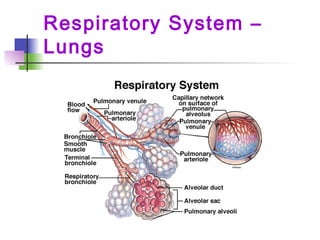
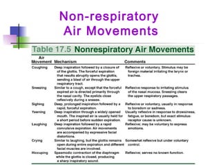
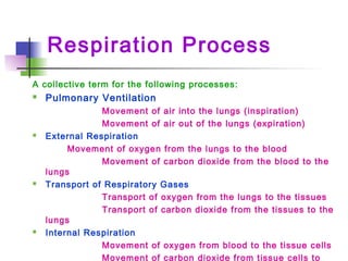
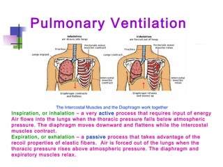
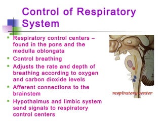
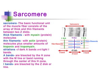
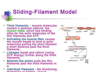
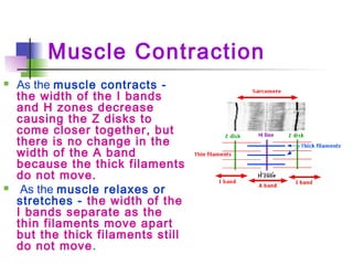
The document provides information about the anatomy and physiology of the respiratory and muscular systems, as well as disorders that can affect these systems. It includes details on the structure and function of lungs, control of breathing, gas exchange, types of muscles, muscle contraction, and common respiratory and muscular disorders. The document appears to be training materials for a Science Olympiad anatomy event, outlining what topics will be covered.
The apparatus used to measure
Volume of air exchanged during breathing
Respiratory rate
The record is called a spirogram
Upward deflection inhalation
Downward deflection exhalation
The apparatus used to measure
Volume of air exchanged during breathing
Respiratory rate
The record is called a spirogram
Upward deflection inhalation
Downward deflection exhalation
Group project for 12 Honors about the orginization Science Olympiad; the goal was to make a presentation about starting a team or two in the middle school
Digestion & Respiration by Jasveer RanaJasveer Rana
What is Digestion???
The chemical breakdown of complex biological molecules into their component parts.
Lipids to fatty acids
Proteins to individual amino acids
Carbohydrates into simple sugars
Respiratory System – Functions
Basic functions of the respiratory system are:
1. provides oxygen to the blood stream and
removes carbon dioxide
2. enables sound production or vocalization as
expired air passes over the vocal chords
3. enables protective and reflexive non-
breathing air movements such as coughing
and sneezing, to keep the air passages clear
4. control of Acid-Base balance
5. control of blood pH
Design of artificial respiratory modelShîvãm Gûptå
Design of Artificial Respiratory Model.. Know about the respiratory system.
The respiratory system consists of the upper respiratory tract (nasal passages), the airway conduction system (larynx, trachea, bronchi, bronchioles and terminal bronchioles), and the lower respiratory tract (alveolar ducts and alveoli). Not all segments of the respiratory system mature at the same pace. The olfactory epithelium matures earliest by PND 7. The lung, however, is not considered mature until PND 21, when alveolarization and microvascular maturation are complete. This chapter will discuss the embryological development (briefly), adult histomorphology, and postnatal histologic development of each major component of the respiratory system.
respiratory system, respiratory mechanism and volumes.pptxGarimaMittal46
respiratory system, respiratory mechanism and volumes and capacities, Human Respiratory system, gaseous exchange, pulmonary system,
submitted by Garima Mittal
1. 20112011 ANATOMYANATOMY (B)(B)
Karen LancourKaren Lancour Patty PalmiettoPatty Palmietto
National Bio RulesNational Bio Rules National EventNational Event
Committee ChairmanCommittee Chairman Supervisor - A & PSupervisor - A & P
karenlancour@charter.netkarenlancour@charter.net
2. Event Rules – 2011
DISCLAIMER
This presentation was prepared using
draft rules. There may be some
changes in the final copy of the rules.
The rules which will be in your Coaches
Manual and Student Manuals will be the
official rules.
3. Event Rules – 2011
BE SURE TO CHECK THE 2011
EVENT RULES FOR EVENT
PARAMETERS AND TOPICS FOR
EACH COMPETITION LEVEL
The Skeletal Muscle List can be
found at www.soinc.org under Event
Information
4. TRAINING MATERIALS
Training Power PointTraining Power Point – content overview– content overview
Training HandoutTraining Handout - content information- content information
Sample TournamentSample Tournament – sample problems with key– sample problems with key
Event Supervisor GuideEvent Supervisor Guide – prep tips , event– prep tips , event
needs, and scoring tipsneeds, and scoring tips
Internet Resource & Training MaterialsInternet Resource & Training Materials – on the– on the
Science Olympiad website atScience Olympiad website at www.soinc.orgwww.soinc.org
under Event Informationunder Event Information
Biology-Earth Science CDBiology-Earth Science CD , as well as the, as well as the DivisionDivision
B and Division C Test PacketsB and Division C Test Packets are available fromare available from
SO store atSO store at www.soinc.orgwww.soinc.org
5. ANATOMY
Event ContentEvent Content:: 20112011
BASIC ANATOMY (Structure and function)BASIC ANATOMY (Structure and function)
Respiratory System (new)Respiratory System (new)
Muscular SystemMuscular System
Major disordersMajor disorders
Treatment and prevention of disordersTreatment and prevention of disorders
PROCESS SKILLS -PROCESS SKILLS - observations,observations,
inferences, predictions, calculations, datainferences, predictions, calculations, data
analysis, and conclusions.analysis, and conclusions.
6. Respiratory SystemRespiratory System –
Functions
Basic functions of the respiratory system are:Basic functions of the respiratory system are:
1. provides oxygen to the blood stream and
removes carbon dioxide
2. enables sound production or vocalization
as
expired air passes over the vocal chords
3. enables protective and reflexive non-
breathing air movements such as
coughing
and sneezing, to keep the air passages
clear
10. Respiration Process
A collective term for the following processes:
Pulmonary Ventilation
Movement of air into the lungs (inspiration)
Movement of air out of the lungs (expiration)
External Respiration
Movement of oxygen from the lungs to the blood
Movement of carbon dioxide from the blood to the
lungs
Transport of Respiratory Gases
Transport of oxygen from the lungs to the tissues
Transport of carbon dioxide from the tissues to the
lungs
Internal Respiration
Movement of oxygen from blood to the tissue cells
Movement of carbon dioxide from tissue cells to
11. Pulmonary Ventilation
The Intercostal Muscles and the Diaphragm work together
Inspiration, or inhalation – a very active process that requires input of energy
Air flows into the lungs when the thoracic pressure falls below atmospheric
pressure. The diaphragm moves downward and flattens while the intercostal
muscles contract.
Expiration, or exhalation – a passive process that takes advantage of the
recoil properties of elastic fibers. Air is forced out of the lungs when the
thoracic pressure rises above atmospheric pressure. The diaphragm and
expiratory muscles relax.
12. Patterns of Breathing
Apnea – temporary cessation of breathing (one or more skipped
breaths)
Dyspnea – labored, gasping breathing; shortness of breath
Eupnea – Normal, relaxed, quiet breathing
Hyperpnea – increased rate and depth of breathing in response
to exercise, pain, or other conditions
Hyperventilation – increased pulmonary ventilation in excess of
metabolic demand
Hypoventilation – reduced pulmonary ventilation
Orthopnea – Dyspnea that occurs when a person is lying down
Respiratory arrest – permanent cessation of breathing
Tachypnea – accelerated respiration
14. Measures of Pulmonary
Ventilation
Respiratory volumes – values determined by
using a spirometer
Tidal Volume (TV) – amount of air inhaled or
exhaled with each breath under resting
conditions
Inspiratory Reserve Volume (IRV) – amount of
air that can be inhaled during forced breathing
in addition to resting tidal volume
Expiratory Reserve Volume (ERV) – amount of
air that can be exhaled during forced breathing
in addition to tidal volume
Residual Volume (RV) – Amount of air remaining
in the lungs after a forced exhalation.
15. Formulas – CapacitiesFormulas – Capacities
Vital Capacity – maximum amount of air
that can be expired after taking the
deepest breath possible (VC = TV + IRV +
ERV)
Inspiratory Capacity – maximum volume of
air that can be inhaled following exhalation
of resting tidal volume (IC = TV + IRV)
Functional Residual Capacity – volume of
air remaining in the lungs following
exhalation of resting volume (FRC = ERV +
RV)
16. Control of Respiratory
System
Respiratory control centers –
found in the pons and the
medulla oblongata
Control breathing
Adjusts the rate and depth of
breathing according to oxygen
and carbon dioxide levels
Afferent connections to the
brainstem
Hypothalmus and limbic system
send signals to respiratory
control centers
17. Effects of Exercise on
Respiratory System
During exercise the muscle cells use up more oxygen and produce
increased amounts of carbon dioxide.
The lungs and heart have to work harder to supply the extra oxygen and
remove the carbon dioxide.
Your breathing rate increases and you breathe more deeply. Heart rate also
increases in order to transport the oxygenated blood to the muscles.
Muscle cell respiration increases - more oxygen is used up and levels of
carbon dioxide rise.
The brain detects increasing levels of carbon dioxide - a signal is sent to
the lungs to increase breathing.
Breathing rate and the volume of air in each breath increase - This means
that more gaseous exchange takes place.
The brain also tells the heart to beat faster so that more blood is pumped to
the lungs for gaseous exchange.
More oxygenated blood is gets to the muscles and more carbon dioxide is
removed.
18. Disorders of the
Respiratory System
Clinical Disorders and Diseases of the Respiratory System
Hypoxia – deficiency of oxygen in a tissue or the inability to use oxygen
Oxygen toxicity – excess oxygen, causing the build up of peroxides and free
radicals
Chronic Obstructive Pulmonary Diseases – long-term obstruction of airflow and a
substantial reduction in pulmonary ventilation
Chronic Bronchitis – cilia are immobilized and reduced in number; goblet
cells increase their production of mucus → mucus clogs the airways and
breeds infection
Emphysema – alveolar walls break down and the surface area of the lungs is
reduced
Asthma – allergens trigger the release of histamine and other inflammatory
chemicals that cause intense bronchoconstriction
Lung Cancer – malignant tumor of the lungs
Acute Rhinitis – the common cold
Laryngitis – inflammation of the vocal folds
Pneumonia – lower respiratory infection that causes fluid build up in the lungs
Sleep Apnea – Cessation of breathing for 10 seconds or longer during sleep
Tuberculosis – pulmonary infection with Mycobacterium tuberculosis; reduces
lung compliance
19. MUSCULAR SYSTEM
Muscle Function:
Stabilizing joints
Maintaining posture
Producing movement
Moving substances within the body
Stabilizing body position and regulating
organ volume
Producing heat– muscle contraction
generates 85% of the body’s heat
20. Characteristics of Muscle
Tissue
Excitability- receive and respond to
stimuli
Contractility- ability to shorten and
thicken
Extensibility- ability to stretch
Elasticity- ability to return to its
original shape after contraction or
extension
21. Types of Muscle
Skeletal
Muscle
Smooth Muscle Cardiac Muscle
Location
Attached to
bone
On hollow organs,
glands and blood
vessels
Heart
Function
Move the
whole body
Compression of tubes
& ducts
Heart
contraction to
propel blood
Nucleus
Multiple,
peripheral
Single, central Central & single
Control voluntary involuntary involuntary
Striations yes no yes
Cell Shape Cylindrical Spindle-shaped Branched
23. Skeletal Muscles
Nearly 650 muscles are attached to the skeleton.
See muscle list for competitions.
Skeletal muscles- work in pairs: one muscle moves
the bone in one direction and the other moves it
back again.
Most muscles- extend from one bone across a joint
to another bone with one bone being more
stationary than another in a given movement.
Muscle movement- bends the skeleton at moveable
joints.
Tendons - made of dense fibrous connective tissue
shaped like heavy cords anchor muscles firmly to
bone.
Tendon injury- though very strong and secure to
muscle, may be injured.
24. Skeletal Muscles
origin - Attachment to the more stationary bone by
tendon closest to the body or muscle head or
proximal
insertion - attachment to the more moveable bone
by tendon at the distal end
During movement, the origin remains stationary
and the insertion moves.
The force producing the bending is always a pull
of contraction. Reversing the direction is produced
by the contraction of a different set of muscles.
As one group of muscles contracts, the other
group stretches and then they reverse actions.
27. Skeletal
Muscle
Anatomy
Each muscle- has thousands of muscle fibers in
a bundle running from origin to insertion bound
together by connective tissue through which run
blood vessels and nerves.
Each muscle fiber - contains many nuclei, an
extensive endoplasmic reticulum or
sarcoplasmic reticulum, many thick and thin
myofibrils running lengthwise the entire length
of the fiber, and many mitochondria for energy
28. Sarcomere
sacromere -The basic functional unit
of the muscle fiber consists of the
array of thick and thin filaments
between two Z disks.
thick filaments - with myosin (protein)
molecules
thin filaments - with actin (protein)
molecules plus smaller amounts of
troponin and tropomysin.
striations -of dark A bands and light I
bands.
A bands- are bisected by the H zone
with the M line or band running
through the center of this H zone.
I bands- are bisected by the Z disk or
line.
29. Sliding-Filament Model
Thick filaments, - myosin molecules
contain a globular subunit, the
myosin head, which has binding
sites for the actin molecules of the
thin filaments and ATP.
Activating the muscle fiber causes
the myosin heads to bind to actin
molecules pulling the short filament
a short distance past the thick
filaments.
Linkages break and reform (using
ATP energy) further along the thick
filaments.
Ratchet-like action pulls the thin
filaments past the thick filaments in
a.
Individual filaments - No shortening,
30. Muscle Contraction
As the muscle contracts -
the width of the I bands
and H zones decrease
causing the Z disks to
come closer together, but
there is no change in the
width of the A band
because the thick filaments
do not move.
As the muscle relaxes or
stretches - the width of the
I bands separate as the
thin filaments move apart
but the thick filaments still
do not move.
31. Muscle and Tendon
Injuries
Strains – injuries from overexertion or
trauma which involve stretching or tearing
of muscle fibers. They often are
accompanied by pain and inflammation of
the muscle and tendon.
Sprain - the injury near a joint and involves
a ligament
Cramps – painful muscle spasms or
involuntary twitches.
Stress-induced muscle tension – may
cause back pain and headaches.
32. Effects of Exercise on
Muscular System
Exercise helps muscles become more effective and efficient.
Tendons will become thicker and stronger
High intensity exercise for short duration produces strength,
size and power gains in muscles
Low intensity exercise for long durations will give endurance
benefits
Trained muscles have better tone or state of readiness to
respond
Exercise promotes good posture enabling muscles to work
effectively and helps prevent injury
33. Muscular Disorders
Poliomyelitis – viral infection of the nerves that
control skeletal muscle movement.
Muscular Dystrophies – most common caused by
mutation of gene for the protein dystrophin which
helps in attaching and organizing the filaments in
the sacromere. Duchenne Muscular Dystrophy and
Becker muscular dystrophy are the two most
common types. The gene for dystrophin is on the X
chromosome so the disorder is sex-linked.
Myasthenia gravis – autoimmune disease affecting
the neuromuscular junction. affecting the ability of
the impulse to cause the muscle contraction.
Administering an inhibitor of acetylcholinesterase
can temporarily restore contractibility.