7. FRET is a non-radiative transfer of energy from
excited donor chromophore molecule to
an acceptor chromophore molecule
8. FRET is proportional to:
• Donor quantum yield
• Acceptor extinction coefficient
• Overlap between donor emission and acceptor absorption spectra
FRET is inversely proportional to:
• Sixth power of the distance between donor and acceptor (1/R6). Thus, R < 10 nm.
http://microscopy.berkeley.edu/courses/TLM/fluor_techniques/fret.html
http://bio.physics.illinois.edu/newTechnique.html
9. How to measure FRET?
FRET results in:
Decrease of donor fluorescence intensity
Increase of acceptor fluorescence intensity (exc at donor wavelength)
Decrease of donor fluorescence lifetime
Common approaches of FRET imaging are:
1. Filter-FRET or spectral imaging FRET
2. Donor dequenching by acceptor photobleaching
3. Fluorescence Lifetime Imaging Microscopy
11. Donor dequenching by acceptor photobleaching
SYFP2-mStrawberry fusion.
J. Goedhart, U. Amsterdam
CFP-Munc18-1 and cYFP-syntaxin1A interaction.
Liu et al., JBC 2004
12. Fluorescence lifetime imaging microscopy (FLIM)
- Absolute value
- Quantitative
- Concentration-independent
- Special expensive equipment
J. Goedhart, U. Amsterdam
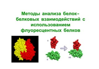
14. Bimolecular fluorescence complementation (split FPs)
Fluorescent protein is separated onto two fragments, which develop
fluorescence only after their association. This association is facilitated by the
interaction between the proteins of interest that are fused to FP fragments.
Kerppola TM. Nat. Rev. Mol. Cell. Biol. 2006, 7:449
18. Principle of the method
Detection of fluorescence fluctuations in a small volume at low
concentrations of fluorophore(s). Further mathematical analysis of
autocorrelations and cross-correlations.
http://www.leica-microsystems.com
20. FRET vs BiFC vs FCCS
FRET
BiFC
FCCS
Detection
Real time
Reversible
Accumulative
Irreversible
Real time
Reversible
Image
Yes
Yes
No
Sensitivity
Low
High
High
FP concentration
High
High
Low
Important
Not important
Relative orientation of FPs Important
Key properties of FPs
Monomer
Assembling
Spectral overlap Maturation rate
Brightness
Photostability
21. Fluorescent Timers - proteins that changes color with
time
Mid-age
organelles
5
Old
organelles
4
Young
organelles
3
2
1
500
600
Wavelength, nm
700
DsRed-E5 – tetrameric
green-to-red timer (Terskikh
et al., Science 2000)
Early
expression
Late
expression
22. DsRed-E5 Fluorescent Timer in C. elegans
DIC
FITC
2h
5h
10h
50h
Terskikh et al., Science 2000
rhodamine
overlay
24. Intracellular
trafficking of lysosomal
membrane protein LAMP-2A
with Fluorescent Timer
Golgi ->
plasma membrane ->
early and recycling endosomes ->
late endosomes and lysosomes.
Subach et al. Nat Chem Biol. 2009.
28. Application of fluorescent proteins for drug discovery
Cell transfection with
fluorescent protein
genes linked to
genes of interest
Transfer of visible
targets to mice
Stably transfected tumor cells
Discovery and evaluation
of candidate drugs
Target visualization
Drug treatment
control
control
drug1
drug2
treated
29. Whole body imaging of carcinogenesis and metastasis
Glioma U87-RFP and GFP
Pancreas cancer XPA1-RFP
Prostate cancer PC-3-RFP
Breast cancer MDA-MB-435-GFP
Real-time non-invasive monitoring of tumor progression, evaluation of drug
treatment efficiency
From Anticancer Inc.