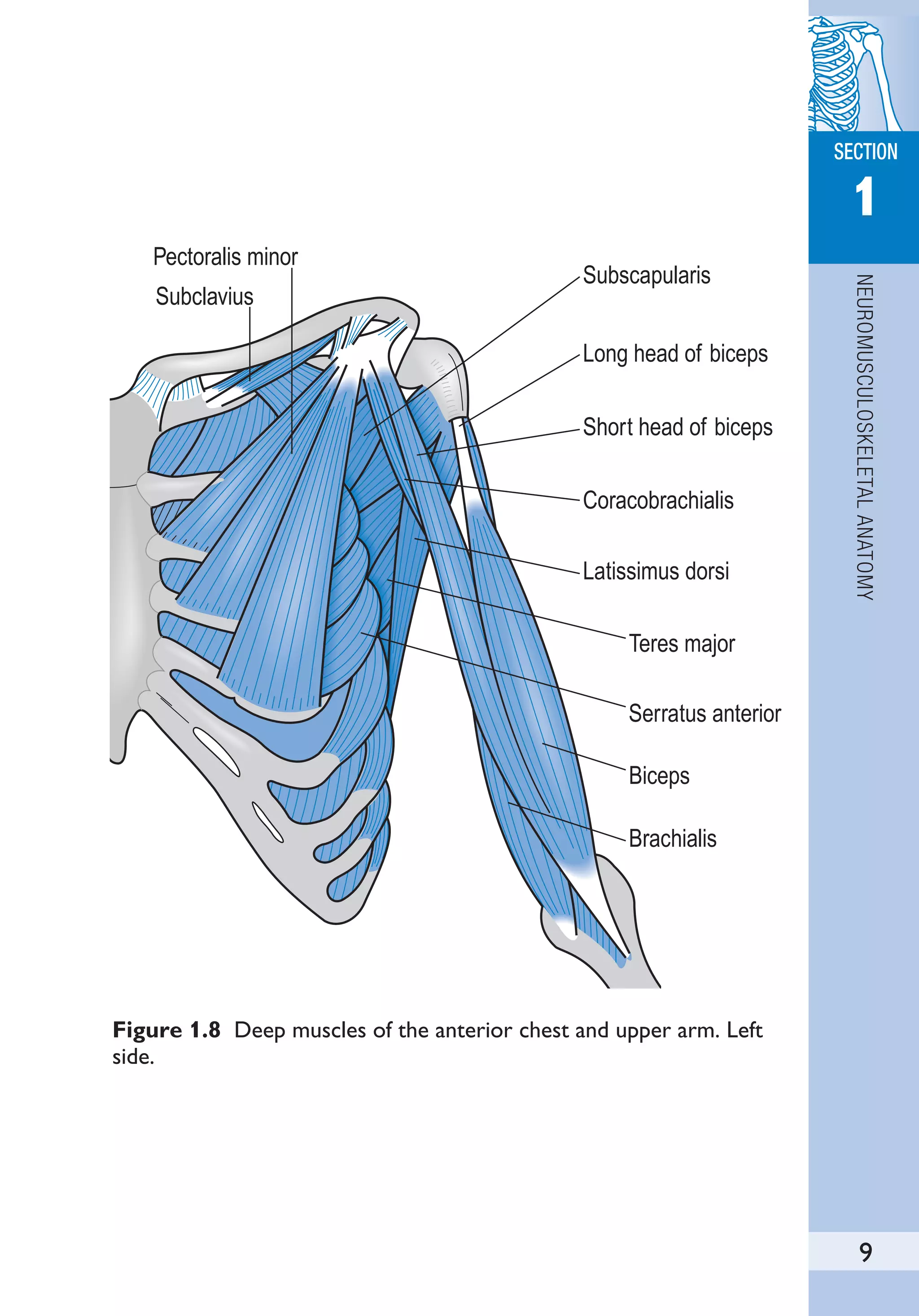
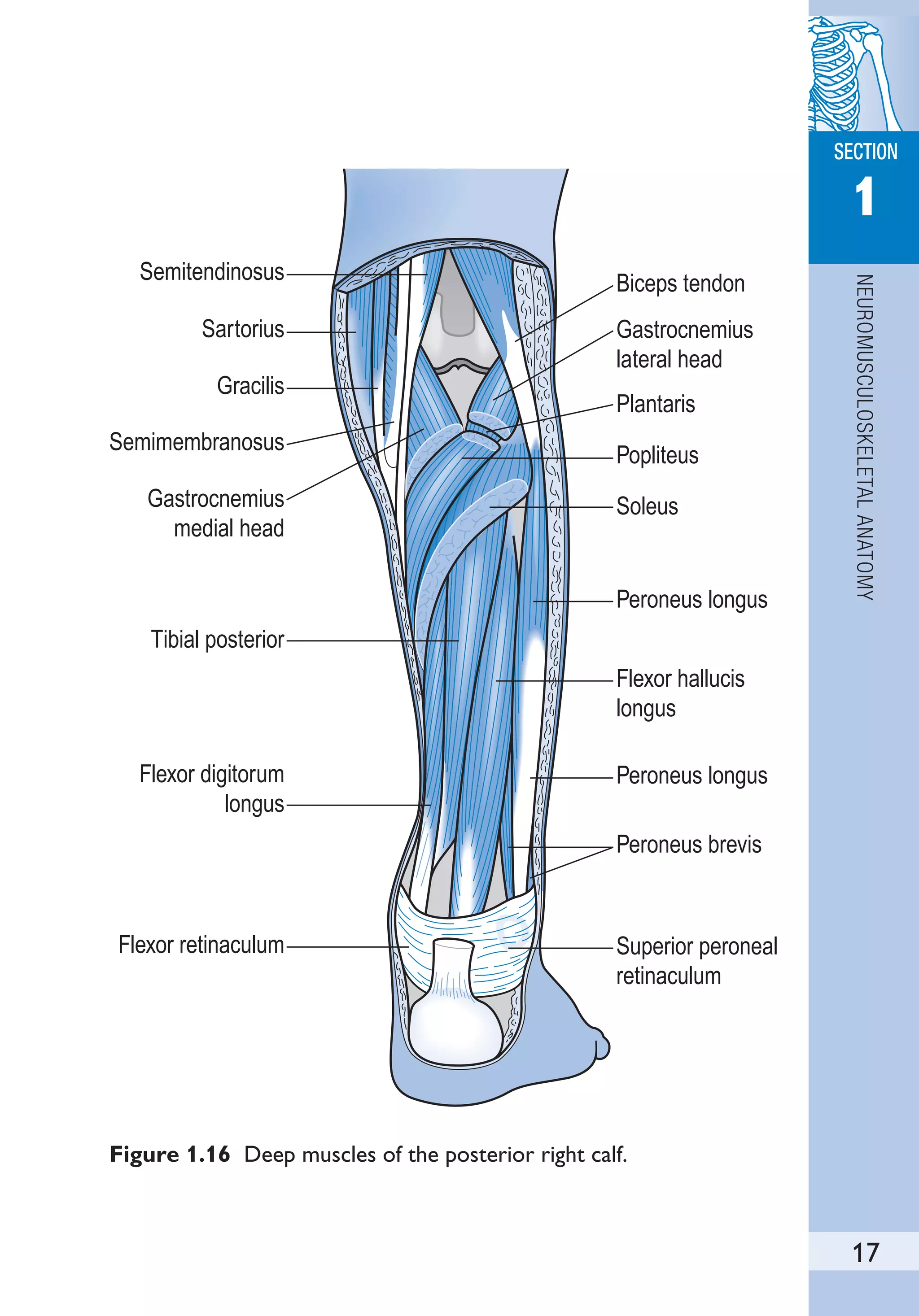
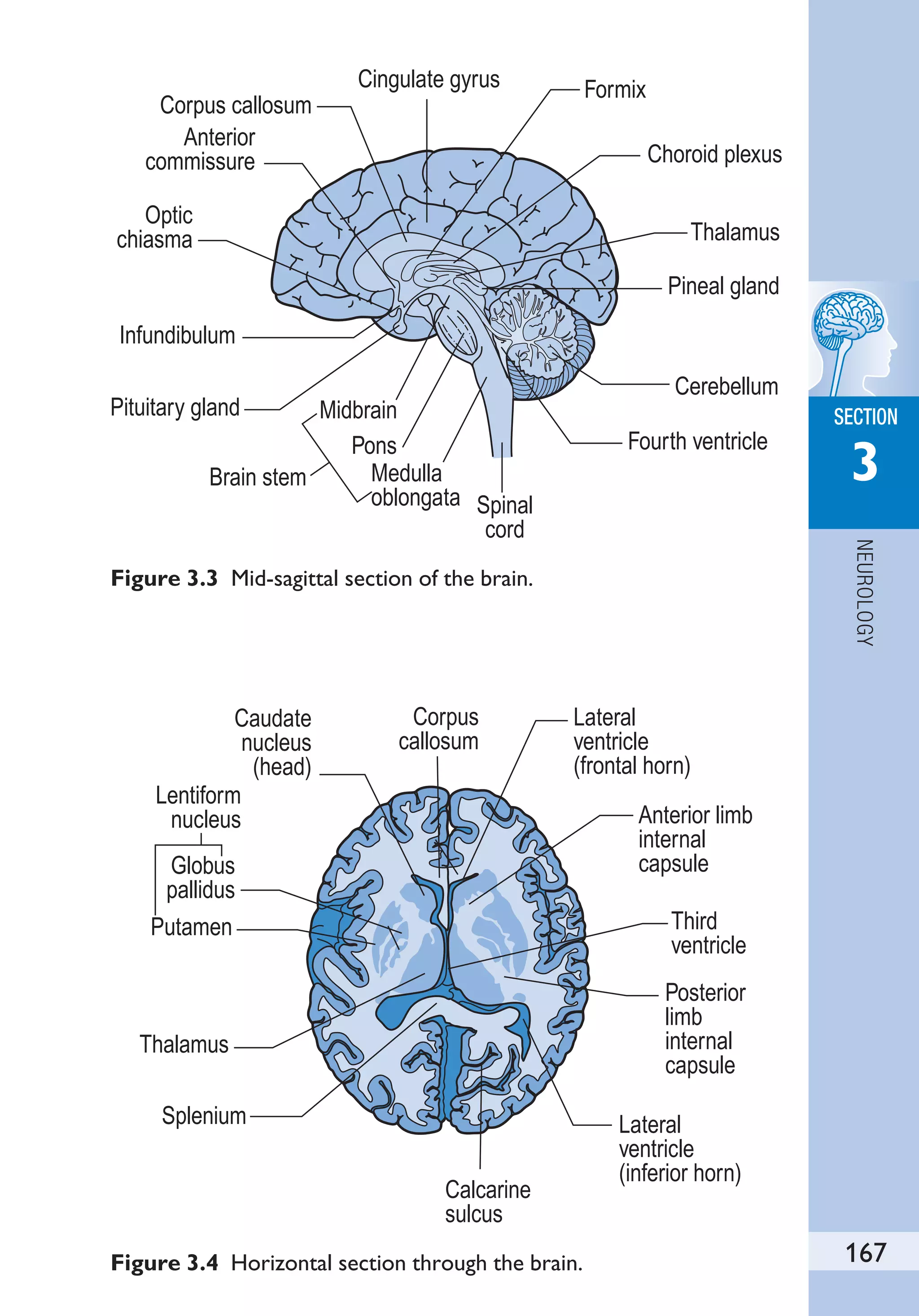
This document provides illustrations and descriptions of key neuromusculoskeletal anatomy including:
- Muscles of the neck, back, and trunk such as the scalenes, erector spinae, and gluteals.
- Brachial and lumbosacral plexuses and their peripheral nerve innervation patterns.
- Dermatome, myotome, and reflex maps.
- Descriptions of common anatomical landmarks such as joints and locations for palpating pulses.


![MUSCULOSKELETAL
SECTION
2
130
and arm while palpating radial pulse. Patient takes a deep
breath and holds it.
Positive sign: disappearance of radial pulse.
Homan’s test
Tests: deep vein thrombophlebitis.
Procedure: patient supine. Passive dorsiflexion of ankle with
knee extended.
Positive sign: pain in the calf.
Provocation elevation test
Tests: thoracic outlet syndrome.
Procedure: patient standing with arms above head. Opens and
closes hands 15 times.
Positive sign: fatigue, cramp, tingling.
Neurodynamic tests (from Petty & Moore 2001, with
permission)
Upper limb neurodynamic tests
When conducting the upper limb neurodynamic tests (ULNT)
the sequence of the test movements is relatively unimportant
and may be adapted to suit the patient’s condition. However,
if the tests are to be of value as an assessment tool, the order
used for a particular patient must be the same each time the
patient is tested.
ULNT 1
ULNT 1 (Fig. 2.10) consists of:
●
Fixing shoulder to prevent shoulder elevation during
abduction [1]
●
Shoulder joint abduction [2]
●
Wrist and finger extension [3]
●
Forearm supination [3]
●
Shoulder lateral rotation [4]
●
Elbow extension [5]
Sensitizing test: cervical lateral flexion away from the symp-
tomatic side [6].](https://image.slidesharecdn.com/184-thephysiotherapistspocketbook-230416200842-d5fddb13/75/184-The-Physiotherapist-s-Pocket-Book-pdf-143-2048.jpg)
![MUSCULOSKELETAL
SECTION
2
131
1 2
3 4
5 6
Figure 2.10 (1–6) Upper limb neurodynamic test 1.
Desensitizing test: cervical lateral flexion towards the sympto-
matic side.
ULNT 2a
ULNT 2a (Fig. 2.11) consists of:
●
Shoulder girdle depression [1, 2]
●
Elbow extension [3]
●
Lateral rotation of whole arm [4]](https://image.slidesharecdn.com/184-thephysiotherapistspocketbook-230416200842-d5fddb13/75/184-The-Physiotherapist-s-Pocket-Book-pdf-144-2048.jpg)
![MUSCULOSKELETAL
SECTION
2
132
Figure 2.11 (1–6) Upper limb neurodynamic test 2a. Median
nerve bias.
1 2
3 4
5 6
●
Wrist, finger and thumb extension [5]
●
Abduction of shoulder [6]
Sensitizing test: cervical lateral flexion away from the symp-
tomatic side.](https://image.slidesharecdn.com/184-thephysiotherapistspocketbook-230416200842-d5fddb13/75/184-The-Physiotherapist-s-Pocket-Book-pdf-145-2048.jpg)
![MUSCULOSKELETAL
SECTION
2
133
Desensitizing tests: cervical lateral flexion towards the symp-
tomatic side or release of the shoulder girdle depression.
ULNT 2b
ULNT 2b (Fig. 2.12) consists of:
●
Shoulder girdle depression [1]
●
Elbow extension [2]
●
Medial rotation of whole arm [3]
●
Wrist and finger flexion [4]
●
Shoulder abduction
Sensitizing test: cervical lateral flexion away from the symp-
tomatic side.
Desensitizing tests: cervical lateral flexion towards the
symptomatic side or release of the shoulder girdle
depression.
1 2
3 4
Figure 2.12 (1–4) Upper limb neurodynamic test 2b. Radial nerve
bias.](https://image.slidesharecdn.com/184-thephysiotherapistspocketbook-230416200842-d5fddb13/75/184-The-Physiotherapist-s-Pocket-Book-pdf-146-2048.jpg)
![MUSCULOSKELETAL
SECTION
2
134
ULNT 3
ULNT 3 (Fig. 2.13) consists of:
●
Shoulder girdle depression [1]
●
Wrist and finger extension [1]
●
Forearm pronation [2]
●
Elbow flexion [3]
●
Shoulder lateral rotation [4]
●
Shoulder abduction [5]
1
3
4
2
5
Figure 2.13 (1–5) Upper limb neurodynamic test 3. Ulnar nerve bias.](https://image.slidesharecdn.com/184-thephysiotherapistspocketbook-230416200842-d5fddb13/75/184-The-Physiotherapist-s-Pocket-Book-pdf-147-2048.jpg)
![MUSCULOSKELETAL
SECTION
2
135
Sensitizing test: cervical lateral flexion away from the symp-
tomatic side.
Desensitizing tests: cervical lateral flexion towards the symp-
tomatic side or release of the shoulder girdle depression.
For all the upper limb neurodynamic tests you may wish
to place the patient’s head in contralateral cervical flexion
before you do the test and then instruct them to bring their
head back to midline at the end of the sequence.
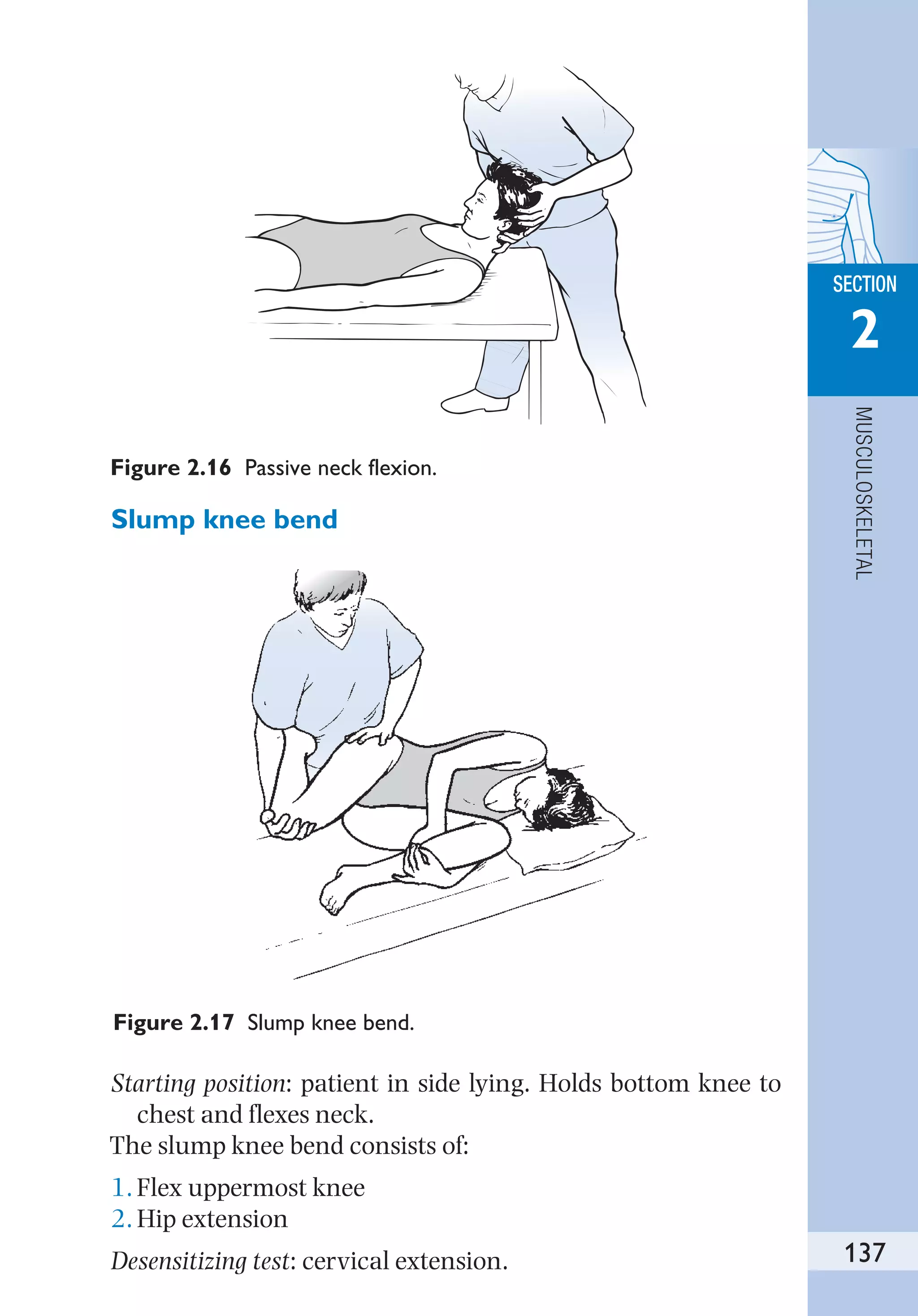
Slump test (Fig. 2.14)
Starting position: patient sits upright with knee crease at the
edge of plinth and hands behind back [1].
The slump test consists of:
●
Spinal slump [2]
●
Neck flexion [3]
1 2 3
4 5 6
Figure 2.14 (1–6) Slump test.](https://image.slidesharecdn.com/184-thephysiotherapistspocketbook-230416200842-d5fddb13/75/184-The-Physiotherapist-s-Pocket-Book-pdf-148-2048.jpg)
![MUSCULOSKELETAL
SECTION
2
136
●
Knee extension [4]
●
Release neck flexion [5]
The steps can be performed in any order.
Additional movements: add dorsiflexion or plantarflexion with
knee extension; bilateral knee extension [6], hip abduction
(obturator nerve bias).
Straight leg raise
Figure 2.15 Straight leg raise.
The test consists of passive hip flexion with the knee
extended.
Sensitizing tests: dorsiflexion, hip adduction, hip medial rota-
tion, neck flexion and spinal lateral flexion.
Additional sensitizing tests: Add ankle dorsiflexion and ever-
sion (tibial nerve bias), plantarflexion and inversion
(superficial peroneal nerve bias), dorsiflexion and inver-
sion (sural nerve bias).
Passive neck flexion
The test consists of passive neck flexion.
Sensitizing tests: straight leg raise, upper limb neurodynamic
tests.](https://image.slidesharecdn.com/184-thephysiotherapistspocketbook-230416200842-d5fddb13/75/184-The-Physiotherapist-s-Pocket-Book-pdf-149-2048.jpg)


![APPENDICES
SECTION
7
324
[Hⴙ
] hydrogen ion concentration
HASO hip abduction spinal orthosis
Hb haemoglobin
HC head circumference
Hct haematocrit
HD haemodialysis
HDU high dependency unit
HF heart failure
HFCWO high-frequency chest wall oscillation
HFJV high-frequency jet ventilation
HFO high-frequency oscillation
HFOV high-frequency oscillatory ventilation
HFPPV high-frequency positive pressure ventilation
HH hiatus hernia/home help
HI head injury
HIV human immunodeficiency virus
HLA human leukocyte antigen
HLT heart–lung transplantation
HME heat and moisture exchanger
HPC history of presenting condition
HPOA hypertrophic pulmonary osteoarthropathy
HR heart rate
HRR heart rate reserve
HT hypertension
IABP intra-aortic balloon pump
ICC intercostal catheter
ICD intercostal drain
ICP intracranial pressure
ICU intensive care unit
IDC indwelling catheter
IDDM insulin-dependent diabetes mellitus
Ig immunoglobulin
IHD ischaemic heart disease
ILD interstitial lung disease
IM intramedullary
IM/i.m. intramuscular
IMA internal mammary artery
IMV intermittent mandatory ventilation
INR international normalized ratio](https://image.slidesharecdn.com/184-thephysiotherapistspocketbook-230416200842-d5fddb13/75/184-The-Physiotherapist-s-Pocket-Book-pdf-337-2048.jpg)


