The document outlines key topics in human development and inheritance covered in Chapter 20, including:
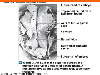
1. The three stages of prenatal development - embryological, fetal, and prenatal - and the major events of each stage such as organogenesis in the first trimester.
2. The process of fertilization and how it produces a zygote with 46 chromosomes through the fusion of a sperm and egg.
3. The formation and importance of the placenta as an endocrine organ that secretes hormones to maintain the corpus luteum and pregnancy.
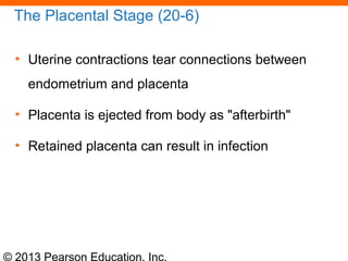
4. The three stages of labor - dilation, expulsion, and placental - and events that occur in each like amniotic rupture and