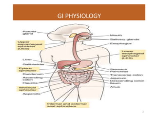
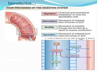
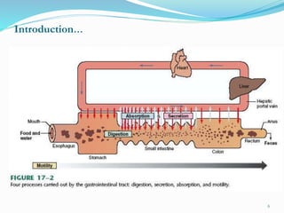
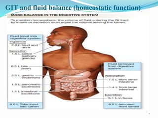
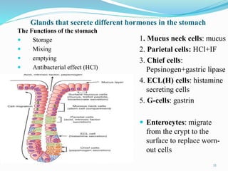
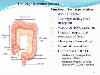
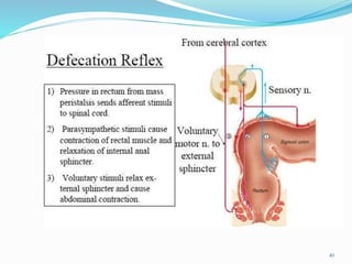
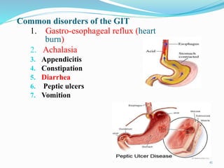
GIT Physiolagy
Dr.Mowlid A(MD)
1
GI PHYSIOLOGY
2
Introduction to GIT
Food is required by the body for the:
o Production of energy.
o Growth & repair of tissues.
 Each day an average adult man consumes
o 1 kg of solid food &
o 1- 2 liters of fluid.
 Majority of this material is in a form that cannot be
used immediately by the body for cellular metabolism.
• It must be broken down into simple
molecules which can be absorbed into the
bloodstream for distribution to the tissues.
:.The GIS performs this task.
3
The major functions of the digestive system
The ingestion & mastication (chewing) of food;
 Transport of food through the GI tract
• at a rate that allow optimal digestion & absorption;
 The secretion of fluid, salts, and digestive enzymes;
• ( Exocrine + Endocrine secretions)
 Digestion (mechanical & chemical);
Supplies energy
 Absorption of end products of digestion;
 Excretion: Removal of indigestible remains, hydrophobic
substances from the body.
 Host defense.
4
Introduction…
5
Introduction…
6
GIT and fluid balance (homeostatic function)
7
Microbial defense by the GIT
Mouth
 Saliva contains lysozymes and
IgA that attacks microbes in the
mouth
Stomach
 HCl secreted in the stomach has
bactericidal actions
Small intestine:
 Payer's patches lymph tissue
attack microbes in the SI
Macrophages:
 In SI, developed from Monocytes
and destroy microbes by
Phagocytosis
8
Anatomically & functionally, the digestive system can be divided into:
Tubular GIT
(Alimentary Canal)
1. Mouth
2. Pharynx
3. Esophagus
4. Stomach
5. Small intestine
6. Large intestine
7. Rectum
8. Anus
Accessories
Organs
1. Teeth,
tongue
2. Salivary
glands
3. Pancreas
4. Liver &
5. Gallbladder
9
Functional layers of the GIT (Esophagus-Anus)
- From the lumen outward they are:
1. The mucosa- Protection, secretion & absorption from the luminal
surfaces
2. The sub mucosa,
 Contains blood vessels, lymph nodes and some nerve fibers
3. The muscularis externa-
 Contraction of the circular muscle decreases the diameter of the lumen
 Contraction of the longitudinal muscle, decreases the length of the GIT
4. The serosa or fibrosa
 Outer most protective layer, consists of connective tissues
10
11
Neuronal plexuses in the wall of GIT
A. Submucosal (Messiner’s) plexus
 Between submucosa and circular muscle of muscularies externa layer
 Involved in GIT secretions & blood flow
B. Myenteric (Aurebach’s) plexus
 Between the circular & longitudinal smooth layers of muscularies externa
 It is involved in GIT motility
These two plexuses together with other neurons of the GIT constitute the Enteric
Nervous System (ENS)
 The ENS
 Integrate the motor & secretory activities of the GI system
 Control most of the GI functions even in the absence of ANS innervations
12
Enteric nervous system (ENS)…
13
Autonomic (PSN and SNS) connection to the GIT
A. Parasympathetic
 Is cholinergic (Ach)
 Cranial:
 All are in vagus nerve
 Innervate Esophagus, stomach, SI,
and the proximal large intestine
 Sacral:
 Innervates distal LI, sigmoid, rectum,
and anus
 PNS is excitatory to the GIT &
causes increased motility
 However, at the sphincters, PNS has
a dilatatory action
B. Sympathetic fibers
 Are adrenergic (NE & Epinephrine)
 The sympathetics is inhibitory to the
GIT- decreases motility
 At sphincters, it causes constriction
14
Blood flow to the GIT
 GIT blood flow is classified under
splanchnic circulation
 Which includes: Gut, Spleen, pancreas
etc…
 Venous blood collected from these regions
pass through the portal circulation and end in
the liver
 liver sinusoids > hepatic vein > inferior
vena cava
 Advantage of portal circulation:
 Potentially harmful agents that come
with the blood are destroyed by
macrophages of the liver (Reticulo-
endothelial cells)
 blood flow to the GIT is controlled by
Metabolic, Decreased O2-tension, Neural
15
1. Motility in the Esophagus
 Upper 1/3 of esophagus is skeletal muscle
 Lower 2/3 of esophagus is smooth muscles
Swallowing reflex (deglutition)
It is the movement of food from the
mouth into the stomach
1. Oral /Voluntary phase
2. Pharyngeal phase
Involuntary from pharynx into the
esophagus
 Cause of primary peristalsis
3. Esophageal phase
 Involuntary
 Distention of the esophageal walls
causes “secondary peristalsis”
16
Motility in the Esophagus…
Swallowing reflex…
• Secondary peristalsis is caused by
stimulation of solid food on the
walls of the esophagus
• It is powerful enough to push the
bolus towards the stomach
• Turning “upside-down” can not
cause the food to move towards the
mouth unless pathological
• Gravity plays a role in swallowing,
but not very potent
17
Stomach
Structure
 Cardiac sphincter (LES)
 Lesser curvature
 Greater curvature
 Cardia
 Fundus
 Corpus (body)
 Pyloric antrum
 Pyloric sphincter
 Oblique muscle
The four regions of the stomach are cardia , fundus , body and pylorus.
18
Stomach…
 Muscular region is
made up of
 Circular muscle
 Longitudinal muscle
 Oblique muscle
 Having oblique
muscle is responsible
for its movement in
every directions
19
Stomach…
 Stomach has pacemaker region
on its body part made up of
cells called cells of Cajal
 The pacemaker controls the
basic electrical rhythm (BER)
of the stomach
20
Motility in the stomach
 Motility of the stomach contents are
accomplished by:
 A. Mixing processes
- Are beneficial to mix the bolus
with gastric juice (chyme)
Pacemaker cells (cells of Cajal)
causing slow waves (BER)
- Slow waves result in tonic, weak
contractions of the stomach walls
(3 slow waves/min)
B. Peristaltic waves
 Orad contraction
 Caudad relaxation
21
Motility in Small intestine
A. Segmentation
 2. Peristalsis (propulsive) motility
22
Migrating Motility Complex (MMC) in small intestine
Migrating Motility Complex (MMC)
 Is a strong peristaltic wave that sweeps over the intestinal wall
after most meals are absorbed
 Important in completely pushing and sweeping the contents of
the small intestine into the large intestine
 Therefore, given the name ‘the house keeper’
 It also prevents the intestine from colonic bacteria
 It can sweep heavy and indigestible substances (e.g. coin) to
colon
23
Motility in Large intestine
 Two types
A. Haustral movements
 large circular constrictions (haustrations) that are
powerful enough to close the diameter to a
narrow loop
B. Mass (propulsive) mov’t
 Mass movements occur seldom (1-3/day
or once after a breakfast)
 Local reflexes such as Gastro-colic or
duodeno-colic reflex initiate mass
movement
 Reflex contraction of the rectum
 Relaxation of the internal anal sphincter
24
GI Secretions
25
Mouth(oral cavity)
 Oral cavity
 Lips
 Muscles/cheeks
 Tongue
 Teeth
 Responsible for mechanical
(physical) digestion
26
Secretion in Mouth
Types of Salivary glands and their secretions
Parotid (25%)
 the largest, just below and in front
of the ears
 Secrete mainly serous watery fluid
rich in ptyaline (salivary amylase)
Submandibular (70%)
 Produce both serous and mucous
fluid
Sublingual (~5%)
 Secrete mainly thick mucous with
little serous fluid
The three principal pair glands of salivation are:
27
Salivary secretion…
Composition and function of saliva
Composition : Total secretion = about 1-1.5 L/day
 H2O (99.5%): facilitates taste and dissolution
 Electrolytes ( 0.5%): Na+, Cl-, K+, HCO3
-, etc
 Mucin (proteoglycan) for lubrication
 Enzymes: salivary amylase (ptyalin), lingual lipase
 Lysozymes (an enzyme that has a weak antibacterial action)
 IgA (prevent bacterial infection) etc… defense
Functions
 Lubrication
 Speech
 Mixing
 Bacterial attack, etc…
28
Three phases and Reflex control of salivary
secretion
 Cephalic (brain) phase
 Triggered by thought, smell, or sight of food
 Oral phase
 Triggered by food that stimulate touch & test receptors
in the mouth
 Gastric phase
 Triggered by substances which stimulate the gastric
mucosa (acids or sour tastes) in the stomach
 Salivation can also be controlled by higher centers like
hypothalamus which has nerve connections with salivatory
nuclei in the Medulla oblongata (MO)
29
Esophageal secretion
 The esophagus
wall contains
mucus cells that
produce mucous.
 Mucous is used for
lubrication and
thus creates easy
swallowing
30
Glands that secrete different hormones in the stomach
1. Mucus neck cells: mucus
2. Parietal cells: HCl+IF
3. Chief cells:
Pepsinogen+gastric lipase
4. ECL(H) cells: histamine
secreting cells
5. G-cells: gastrin
 Enterocytes: migrate
from the crypt to the
surface to replace worn-
out cells
The Functions of the stomach
 Storage
 Mixing
 emptying
 Antibacterial effect (HCl)
31
Function of HCl in the stomach
1. Kills most of the bacteria in food
2. Stimulates hunger – important in appetite
regulation
3. Activates the pro-enzyme Pepsinogen into its
active pepsin that denatures peptide bonds to
produce amino acids
4. Enhances motility of the stomach etc…
32
Emptying and delaying of stomach contents
Thus, liquids empty faster than solids
Moreover, the following steps take
place for organic chyme when emptying
stomach
CHO > protein > fat
33
Absorption in the stomach
 An insignificant
absorptive functions takes
place in the stomach,
these are:
 Alcohol
 Certain drugs (aspirin,
morphine etc.)
 Small quantities of H2O
Organic nutrients
(glucose, amino acids
and FFA etc… are not
absorbed)
• The following effects
protect the stomach wall
from acidic attacks (i.e.
pH<3)
a. Production of thick
mucous
b. HCO3
- secretion
(buffers the acid)
34
Pancreas
 The pancreatic
secretion contains:
1. Bicarbonate (HCO3
-)
2. Electrolytes (Na+, K+,
Cl- etc.)
3. Digestive enzymes:
(inactive pro-enzymes)
a. Pancreatic Amylase
b. Pancreatic lipase
c. Proteases
d. Nucleases
35
Bile Production in the liver
Bile
 Is an alkaline fluid (pH 8)
 Secreted in the liver
 Functions in emulsification
and absorption of fat in the
duodenum
 Emulsification means changing
greater fat globules into smaller
fat-droplets called micelles
 Excretion of several toxic end
products from the blood
These include: Bilirubin and Excess
cholesterol
Gall stone formation
36
The Small intestine (SI)
 SI is specialized for
completion of digestion
and absorption of nutrients
a. Duodenum: ~ 25 cm,
mainly secretory, mucous,
hormones, enzymes
b. Jejunum : ~ 1.5 m mainly
absorptive
c. Ileum : ~1.7 m mainly
absorptive
37
Small intestine: secretions and villi
Brush border (microvilli)
• Increase the absorptive surface
area to a great extent in the small
intestine
• Site of digestion and absorption
38
Function of the large intestine
1. Water absorption
2. Electrolyte mainly NaCl
absorption
2. Mucous & HCO3
- Secretion
3. Storage, transport, and
evacuation of feces
4. Absorption of some drugs
5. Microbial fermentation
• The microbes in the LI
– Produce enzymes capable of
digesting cellulose
– Stimulates synthesis of some
vitamins (Vit. K, and B-groups)
The large intestine (colon)
39
40
Common disorders of the GIT
1. Gastro-esophageal reflux (heart
burn)
2. Achalasia
3. Appendicitis
4. Constipation
5. Diarrhea
6. Peptic ulcers
7. Vomition
41

10. GIT 2014.pptx

  • 1.
  • 2.
  • 3.
    Introduction to GIT Foodis required by the body for the: o Production of energy. o Growth & repair of tissues.  Each day an average adult man consumes o 1 kg of solid food & o 1- 2 liters of fluid.  Majority of this material is in a form that cannot be used immediately by the body for cellular metabolism. • It must be broken down into simple molecules which can be absorbed into the bloodstream for distribution to the tissues. :.The GIS performs this task. 3
  • 4.
    The major functionsof the digestive system The ingestion & mastication (chewing) of food;  Transport of food through the GI tract • at a rate that allow optimal digestion & absorption;  The secretion of fluid, salts, and digestive enzymes; • ( Exocrine + Endocrine secretions)  Digestion (mechanical & chemical); Supplies energy  Absorption of end products of digestion;  Excretion: Removal of indigestible remains, hydrophobic substances from the body.  Host defense. 4
  • 5.
  • 6.
  • 7.
    GIT and fluidbalance (homeostatic function) 7
  • 8.
    Microbial defense bythe GIT Mouth  Saliva contains lysozymes and IgA that attacks microbes in the mouth Stomach  HCl secreted in the stomach has bactericidal actions Small intestine:  Payer's patches lymph tissue attack microbes in the SI Macrophages:  In SI, developed from Monocytes and destroy microbes by Phagocytosis 8
  • 9.
    Anatomically & functionally,the digestive system can be divided into: Tubular GIT (Alimentary Canal) 1. Mouth 2. Pharynx 3. Esophagus 4. Stomach 5. Small intestine 6. Large intestine 7. Rectum 8. Anus Accessories Organs 1. Teeth, tongue 2. Salivary glands 3. Pancreas 4. Liver & 5. Gallbladder 9
  • 10.
    Functional layers ofthe GIT (Esophagus-Anus) - From the lumen outward they are: 1. The mucosa- Protection, secretion & absorption from the luminal surfaces 2. The sub mucosa,  Contains blood vessels, lymph nodes and some nerve fibers 3. The muscularis externa-  Contraction of the circular muscle decreases the diameter of the lumen  Contraction of the longitudinal muscle, decreases the length of the GIT 4. The serosa or fibrosa  Outer most protective layer, consists of connective tissues 10
  • 11.
  • 12.
    Neuronal plexuses inthe wall of GIT A. Submucosal (Messiner’s) plexus  Between submucosa and circular muscle of muscularies externa layer  Involved in GIT secretions & blood flow B. Myenteric (Aurebach’s) plexus  Between the circular & longitudinal smooth layers of muscularies externa  It is involved in GIT motility These two plexuses together with other neurons of the GIT constitute the Enteric Nervous System (ENS)  The ENS  Integrate the motor & secretory activities of the GI system  Control most of the GI functions even in the absence of ANS innervations 12
  • 13.
  • 14.
    Autonomic (PSN andSNS) connection to the GIT A. Parasympathetic  Is cholinergic (Ach)  Cranial:  All are in vagus nerve  Innervate Esophagus, stomach, SI, and the proximal large intestine  Sacral:  Innervates distal LI, sigmoid, rectum, and anus  PNS is excitatory to the GIT & causes increased motility  However, at the sphincters, PNS has a dilatatory action B. Sympathetic fibers  Are adrenergic (NE & Epinephrine)  The sympathetics is inhibitory to the GIT- decreases motility  At sphincters, it causes constriction 14
  • 15.
    Blood flow tothe GIT  GIT blood flow is classified under splanchnic circulation  Which includes: Gut, Spleen, pancreas etc…  Venous blood collected from these regions pass through the portal circulation and end in the liver  liver sinusoids > hepatic vein > inferior vena cava  Advantage of portal circulation:  Potentially harmful agents that come with the blood are destroyed by macrophages of the liver (Reticulo- endothelial cells)  blood flow to the GIT is controlled by Metabolic, Decreased O2-tension, Neural 15
  • 16.
    1. Motility inthe Esophagus  Upper 1/3 of esophagus is skeletal muscle  Lower 2/3 of esophagus is smooth muscles Swallowing reflex (deglutition) It is the movement of food from the mouth into the stomach 1. Oral /Voluntary phase 2. Pharyngeal phase Involuntary from pharynx into the esophagus  Cause of primary peristalsis 3. Esophageal phase  Involuntary  Distention of the esophageal walls causes “secondary peristalsis” 16
  • 17.
    Motility in theEsophagus… Swallowing reflex… • Secondary peristalsis is caused by stimulation of solid food on the walls of the esophagus • It is powerful enough to push the bolus towards the stomach • Turning “upside-down” can not cause the food to move towards the mouth unless pathological • Gravity plays a role in swallowing, but not very potent 17
  • 18.
    Stomach Structure  Cardiac sphincter(LES)  Lesser curvature  Greater curvature  Cardia  Fundus  Corpus (body)  Pyloric antrum  Pyloric sphincter  Oblique muscle The four regions of the stomach are cardia , fundus , body and pylorus. 18
  • 19.
    Stomach…  Muscular regionis made up of  Circular muscle  Longitudinal muscle  Oblique muscle  Having oblique muscle is responsible for its movement in every directions 19
  • 20.
    Stomach…  Stomach haspacemaker region on its body part made up of cells called cells of Cajal  The pacemaker controls the basic electrical rhythm (BER) of the stomach 20
  • 21.
    Motility in thestomach  Motility of the stomach contents are accomplished by:  A. Mixing processes - Are beneficial to mix the bolus with gastric juice (chyme) Pacemaker cells (cells of Cajal) causing slow waves (BER) - Slow waves result in tonic, weak contractions of the stomach walls (3 slow waves/min) B. Peristaltic waves  Orad contraction  Caudad relaxation 21
  • 22.
    Motility in Smallintestine A. Segmentation  2. Peristalsis (propulsive) motility 22
  • 23.
    Migrating Motility Complex(MMC) in small intestine Migrating Motility Complex (MMC)  Is a strong peristaltic wave that sweeps over the intestinal wall after most meals are absorbed  Important in completely pushing and sweeping the contents of the small intestine into the large intestine  Therefore, given the name ‘the house keeper’  It also prevents the intestine from colonic bacteria  It can sweep heavy and indigestible substances (e.g. coin) to colon 23
  • 24.
    Motility in Largeintestine  Two types A. Haustral movements  large circular constrictions (haustrations) that are powerful enough to close the diameter to a narrow loop B. Mass (propulsive) mov’t  Mass movements occur seldom (1-3/day or once after a breakfast)  Local reflexes such as Gastro-colic or duodeno-colic reflex initiate mass movement  Reflex contraction of the rectum  Relaxation of the internal anal sphincter 24
  • 25.
  • 26.
    Mouth(oral cavity)  Oralcavity  Lips  Muscles/cheeks  Tongue  Teeth  Responsible for mechanical (physical) digestion 26
  • 27.
    Secretion in Mouth Typesof Salivary glands and their secretions Parotid (25%)  the largest, just below and in front of the ears  Secrete mainly serous watery fluid rich in ptyaline (salivary amylase) Submandibular (70%)  Produce both serous and mucous fluid Sublingual (~5%)  Secrete mainly thick mucous with little serous fluid The three principal pair glands of salivation are: 27
  • 28.
    Salivary secretion… Composition andfunction of saliva Composition : Total secretion = about 1-1.5 L/day  H2O (99.5%): facilitates taste and dissolution  Electrolytes ( 0.5%): Na+, Cl-, K+, HCO3 -, etc  Mucin (proteoglycan) for lubrication  Enzymes: salivary amylase (ptyalin), lingual lipase  Lysozymes (an enzyme that has a weak antibacterial action)  IgA (prevent bacterial infection) etc… defense Functions  Lubrication  Speech  Mixing  Bacterial attack, etc… 28
  • 29.
    Three phases andReflex control of salivary secretion  Cephalic (brain) phase  Triggered by thought, smell, or sight of food  Oral phase  Triggered by food that stimulate touch & test receptors in the mouth  Gastric phase  Triggered by substances which stimulate the gastric mucosa (acids or sour tastes) in the stomach  Salivation can also be controlled by higher centers like hypothalamus which has nerve connections with salivatory nuclei in the Medulla oblongata (MO) 29
  • 30.
    Esophageal secretion  Theesophagus wall contains mucus cells that produce mucous.  Mucous is used for lubrication and thus creates easy swallowing 30
  • 31.
    Glands that secretedifferent hormones in the stomach 1. Mucus neck cells: mucus 2. Parietal cells: HCl+IF 3. Chief cells: Pepsinogen+gastric lipase 4. ECL(H) cells: histamine secreting cells 5. G-cells: gastrin  Enterocytes: migrate from the crypt to the surface to replace worn- out cells The Functions of the stomach  Storage  Mixing  emptying  Antibacterial effect (HCl) 31
  • 32.
    Function of HClin the stomach 1. Kills most of the bacteria in food 2. Stimulates hunger – important in appetite regulation 3. Activates the pro-enzyme Pepsinogen into its active pepsin that denatures peptide bonds to produce amino acids 4. Enhances motility of the stomach etc… 32
  • 33.
    Emptying and delayingof stomach contents Thus, liquids empty faster than solids Moreover, the following steps take place for organic chyme when emptying stomach CHO > protein > fat 33
  • 34.
    Absorption in thestomach  An insignificant absorptive functions takes place in the stomach, these are:  Alcohol  Certain drugs (aspirin, morphine etc.)  Small quantities of H2O Organic nutrients (glucose, amino acids and FFA etc… are not absorbed) • The following effects protect the stomach wall from acidic attacks (i.e. pH<3) a. Production of thick mucous b. HCO3 - secretion (buffers the acid) 34
  • 35.
    Pancreas  The pancreatic secretioncontains: 1. Bicarbonate (HCO3 -) 2. Electrolytes (Na+, K+, Cl- etc.) 3. Digestive enzymes: (inactive pro-enzymes) a. Pancreatic Amylase b. Pancreatic lipase c. Proteases d. Nucleases 35
  • 36.
    Bile Production inthe liver Bile  Is an alkaline fluid (pH 8)  Secreted in the liver  Functions in emulsification and absorption of fat in the duodenum  Emulsification means changing greater fat globules into smaller fat-droplets called micelles  Excretion of several toxic end products from the blood These include: Bilirubin and Excess cholesterol Gall stone formation 36
  • 37.
    The Small intestine(SI)  SI is specialized for completion of digestion and absorption of nutrients a. Duodenum: ~ 25 cm, mainly secretory, mucous, hormones, enzymes b. Jejunum : ~ 1.5 m mainly absorptive c. Ileum : ~1.7 m mainly absorptive 37
  • 38.
    Small intestine: secretionsand villi Brush border (microvilli) • Increase the absorptive surface area to a great extent in the small intestine • Site of digestion and absorption 38
  • 39.
    Function of thelarge intestine 1. Water absorption 2. Electrolyte mainly NaCl absorption 2. Mucous & HCO3 - Secretion 3. Storage, transport, and evacuation of feces 4. Absorption of some drugs 5. Microbial fermentation • The microbes in the LI – Produce enzymes capable of digesting cellulose – Stimulates synthesis of some vitamins (Vit. K, and B-groups) The large intestine (colon) 39
  • 40.
  • 41.
    Common disorders ofthe GIT 1. Gastro-esophageal reflux (heart burn) 2. Achalasia 3. Appendicitis 4. Constipation 5. Diarrhea 6. Peptic ulcers 7. Vomition 41