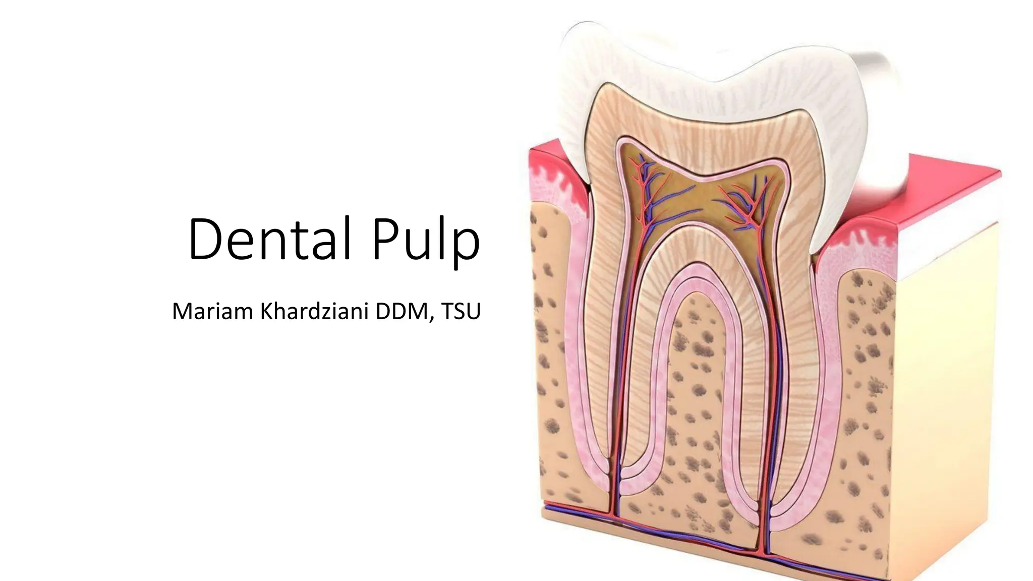
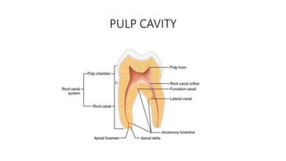
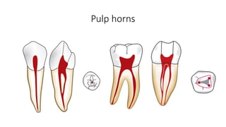
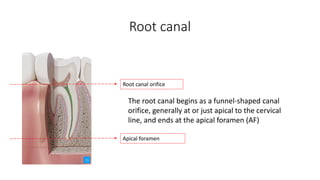
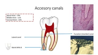
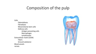
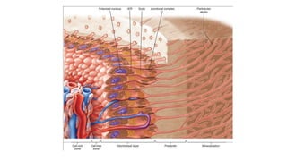
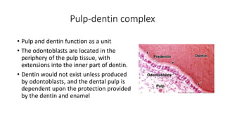
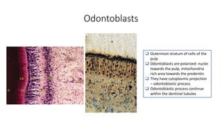
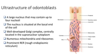
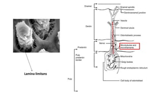
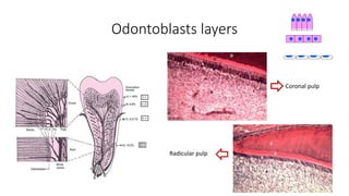
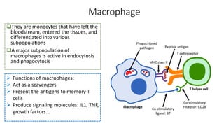
Dental Pulp
Mariam Khardziani DDM, TSU
PULP CAVITY
Pulp horns
Root canal
The root canal begins as a funnel-shaped canal
orifice, generally at or just apical to the cervical
line, and ends at the apical foramen (AF)
Root canal orifice
Apical foramen
Accesory canals
Furcation (chamber) canal
Lateral canal
Apical delta Δ
Apical third – 74%
Middle third – 11%
Cervical third – 15%
Accessory canals
According to the glossary of American Association of Endodontists,
lateral and accessory canals are differentiated as
 Lateral canal is a canal that is located at approximately at right
angles to main root canal
 Accessory canal is the one that branches off from main root canal,
usually in the apical region of the root.
 Apical deltas are multiple accessory canals that branch out from the
main canal at or near the root apex
Composition of the pulp
Cells
Odontoblasts
Fibroblasts
Mesenchymal stem cells
Defense cells
Antigen presenting cells
Macrophages
Lymphocytes
Extracellular matrix (ECM)
Fibers
Ground substance
Blood vessels
nerves
Pulp-dentin complex
• Pulp and dentin function as a unit
• The odontoblasts are located in the
periphery of the pulp tissue, with
extensions into the inner part of dentin.
• Dentin would not exist unless produced
by odontoblasts, and the dental pulp is
dependent upon the protection provided
by the dentin and enamel
Odontoblasts
 Outermost stratum of cells of the
pulp
 Odontoblasts are polarized: nuclei
towards the pulp, mitochondria
rich area towards the predentin
 They have cytoplasmic projection
– odontoblastic process
 Odontoblastic process continue
within the dentinal tubules
Ultrastructure of odontoblasts
 A large nucleus that may contain up to
four nucleoli
 The nucleus is situated at the basal end
of the cell
 Well-developed Golgi complex, centrally
located in the supranuclear cytoplasm
 Numerous mitochondria and ribosomes
 Prominent RER (rough endoplasmic
reticulum)
What does the odontoblasts synthesize?
• Type I collagen
• Type V collagen
• Proteoglycans
• Dentin sialoprotein
• Phosphophoryn (involved in mineralization)
• Alkaline phosphatase (involved in mineralization)
• Acid phosphatase (lysosomal enzyme, which is involved in digesting
the proteins from predentin)
Odontoblast process
Odontoblasts layers
Coronal pulp
Radicular pulp
Cell-poor zone
 Immediately subjacent to the odontoblast layer in the coronal pulp
 Approximately 40 μm in width
 So called cell-free layer of Weil
 Contains blood capillaries, unmyelinated nerve fibers, and the
slender cytoplasmic processes of fibroblasts
Cell-rich zone
 This stratum contains a relatively high proportion of fibroblasts
compared with the more central region of the pulp
 It is much more prominent in the coronal pulp than in the radicular
pulp
 Besides fibroblasts, the cell-rich zone contains a variable number of
immune cells like macrophages and dendritic cells but also
undifferentiated mesenchymal stem cells
Pulp proper
 central mass of the pulp
 loose connective tissue
 larger blood vessels
 nerves
 fibroblasts
Fibroblasts
 Most numerous cells of the pulp
 They are tissue specific
 Fibroblasts synthesize collagen type I and III, as well as proteoglycans and GAGs
 This cells also phagocytose and digest collagen
Defense cells
Macrophage
They are monocytes that have left the
bloodstream, entered the tissues, and
differentiated into various
subpopulations
A major subpopulation of
macrophages is active in endocytosis
and phagocytosis
 Functions of macrophages:
 Act as a scavengers
 Present the antigens to memory T
cells
 Produce signaling molecules: IL1, TNF,
growth factors…
Dendritic cells
• Accessory cells of immune system
• Similar cells to Langerhans cells in the epidermis and mucous
membrane
• They are antigen presenting cells
• In the normal pulp they are mostly
• Located in the periphery of the coronal pulp close to the predentin
Lymphocytes
In normal pulp T cells are
found, mostly T8 lymphocytes
(suppresors)
B lymphocytes are scarcely
found uninflamed pulp
Mast cells
 Mast cells are seldom found in the normal
pulp tissue
 They are routinely found in chronically
inflamed pulps.
 The granules of mast cells contain heparin,
an anticoagulant, and histamine
Pulpal interstitium (ECM)
 Interstitial fluid
 Interstitial matrix
• Major structural component – collagen
• The other components are proteoglycans, hyaluronan and elastic
fibers
Interstitium
Glycosaminoglycans (GAGs)
Extracellular matrix (ECM) = Ground
substance
In the pulp, the principal proteoglycans include:
 hyaluronic acid (strong affinity for water)
 dermatan sulfate
 heparan sulfate
 chondroitin sulfate (active during dentinogenesis)
 Ground substance also acts as a molecular sieve in that it excludes large
proteins
 Cell metabolites, nutrients, and wastes pass through the ground
substance between cells and blood vessels
Mariam.khardziani@tsu.ge

1. pulp structure, DENTAL PULP, PULP CHARACTHERISTICS

  • 1.
  • 2.
  • 3.
  • 4.
    Root canal The rootcanal begins as a funnel-shaped canal orifice, generally at or just apical to the cervical line, and ends at the apical foramen (AF) Root canal orifice Apical foramen
  • 5.
    Accesory canals Furcation (chamber)canal Lateral canal Apical delta Δ Apical third – 74% Middle third – 11% Cervical third – 15%
  • 6.
    Accessory canals According tothe glossary of American Association of Endodontists, lateral and accessory canals are differentiated as  Lateral canal is a canal that is located at approximately at right angles to main root canal  Accessory canal is the one that branches off from main root canal, usually in the apical region of the root.  Apical deltas are multiple accessory canals that branch out from the main canal at or near the root apex
  • 7.
    Composition of thepulp Cells Odontoblasts Fibroblasts Mesenchymal stem cells Defense cells Antigen presenting cells Macrophages Lymphocytes Extracellular matrix (ECM) Fibers Ground substance Blood vessels nerves
  • 9.
    Pulp-dentin complex • Pulpand dentin function as a unit • The odontoblasts are located in the periphery of the pulp tissue, with extensions into the inner part of dentin. • Dentin would not exist unless produced by odontoblasts, and the dental pulp is dependent upon the protection provided by the dentin and enamel
  • 10.
    Odontoblasts  Outermost stratumof cells of the pulp  Odontoblasts are polarized: nuclei towards the pulp, mitochondria rich area towards the predentin  They have cytoplasmic projection – odontoblastic process  Odontoblastic process continue within the dentinal tubules
  • 11.
    Ultrastructure of odontoblasts A large nucleus that may contain up to four nucleoli  The nucleus is situated at the basal end of the cell  Well-developed Golgi complex, centrally located in the supranuclear cytoplasm  Numerous mitochondria and ribosomes  Prominent RER (rough endoplasmic reticulum)
  • 12.
    What does theodontoblasts synthesize? • Type I collagen • Type V collagen • Proteoglycans • Dentin sialoprotein • Phosphophoryn (involved in mineralization) • Alkaline phosphatase (involved in mineralization) • Acid phosphatase (lysosomal enzyme, which is involved in digesting the proteins from predentin)
  • 14.
  • 15.
  • 16.
    Cell-poor zone  Immediatelysubjacent to the odontoblast layer in the coronal pulp  Approximately 40 μm in width  So called cell-free layer of Weil  Contains blood capillaries, unmyelinated nerve fibers, and the slender cytoplasmic processes of fibroblasts
  • 17.
    Cell-rich zone  Thisstratum contains a relatively high proportion of fibroblasts compared with the more central region of the pulp  It is much more prominent in the coronal pulp than in the radicular pulp  Besides fibroblasts, the cell-rich zone contains a variable number of immune cells like macrophages and dendritic cells but also undifferentiated mesenchymal stem cells
  • 18.
    Pulp proper  centralmass of the pulp  loose connective tissue  larger blood vessels  nerves  fibroblasts
  • 19.
    Fibroblasts  Most numerouscells of the pulp  They are tissue specific  Fibroblasts synthesize collagen type I and III, as well as proteoglycans and GAGs  This cells also phagocytose and digest collagen
  • 20.
  • 21.
    Macrophage They are monocytesthat have left the bloodstream, entered the tissues, and differentiated into various subpopulations A major subpopulation of macrophages is active in endocytosis and phagocytosis  Functions of macrophages:  Act as a scavengers  Present the antigens to memory T cells  Produce signaling molecules: IL1, TNF, growth factors…
  • 22.
    Dendritic cells • Accessorycells of immune system • Similar cells to Langerhans cells in the epidermis and mucous membrane • They are antigen presenting cells • In the normal pulp they are mostly • Located in the periphery of the coronal pulp close to the predentin
  • 23.
    Lymphocytes In normal pulpT cells are found, mostly T8 lymphocytes (suppresors) B lymphocytes are scarcely found uninflamed pulp Mast cells  Mast cells are seldom found in the normal pulp tissue  They are routinely found in chronically inflamed pulps.  The granules of mast cells contain heparin, an anticoagulant, and histamine
  • 24.
    Pulpal interstitium (ECM) Interstitial fluid  Interstitial matrix • Major structural component – collagen • The other components are proteoglycans, hyaluronan and elastic fibers Interstitium Glycosaminoglycans (GAGs)
  • 25.
    Extracellular matrix (ECM)= Ground substance In the pulp, the principal proteoglycans include:  hyaluronic acid (strong affinity for water)  dermatan sulfate  heparan sulfate  chondroitin sulfate (active during dentinogenesis)  Ground substance also acts as a molecular sieve in that it excludes large proteins  Cell metabolites, nutrients, and wastes pass through the ground substance between cells and blood vessels
  • 26.